. 2022 May 2;10(1):67.
doi: 10.1186/s40478-022-01366-z.

Acute axon damage and demyelination are mitigated by 4-aminopyridine (4-AP) therapy after experimental traumatic brain injury

Affiliations

Acute axon damage and demyelination are mitigated by 4-aminopyridine (4-AP) therapy after experimental traumatic brain injury

Kryslaine L Radomski et al. Acta Neuropathol Commun. .

Abstract

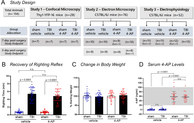
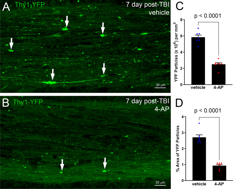
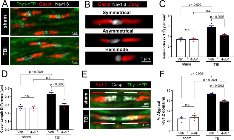
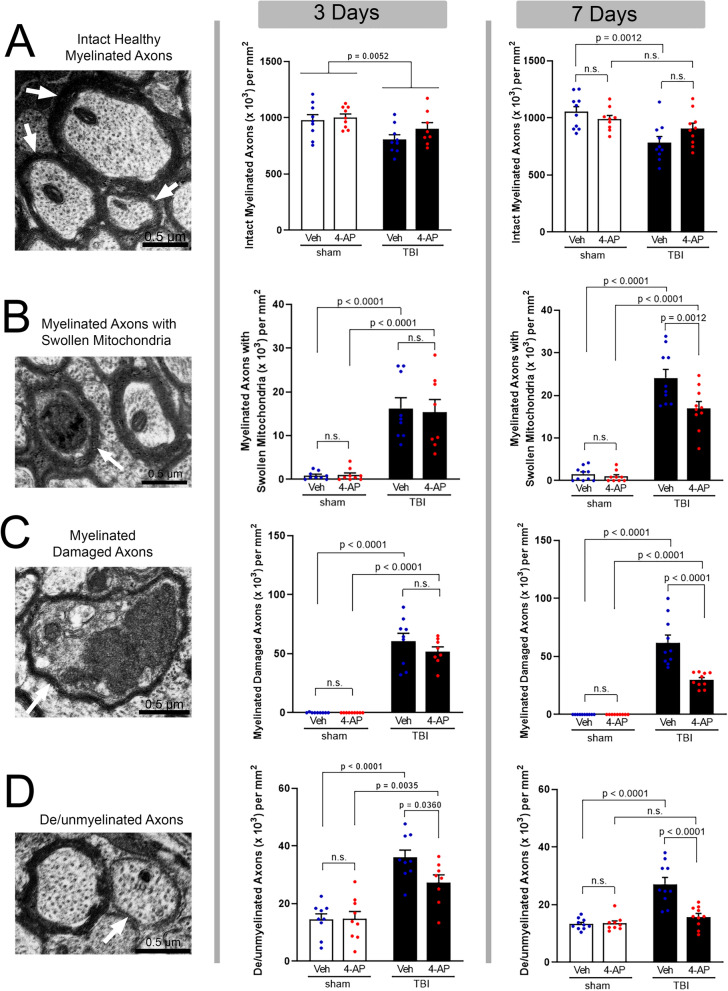
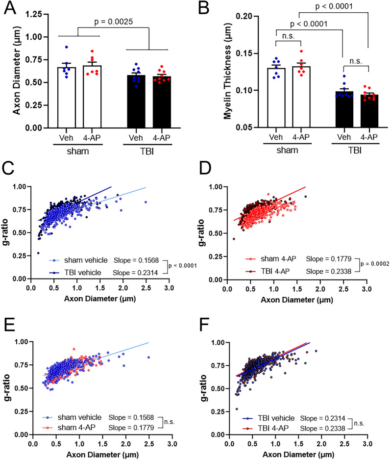
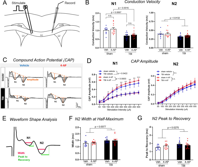
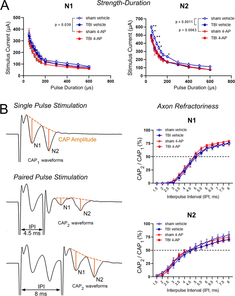
Damage to long axons in white matter tracts is a major pathology in closed head traumatic brain injury (TBI). Acute TBI treatments are needed that protect against axon damage and promote recovery of axon function to prevent long term symptoms and neurodegeneration. Our prior characterization of axon damage and demyelination after TBI led us to examine repurposing of 4-aminopyridine (4-AP), an FDA-approved inhibitor of voltage-gated potassium (Kv) channels. 4-AP is currently indicated to provide symptomatic relief for patients with chronic stage multiple sclerosis, which involves axon damage and demyelination. We tested clinically relevant dosage of 4-AP as an acute treatment for experimental TBI and found multiple benefits in corpus callosum axons. This randomized, controlled pre-clinical study focused on the first week after TBI, when axons are particularly vulnerable. 4-AP treatment initiated one day post-injury dramatically reduced axon damage detected by intra-axonal fluorescence accumulations in Thy1-YFP mice of both sexes. Detailed electron microscopy in C57BL/6 mice showed that 4-AP reduced pathological features of mitochondrial swelling, cytoskeletal disruption, and demyelination at 7 days post-injury. Furthermore, 4-AP improved the molecular organization of axon nodal regions by restoring disrupted paranode domains and reducing Kv1.2 channel dispersion. 4-AP treatment did not resolve deficits in action potential conduction across the corpus callosum, based on ex vivo electrophysiological recordings at 7 days post-TBI. Thus, this first study of 4-AP effects on axon damage in the acute period demonstrates a significant decrease in multiple pathological hallmarks of axon damage after experimental TBI.

Keywords: 4-aminopyridine; Demyelination; Electrophysiology; Kv1.2; Traumatic brain injury; White matter.


Conflict of interest statement

The authors declare no competing financial interests.

Figures

Fig. 1
Fig. 1
Experimental study design and key assessments of group allocation. A Graphical representation of experimental timeline and treatment groups. Three sets of experiments were conducted according to pre-determined study designs. For each study, the strain and number of mice used is shown along with the study endpoint and method of analysis of corpus callosum axons. Excl. = number of mice excluded based on pre-determined criteria (see Materials and Methods for definition). B TBI increased the time to right from supine to prone position immediately after surgery (righting reflex) as compared with respective sham groups. This confirms that mice randomized for allocation to the TBI vehicle and TBI 4-AP groups exhibited similar righting times prior to initiating 4-AP treatment. C 4-AP treatment did not cause a significant change in body weight over the period of the experiment. D 4-AP serum drug level concentrations are within the clinical therapeutic target range (20–100 ng/ml) in cardiac blood collected approximately 30–60 min after last 4-AP injection. Graphs show the mean ± SEM with each mouse as an individual data point. One-way ANOVA with Tukey’s multiple comparisons test: B F3,145 = 211.3, p < 0.0001, C F3,145 = 1.057, p = 0.3692. Two-way ANOVA with Holm-Sidak post hoc test: D Interaction: F1,185 = 0.1281, p = 0.7213; Injury F1,185 = 0.0183, p = 0.8927; Drug; F1,185 = 332.8, p < 0.0001
Fig. 2
Fig. 2
4-AP treatment shows potent axon protection using YFP-filled swellings to screen for damaged axons. A–B Thy1-YFP-16 mice were given vehicle (A) or 4-AP (B) treatment on days 1–7 after TBI. Confocal images of axons in the corpus callosum illustrate YFP (green) fluorescence accumulation in axonal swellings (white arrows), which is a sensitive indicator of axon damage. Adjacent YFP-positive axons have YFP diffusely distributed along normal appearing axons. C–D ImageJ particle analysis of YFP accumulation in axonal swellings. 4-AP treatment reduced the density (C) and the percent area occupied by axon swellings (D). Bars represent mean ± SEM with an individual data point shown for each mouse. Unpaired Student’s t-tests. See Fig. 1 for mouse sample numbers and Table 1 for statistical details
Fig. 3
Fig. 3
4-AP treatment improves the molecular organization of excitable axonal domains that are disrupted by TBI. Confocal imaging analysis of node of Ranvier complexes in individual corpus callosum axons of Thy1-YFP-16 mice at 7 days post-TBI or sham procedures. (A) Immunostaining along YFP-labeled axons (green) detected clustering of voltage-gated sodium channels (Nav1.6, white) at the node and Caspr (red) in the paranode region, where myelin attaches to the axon. B-D TBI disrupted paranode domain organization, which was normalized by 4-AP treatment. (B) “Symmetrical” Caspr paranodes exhibit paired Caspr bands of approximately symmetrical length, while “Asymmetrical” paranodes exhibit uneven Caspr domain lengths. Single Caspr paranodes with a missing domain counterpart on the opposite side of a node were classified as “Heminodes”. C TBI increased the number of Caspr heminodes while acute 4-AP treatment post-TBI resulted in heminode numbers similar to sham levels. D TBI increased the asymmetry of Caspr domains in TBI mice compared to sham. 4-AP treatment significantly improved paranode organization following TBI. E–F Kv1.2 channel (red) and Caspr (white) immunostaining along individual YFP (green) axons in single confocal optical slices. E In injured mice, Kv1.2 channels mislocalize from the juxtaparanode domain into Caspr-labeled paranode domain. F Quantification of atypical Kv1.2 domains that overlapped with Caspr domains and/or were asymmetrical in length. Acute 4-AP treatment reduced the percentage of atypical Kv1.2 domains after TBI, but the distribution patterns of Kv1.2 channels was not fully normalized to sham levels. C, D, F Bars represent mean ± SEM with an individual data point shown for each mouse. Two-way ANOVA for main effect of injury or drug with Holm-Sidak’s multiple comparisons test for significance of post-hoc pair effects. See Fig. 1 for mouse sample numbers and Table 1 for statistical details
Fig. 4
Fig. 4
4-AP benefit requires continued treatment during the first week post-TBI to protect against multiple features of axon damage. Electron microscopy imaging of axon and myelin ultrastructural features (left) with classification of axon pathology at 3 days (middle) or 7 days (right) after TBI or sham procedure. Left panels: Examples of axon and myelin ultrastructural features. A Intact healthy myelinated axons (arrows). B Swollen mitochondrion (i.e., occupying over 50% of the axon cross section; arrow) in contrast to the typical mitochondrion in the adjacent axon. C Damaged myelinated axon (arrow) exhibiting condensed cytoplasm and vesicle/organelle accumulation. D Demyelinated axon (arrow) lacking a myelin sheath but structurally intact with diameter > 0.3 µm, which is typically myelinated. Middle Panels: At the 3-day time point, TBI significantly reduced the number of intact myelinated axons (A; only TBI main effect significant). TBI significantly increased myelinated axons with swollen mitochondria (B) or axon damage (C), and induced demyelination of additional axons (D) in both vehicle and 4-AP groups at 3 days. 4-AP treatment on days 1–3 did not have a significant benefit as compared to vehicle (B) or the number of damaged (C) and demyelinated (D) axons post-TBI. Right Panels: With 4-AP treatment extended to 7 days, the number of intact myelinated axons post-TBI was statistically similar to sham levels (A). More specifically, 4-AP treatment significantly reduced axons exhibiting mitochondrial swelling (B), axon damage (C), or TBI-induced demyelination (D). AD Bars represent mean ± SEM with an individual data point shown for each mouse. Two-way ANOVA for main effects of injury or drug with Holm-Sidak’s multiple comparisons test of significance for post-hoc pair effects. See Fig. 1 for mouse sample numbers and Table 2 for statistical details
Fig. 5
Fig. 5
TBI reduces axon diameter and myelin thickness in 4-AP and vehicle conditions. Morphological analysis of intact myelinated axons in the corpus callosum of mice treated with 4-AP or vehicle on days 1–7 post-TBI or sham condition. A A main effect of TBI results in atrophic axons regardless of vehicle or 4-AP treatment. B Intact axons have thinner myelin after TBI in both vehicle and 4-AP treated mice. CF Scatter plots display axon diameter against g-ratio (inner axonal diameter divided by total outer diameter), since appropriate myelin thickness is related to the diameter of a given axon. The slope of the linear regression was steeper for TBI versus sham mice. This indicates myelin thinning in proportion to axon diameter after TBI. This relationship was not influenced by 4-AP treatment as compared to vehicle (E, sham p = 0.1362; F, TBI p = 0.8818). AB Two-way ANOVA for main effect of injury or drug with Holm-Sidak’s multiple comparisons test of significance for post-hoc pair effects. Bars represent mean ± SEM with an individual data point shown for each mouse. CF Linear regression analysis. Each circle represents a measured axon (50 axons/mouse). See Fig. 1 for mouse sample numbers and Table 3 for statistical details
Fig. 6
Fig. 6
TBI slows the axon compound action potential velocity and amplitude. Electrophysiology recordings were used to directly test the function of axons in the corpus callosum. A Schematic of the position of the stimulating and recording electrodes for measurements of axon compound action potentials (CAP) and conduction velocity in ex vivo brain slices. B TBI reduced the speed of action potential conduction in both faster N1 wave comprised of myelinated axons and slower N2 wave comprised of unmyelinated and potentially demyelinated axons. The 7-day 4-AP treatment regimen did not restore this velocity deficit. C Representative input–output traces show the evoked N1 and N2 CAP waveforms with stimulus intensity pulses ranging from 50 to 500 µA (50 µA increments). Orange lines from CAP peaks to their projected bases indicate the amplitude of the response of fibers at a given stimulus intensity. D CAP amplitude analysis revealed a main injury effect on myelinated axons based on the reduced amplitude of only the N1 wave. N1 CAP amplitude difference was significant with post-hoc comparison for the sham versus TBI mice with 4-AP treatment. E Schematic of complementary spike waveform parameters. The width is dependent on the conduction velocity distribution among contributing axons. The time from peak to recovery represents the time for membrane repolarization among the slowest conducting axons comprising each waveform. The CAP width (F) and time from peak to recovery (G) indicated that TBI prolonged the recovery of the N2 waveform in both vehicle and 4-AP conditions. The N1 wave shape parameters were not altered by TBI or 4-AP treatment (data not shown). Abbreviations: Cing, cingulum; CC, corpus callosum; LV, lateral ventricles. B, F, G Bars represent mean ± SEM with individual data points for each mouse (n = 11–13 animals per group). D Linear effects model statistical analysis and Holm-Sidak’s multiple comparison test (*p < 0.05, **p < 0.01, ***p < 0.001). Two-way ANOVA for main effect of injury or drug with Holm-Sidak’s multiple comparisons test for post-hoc comparison of pair effects. See Fig. 1 for mouse sample numbers and Table 4 for statistical details
Fig. 7
Fig. 7
TBI alters the axon population excitability without changing the refractory period. A subset of mice had extended studies of additional conduction properties from the ex vivo brain slice preparations as shown in Fig. 6. A The axon intrinsic excitability was probed by measuring the stimulus duration relative to increasing stimulus current. In both N1 and N2 axons, 4-AP treatment shifted the strength-duration curve toward a more excitable state in sham mice, based on eliciting a similar response with a lower stimulus strength at a given stimulus duration. This strength-duration analysis also revealed that TBI induced hyperexcitability of N2 axons in the vehicle condition. The N2 axon hyperexcitability after TBI was similar in the 4-AP and vehicle conditions. B The axon refractory period due to recovery time between action potentials was probed with a paired-pulse protocol. Representative N1 and N2 compound action potential (CAP) waveforms from a single pulse stimulation as compared to a paired-pulse protocol with varying interpulse intervals (IPI). Orange lines from CAP peaks to their projected bases indicate the amplitude of each waveform. Plots of the percent ratio of each second pulse CAP amplitude (CAP2), evoked in the paired-pulse stimulation, divided by the CAP amplitude of the single reference pulse stimulation (CAP1) showed no differences in refractoriness between sham and TBI animals or between vehicle and 4-AP treatments. A–B Linear effects model statistical analysis and Holm-Sidak’s multiple comparison test (*p < 0.05, **p < 0.01). Data is expressed as mean ± SEM with n = 6–7 animals per group

Similar articles

Cited by

References

    1. Agren R, Nilsson J, Arhem P. Closed and open state dependent block of potassium channels cause opposing effects on excitability - a computational approach. Sci Rep. 2019;9:8175. doi: 10.1038/s41598-019-44564-x. - DOI - PMC - PubMed
    1. Andravizou A, Dardiotis E, Artemiadis A, Sokratous M, Siokas V, Tsouris Z, Aloizou AM, Nikolaidis I, Bakirtzis C, Tsivgoulis G, et al. Brain atrophy in multiple sclerosis: mechanisms, clinical relevance and treatment options. Auto Immun Highlights. 2019;10:7. doi: 10.1186/s13317-019-0117-5. - DOI - PMC - PubMed
    1. Balan IS, Saladino AJ, Aarabi B, Castellani RJ, Wade C, Stein DM, Eisenberg HM, Chen HH, Fiskum G. Cellular alterations in human traumatic brain injury: changes in mitochondrial morphology reflect regional levels of injury severity. J Neurotrauma. 2013;30:367–381. doi: 10.1089/neu.2012.2339. - DOI - PMC - PubMed
    1. Bei F, Lee HHC, Liu X, Gunner G, Jin H, Ma L, Wang C, Hou L, Hensch TK, Frank E, et al. Restoration of visual function by enhancing conduction in regenerated axons. Cell. 2016;164:219–232. doi: 10.1016/j.cell.2015.11.036. - DOI - PMC - PubMed
    1. Boscia F, Elkjaer ML, Illes Z, Kukley M. Altered expression of ion channels in white matter lesions of progressive multiple sclerosis: what do we know about their function? Front Cell Neurosci. 2021;15:685703. doi: 10.3389/fncel.2021.685703. - DOI - PMC - PubMed

Publication types

Substances