Review
. 2022 May 3;42(1):15.
doi: 10.1186/s41232-022-00199-6.

Neuroplasticity related to chronic pain and its modulation by microglia

Affiliations
Review

Neuroplasticity related to chronic pain and its modulation by microglia

Shin-Ichiro Hiraga et al. Inflamm Regen. .

Abstract

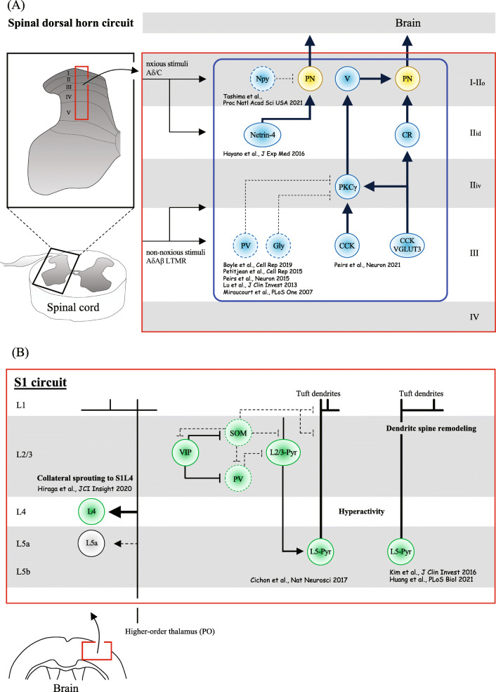
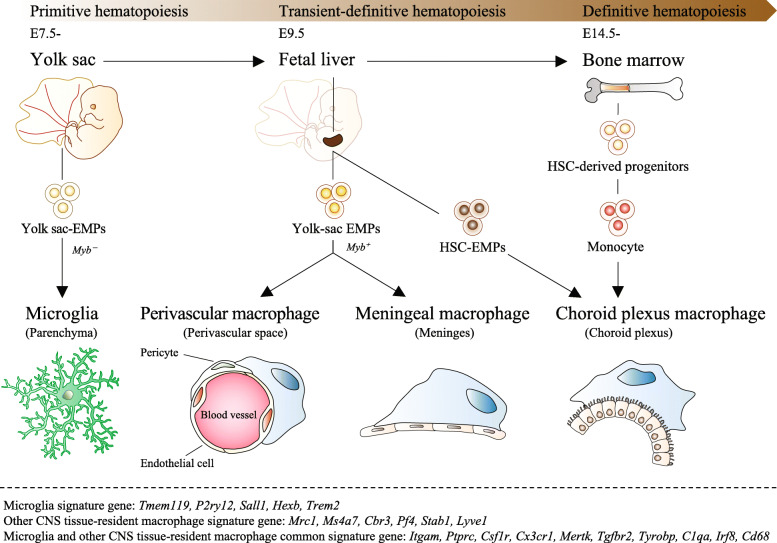
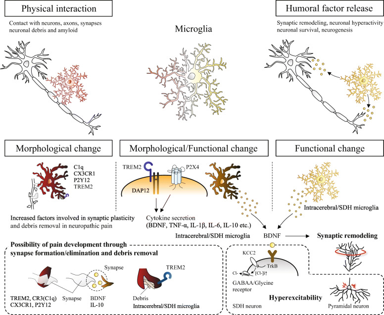
Neuropathic pain is often chronic and can persist after overt tissue damage heals, suggesting that its underlying mechanism involves the alteration of neuronal function. Such an alteration can be a direct consequence of nerve damage or a result of neuroplasticity secondary to the damage to tissues or to neurons. Recent studies have shown that neuroplasticity is linked to causing neuropathic pain in response to nerve damage, which may occur adjacent to or remotely from the site of injury. Furthermore, studies have revealed that neuroplasticity relevant to chronic pain is modulated by microglia, resident immune cells of the central nervous system (CNS). Microglia may directly contribute to synaptic remodeling and altering pain circuits, or indirectly contribute to neuroplasticity through property changes, including the secretion of growth factors. We herein highlight the mechanisms underlying neuroplasticity that occur in the somatosensory circuit of the spinal dorsal horn, thalamus, and cortex associated with chronic pain following injury to the peripheral nervous system (PNS) or CNS. We also discuss the dynamic functions of microglia in shaping neuroplasticity related to chronic pain. We suggest further understanding of post-injury ectopic plasticity in the somatosensory circuits may shed light on the differential mechanisms underlying nociceptive, neuropathic, and nociplastic-type pain. While one of the prominent roles played by microglia appears to be the modulation of post-injury neuroplasticity. Therefore, future molecular- or genetics-based studies that address microglia-mediated post-injury neuroplasticity may contribute to the development of novel therapies for chronic pain.

Keywords: CNS injury; Microglia; Neuroplasticity; Nociplastic pain; PNS injury; Thalamic hemorrhage.


Conflict of interest statement

The authors declare that they have no competing interests.

Figures

Fig. 1
Fig. 1
The spinal dorsal horn and S1 neuroplasticity mechanism inducing chronic pain following PNS or CNS injury. A Synaptic modifications in the dorsal horn of the spinal cord result in a balance alteration of excitatory and inhibitory synaptic transmission in lamina I projection neurons to the brain, which account at least partially for pain sensation evoked by non-noxious stimuli. The continuous lines and arrows indicate allodynia-relevant circuits while dotted lines indicate neuronal activity suppression or synaptic loss in the cause of enhanced pathology. Arrowheads indicate the direction of excitatory inputs. PV, parvalbumin-expressing neurons; SOM, somatostatin-expressing neurons; CCK, cholecystokinin-expressing neurons; VGLUT3, vesicular glutamate transporter 3-expressing neurons; PKCγ, protein kinase C gamma-expressing neurons; CR, calretinin-expressing neurons; Gly, glycine-expressing neurons; Netrin-4, netrin-4-expressing neurons; Npy, neuropeptide Y inhibitory interneurons; PN, projection neuron; LTMR, low-threshold mechanoreceptors; V, vertical cells; IIo, outer lamina II, IIi, inner lamina II; IIid, inner lamina II (dorsal); IIiv, inner lamina II (ventral); Aβ, Aβ myelinated fibers; Aδ, Aδ myelinated fibers; C, C fibers. B Axonal sprouting and circuit functional reorganization of S1 involving both intracortical neurons and projection neurons are at least partially responsible for inducing pain by non-noxious stimuli. The continuous lines and arrows indicate allodynia-relevant circuits, and dotted lines indicate suppressed neuronal activity or synaptic losses in the cause of enhanced pathology. Arrowheads indicate the direction of excitatory inputs. White circle (L5a) indicates decreased neuronal hyperactivity after injury. L, layer; Pyr, pyramidal neurons; PV, parvalbumin-expressing neurons; SOM, somatostatin-expressing interneurons; VIP, vasoactive intestinal polypeptide-expressing interneurons; PO, neurons from the thalamic posterior nucleus
Fig. 2
Fig. 2
The origin of tissue-resident macrophages (microglia, perivascular/ meningeal/ choroid plexus macrophage) in the CNS. Tissue-resident macrophages of the CNS in mammals are derived from three consecutive hematopoietic systems (primitive hematopoiesis, transient-definitive hematopoiesis, definitive hematopoiesis) during embryogenesis. In mouse primitive hematopoiesis, erythro-myeloid progenitors (EMPs) occur in the yolk sac of extraembryonic tissue during the embryonic period (E7-7.5), and primitive macrophages are produced from these EMPs independently of the transcription factor Myb without going through monocytes. Primitive macrophages are then carried to each organ by the circulatory system and engrafted before the formation of the blood-brain barrier. Microglia in the parenchyma of the brain and spinal cord differentiate from these primitive macrophages. Around E9.5, yolk sac-derived EMPs migrate from the yolk sac to the fetal liver and initiate transient-definitive hematopoiesis. Then, in transient-definitive hematopoiesis, monocytes are produced from EMPs in a Myb-dependent manner, and the primitive macrophages previously engrafted in each organ are replaced. Since the blood-brain barrier is formed in the brain and infiltration of these monocytes rarely occurs, only microglia present in the parenchyma of the brain and spinal cord are the main origins of primitive macrophages derived in the yolk sac. Around E10.5, hematopoietic stem cells (HSCs) migrate to the fetal liver to start definitive hematopoiesis. HSCs migrate from the fetal liver to the bone marrow before birth and supply the entire line of blood cells throughout life. Choroid plexus macrophages derive from HSC-EMP or blood monocytes. Below the dotted line, microglia and CNS tissue-resident macrophages, and common gene expression patterns are shown
Fig. 3
Fig. 3
Microglial modulation of neuroplasticity in the spinal dorsal horn and S1 that underlies chronic pain after PNS or CNS injury. TOP: the physical interaction and humoral factor release. BOTTOM: morphological and functional changes in microglia after PNS or CNS injury. Studies showing morphological changes in microglia have not shown specific functions, but have suggested their involvements in the pain development based on the molecular functions. Functional changes in microglia have been suggested to be factors released via TREM2/DAP12 or P2X4 signals. BDNF is one factor shown to be secreted from microglia in the spinal cord and brain after PNS or CNS injury, involved in pain development through synaptic remodeling and inducing hyperexcitability

Similar articles

Cited by

References

    1. Jensen TS, Baron R, Haanpaa M, Kalso E, Loeser JD, Rice ASC, et al. A new definition of neuropathic pain. Pain. 2011;152(10):2204–2205. doi: 10.1016/j.pain.2011.06.017. - DOI - PubMed
    1. Fitzcharles MA, Cohen SP, Clauw DJ, Littlejohn G, Usui C, Hauser W. Nociplastic pain: towards an understanding of prevalent pain conditions. Lancet. 2021;397(10289):2098–2110. doi: 10.1016/S0140-6736(21)00392-5. - DOI - PubMed
    1. Chen G, Zhang YQ, Qadri YJ, Serhan CN, Ji RR. Microglia in pain: detrimental and protective roles in pathogenesis and resolution of pain. Neuron. 2018;100(6):1292–1311. doi: 10.1016/j.neuron.2018.11.009. - DOI - PMC - PubMed
    1. Inoue K, Tsuda M. Microglia in neuropathic pain: cellular and molecular mechanisms and therapeutic potential. Nat Rev Neurosci. 2018;19(3):138–152. doi: 10.1038/nrn.2018.2. - DOI - PubMed
    1. Hiraga SI, Itokazu T, Hoshiko M, Takaya H, Nishibe M, Yamashita T. Microglial depletion under thalamic hemorrhage ameliorates mechanical allodynia and suppresses aberrant axonal sprouting. JCI Insight. 2020;5(3). 10.1172/jci.insight.131801. - PMC - PubMed

LinkOut - more resources