

Incredible affinity of Kattosh with PPAR-γ receptors attenuates STZ-induced pancreas and kidney lesions evidenced in chemicobiological interactions
- PMID: 35502486
- PMCID: PMC9189352
- DOI: 10.1111/jcmm.17339
Incredible affinity of Kattosh with PPAR-γ receptors attenuates STZ-induced pancreas and kidney lesions evidenced in chemicobiological interactions
Abstract
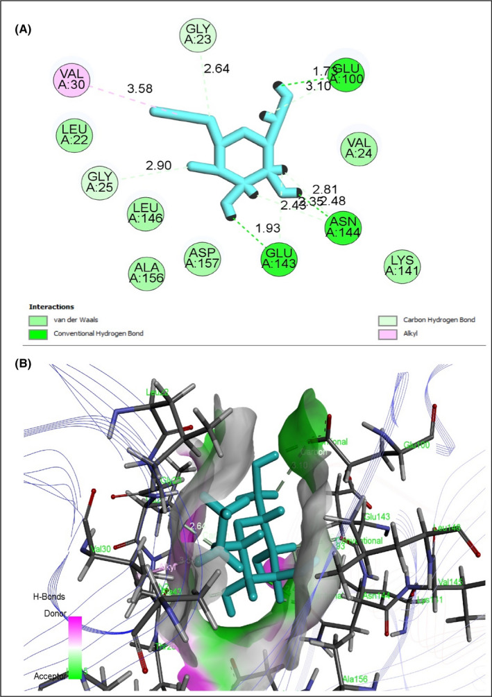
Since ancient times, plants have been used as green bioresources to ensure a healthier life by recovering from different diseases. Kattosh (Lasia spinosa L. Thwaites) is a local plant with various traditional uses, especially for arthritis, constipation and coughs. This research investigated the effect of Kattosh stem extract (LSES) on streptozotocin-induced damage to the pancreas, kidney, and liver using in vitro, in vivo and in silico methods. In vitro phytochemical, antioxidative and anti-inflammatory effects of LSES were accomplished by established methods followed by antidiabetic actions in in vivo randomized controlled intervention in STZ-induced animal models for four weeks. In an in silico study, LSES phytocompounds interacted with antidiabetic receptors of peroxisome proliferator-activated receptor-gamma (PPAR, PDB ID: 3G9E), AMP-activated protein kinase (AMPK, PDB ID: 4CFH) and α-amylase enzyme (PDB ID: 1PPI) to verify the in vivo results. In addition, LSES showed promising in vitro antioxidative and anti-inflammatory effects. In contrast, it showed a decrease in weekly blood glucose level, normalized lipid profile, ameliorated liver and cardiac markers, managed serum AST and ALT levels, and increased glucose tolerance ability in the animal model study. Restoration of pancreatic and kidney damage was reflected by improving histopathological images. In ligand-receptor interaction, ethyl α-d-glucopyranoside of Kattosh showed the highest affinity for the α-amylase enzyme, PPAR, and AMPK receptors. Results demonstrate that the affinity of Kattosh phytocompounds potentially attenuates pancreatic and kidney lesions and could be approached as an alternative antidiabetic source with further clarification.
Keywords: Lasia spinosa; diabetes mellitus; ethyl alpha-D-glucopyranoside; histopathological examination; phytochemical screening; α-amylase.
© 2022 The Authors. Journal of Cellular and Molecular Medicine published by Foundation for Cellular and Molecular Medicine and John Wiley & Sons Ltd.
Conflict of interest statement
The authors declare that they have no conflict of interest.
Figures


Similar articles
-
Natural Compounds of Lasia spinosa (L.) Stem Potentiate Antidiabetic Actions by Regulating Diabetes and Diabetes-Related Biochemical and Cellular Indexes.Pharmaceuticals (Basel). 2022 Nov 25;15(12):1466. doi: 10.3390/ph15121466. Pharmaceuticals (Basel). 2022. PMID: 36558918 Free PMC article.
-
Homalium zeylanicum attenuates streptozotocin-induced hyperglycemia and cellular stress in experimental rats via attenuation of oxidative stress imparts inflammation.J Ethnopharmacol. 2022 Jan 30;283:114649. doi: 10.1016/j.jep.2021.114649. Epub 2021 Sep 16. J Ethnopharmacol. 2022. PMID: 34536517
-
Nephroprotective effect of Combretum micranthum G. Don in nicotinamide-streptozotocin induced diabetic nephropathy in rats: In-vivo and in-silico experiments.J Ethnopharmacol. 2020 Oct 28;261:113133. doi: 10.1016/j.jep.2020.113133. Epub 2020 Jul 14. J Ethnopharmacol. 2020. PMID: 32673708
-
The methanolic extract of Thymus praecox subsp. skorpilii var. skorpilii restores glucose homeostasis, ameliorates insulin resistance and improves pancreatic β-cell function on streptozotocin/nicotinamide-induced type 2 diabetic rats.J Ethnopharmacol. 2019 Mar 1;231:29-38. doi: 10.1016/j.jep.2018.10.028. Epub 2018 Nov 3. J Ethnopharmacol. 2019. PMID: 30399410
-
Hepatorenal protective efficacy of flavonoids from Ocimum basilicum extract in diabetic albino rats: A focus on hypoglycemic, antioxidant, anti-inflammatory and anti-apoptotic activities.Biomed Pharmacother. 2021 Dec;144:112287. doi: 10.1016/j.biopha.2021.112287. Epub 2021 Oct 12. Biomed Pharmacother. 2021. PMID: 34649220
Cited by
-
Pharmacological insights into Chukrasia velutina bark: Experimental and computer-aided approaches.Animal Model Exp Med. 2022 Dec;5(4):377-388. doi: 10.1002/ame2.12268. Animal Model Exp Med. 2022. PMID: 36047481 Free PMC article.
-
Natural Compounds of Lasia spinosa (L.) Stem Potentiate Antidiabetic Actions by Regulating Diabetes and Diabetes-Related Biochemical and Cellular Indexes.Pharmaceuticals (Basel). 2022 Nov 25;15(12):1466. doi: 10.3390/ph15121466. Pharmaceuticals (Basel). 2022. PMID: 36558918 Free PMC article.
-
Upregulation of Antioxidative Gene Expression by Lasia spinosa Organic Extract Improves the Predisposing Biomarkers and Tissue Architectures in Streptozotocin-Induced Diabetic Models of Long Evans Rats.Antioxidants (Basel). 2022 Dec 2;11(12):2398. doi: 10.3390/antiox11122398. Antioxidants (Basel). 2022. PMID: 36552606 Free PMC article.
-
Apoptosis-inducing anti-proliferative and quantitative phytochemical profiling with in silico study of antioxidant-rich Leea aequata L. leaves.Heliyon. 2023 Dec 10;10(1):e23400. doi: 10.1016/j.heliyon.2023.e23400. eCollection 2024 Jan 15. Heliyon. 2023. PMID: 38170014 Free PMC article.
-
Isoflavone Protects the Renal Tissue of Diabetic Ovariectomized Rats via PPARγ.Nutrients. 2022 Jun 21;14(13):2567. doi: 10.3390/nu14132567. Nutrients. 2022. PMID: 35807748 Free PMC article.
References
-
- Thomas M, Tsalamandris C, Macisaac R, Jerums G. Anaemia in diabetes: an emerging complication of microvascular disease. Curr Diabetes Rev. 2005;1:107–126. - PubMed
-
- Saeedi P, Petersohn I, Salpea P, et al. Global and regional diabetes prevalence estimates for 2019 and projections for 2030 and 2045: Results from the International Diabetes Federation Diabetes Atlas. Diabetes Res Clin Pract. 2019;157: 107843. - PubMed
-
- Kumar S, Mittal A, Babu D, Mittal A. Herbal medicines for diabetes management and its secondary complications. Curr Diabetes Rev. 2021;17:437–456. - PubMed
Publication types
MeSH terms
Substances
Grants and funding
LinkOut - more resources
Full Text Sources
