

Immune cells in cardiac repair and regeneration
- PMID: 35502777
- PMCID: PMC9124571
- DOI: 10.1242/dev.199906
Immune cells in cardiac repair and regeneration
Abstract
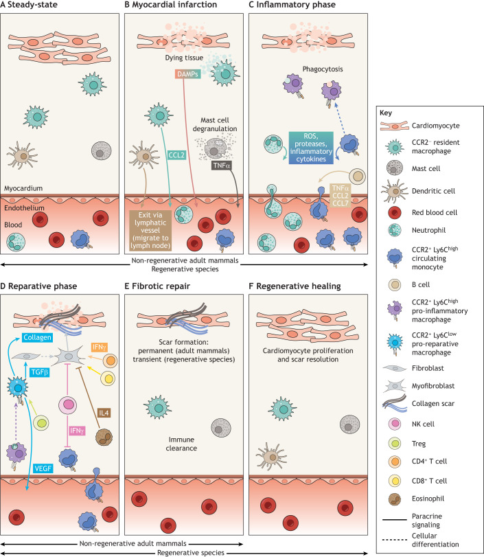
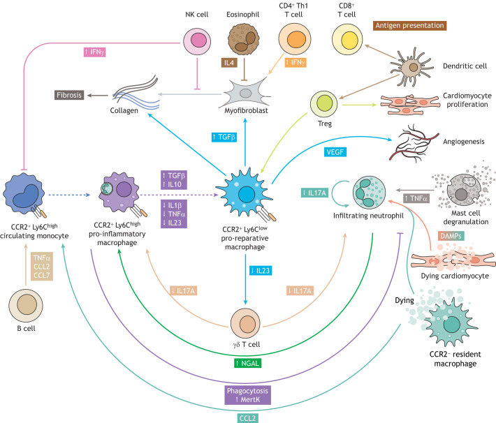
The immune system is fundamental to tissue homeostasis and is the first line of defense following infection, injury or disease. In the damaged heart, large numbers of immune cells are recruited to the site of injury. These cells play an integral part in both repair by scar formation and the initiation of tissue regeneration. They initially assume inflammatory phenotypes, releasing pro-inflammatory cytokines and removing dead and dying tissue, before entering a reparative stage, replacing dead muscle tissue with a non-contractile scar. In this Review, we present an overview of the innate and adaptive immune response to heart injury. We explore the kinetics of immune cell mobilization following cardiac injury and how the different innate and adaptive immune cells interact with one another and with the damaged tissue. We draw on key findings from regenerative models, providing insight into how to support a robust immune response permissible for cardiac regeneration. Finally, we consider how the latest technological developments can offer opportunities for a deeper and unbiased functional understanding of the immune response to heart disease, highlighting the importance of such knowledge as the basis for promoting regeneration following cardiac injury in human patients.
Keywords: Cardiac; Immune system; Muscle; Regeneration.
© 2022. Published by The Company of Biologists Ltd.
Conflict of interest statement
Competing interests P.R.R. is cofounder of and equity holder in OxStem Cardio. F.C.S. declares no competing or financial interests.
Figures
Similar articles
-
The immune system and cardiac repair.Pharmacol Res. 2008 Aug;58(2):88-111. doi: 10.1016/j.phrs.2008.06.007. Epub 2008 Jun 24. Pharmacol Res. 2008. PMID: 18620057 Free PMC article. Review.
-
The neonate versus adult mammalian immune system in cardiac repair and regeneration.Biochim Biophys Acta. 2016 Jul;1863(7 Pt B):1813-21. doi: 10.1016/j.bbamcr.2016.01.011. Epub 2016 Jan 20. Biochim Biophys Acta. 2016. PMID: 26801961 Review.
-
Specific macrophage populations promote both cardiac scar deposition and subsequent resolution in adult zebrafish.Cardiovasc Res. 2020 Jun 1;116(7):1357-1371. doi: 10.1093/cvr/cvz221. Cardiovasc Res. 2020. PMID: 31566660 Free PMC article.
-
The Innate Immune Response in Myocardial Infarction, Repair, and Regeneration.Adv Exp Med Biol. 2017;1003:251-272. doi: 10.1007/978-3-319-57613-8_12. Adv Exp Med Biol. 2017. PMID: 28667562 Review.
-
Role of innate and adaptive immune mechanisms in cardiac injury and repair.Nat Rev Immunol. 2015 Feb;15(2):117-29. doi: 10.1038/nri3800. Nat Rev Immunol. 2015. PMID: 25614321 Free PMC article. Review.
Cited by
-
Development of a hepatic cryoinjury model to study liver regeneration.Development. 2024 Aug 1;151(15):dev203124. doi: 10.1242/dev.203124. Epub 2024 Jul 31. Development. 2024. PMID: 38975841 Free PMC article.
-
Regenerative neurogenesis: the integration of developmental, physiological and immune signals.Development. 2022 Apr 15;149(8):dev199907. doi: 10.1242/dev.199907. Epub 2022 May 3. Development. 2022. PMID: 35502778 Free PMC article.
-
Turning gray selenium and sublimed sulfur into a nanocomposite to accelerate tissue regeneration by isothermal recrystallization.J Nanobiotechnology. 2023 Feb 21;21(1):57. doi: 10.1186/s12951-023-01796-4. J Nanobiotechnology. 2023. PMID: 36803772 Free PMC article.
-
Antigen presentation plays positive roles in the regenerative response to cardiac injury in zebrafish.Nat Commun. 2024 Apr 29;15(1):3637. doi: 10.1038/s41467-024-47430-1. Nat Commun. 2024. PMID: 38684665 Free PMC article.
-
Animal models to study cardiac regeneration.Nat Rev Cardiol. 2024 Feb;21(2):89-105. doi: 10.1038/s41569-023-00914-x. Epub 2023 Aug 14. Nat Rev Cardiol. 2024. PMID: 37580429 Review.
References
-
- Abbate, A., Kontos, M. C., Grizzard, J. D., Biondi-Zoccai, G. G., Van Tassell, B. W., Robati, R., Roach, L. M., Arena, R. A., Roberts, C. S., Varma, A.et al. (2010). Interleukin-1 blockade with anakinra to prevent adverse cardiac remodeling after acute myocardial infarction (Virginia Commonwealth University Anakinra Remodeling Trial [VCU-ART] Pilot study). Am. J. Cardiol. 105, 1371-1377. 10.1016/j.amjcard.2009.12.059 - DOI - PubMed
-
- Adamo, L., Staloch, L. J., Rocha-Resende, C., Matkovich, S. J., Jiang, W., Bajpai, G., Weinheimer, C. J., Kovacs, A., Schilling, J. D., Barger, P. M.et al. (2018). Modulation of subsets of cardiac B lymphocytes improves cardiac function after acute injury. JCI Insight 3, e120137. 10.1172/jci.insight.120137 - DOI - PMC - PubMed
-
- Anzai, A., Anzai, T., Nagai, S., Maekawa, Y., Naito, K., Kaneko, H., Sugano, Y., Takahashi, T., Abe, H., Mochizuki, S.et al. (2012). Regulatory role of dendritic cells in postinfarction healing and left ventricular remodeling. Circulation 125, 1234-1245. 10.1161/CIRCULATIONAHA.111.052126 - DOI - PubMed
Publication types
MeSH terms
Grants and funding
LinkOut - more resources
Full Text Sources
Medical
