Review
. 2022 Apr 15;149(8):dev200425.
doi: 10.1242/dev.200425. Epub 2022 May 3.

Microglia in brain development and regeneration

Affiliations
Review

Microglia in brain development and regeneration

Lindsey C Mehl et al. Development. .

Abstract

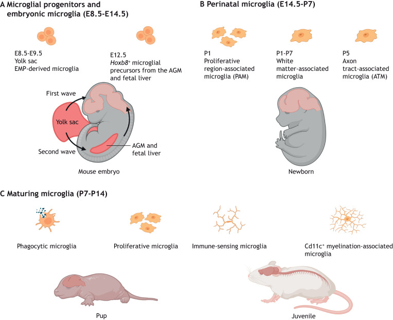
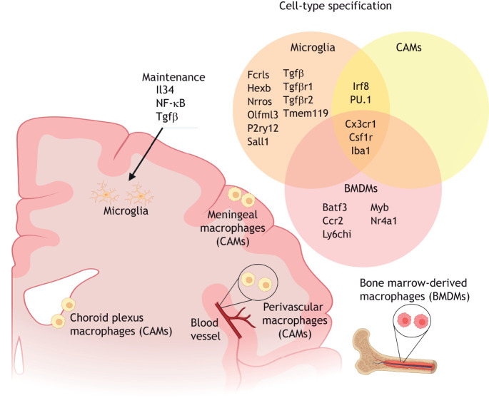
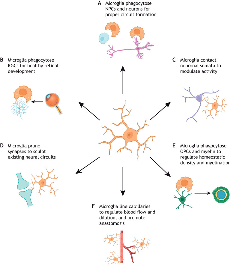
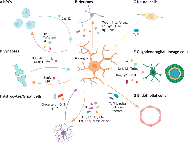
It has recently emerged that microglia, the tissue-resident macrophages of the central nervous system, play significant non-innate immune roles to support the development, maintenance, homeostasis and repair of the brain. Apart from being highly specialized brain phagocytes, microglia modulate the development and functions of neurons and glial cells through both direct and indirect interactions. Thus, recognizing the elements that influence the homeostasis and heterogeneity of microglia in normal brain development is crucial to understanding the mechanisms that lead to early disease pathogenesis of neurodevelopmental disorders. In this Review, we discuss recent studies that have elucidated the physiological development of microglia and summarize our knowledge of their non-innate immune functions in brain development and tissue repair.

Keywords: Brain development; Heterogeneity; Microglia; Neurons; Oligodendrocytes; Synapse.


Conflict of interest statement

Competing interests The authors declare no competing or financial interests.

Figures

Fig. 1.
Fig. 1.
Microglial specification, heterogeneity and functional diversity across stages of CNS development and maturation. (A) During early CNS development, the brain is infiltrated by erythromyeloid progenitors (EMPs) from the yolk sac between E8.5 and E9.5 from the first wave of hematopoiesis. A second wave of yolk sac hematopoiesis generates Hoxb8+ microglial precursors that migrate to the CNS from the aorta-gonad-mesonephros (AGM) and fetal liver from E12.5. (B,C) Proliferative microglial cells are a common feature during the perinatal (E14.5-P7) and early maturation (P7-P14) stages. (B) Specific proliferative microglial clusters, such as the white matter-associated microglia and axon tract-associated microglia, are observed within the first week after birth. (C) Microglial maturation and expansion of the population continue within various brain compartments. Phagocytic microglia, immune-sensing microglia and Cd11c+ myelination-associated microglia can be found in fetal human and juvenile murine brains.
Fig. 2.
Fig. 2.
Microglial identity. Microglia reside in the brain parenchyma and can be distinguished by their expression of the combination of Fcrls, Hexb, Nrros, Olfml3, P2ry12, Sall1, Tgfβ, Tgfβ receptors 1 and 2, and Tmem119. Microglial identity is maintained by signaling through Il34, NF-κB and Tgfβ. In the CNS border compartments, choroid plexus, meningeal and perivascular macrophages, otherwise known as CNS-associated macrophages (CAMs) or border-associated macrophages (BAMs), are distinct from microglia but share the expression of the transcription factors Irf8 and PU.1 in their lineage specification. Bone marrow-derived macrophages (BMDMs), which are not typically localized in the brain, share the common expression of Csf1r and Cx3cr1 with microglia and CAMs, but selectively express Batf3, Ccr2, Ly6chi, Myb and Nr4a1.
Fig. 3.
Fig. 3.
Microglia physically interact with neural cell types to promote healthy brain development. Microglia are capable of modulating brain development through direct contact with various cell types of the CNS. (A) Elimination of NPCs and neurons via phagocytosis is crucial for the formation and function of neuronal circuitry. (B) In particular, microglial phagocytosis of retinal ganglion cells (RGCs) to regulate proper eye development has been well documented. (C) Microglia can also modulate neuronal activity by directly interacting with neuronal somata. (D) Microglia physically interact with neuronal dendrites and routinely prune synapses to direct brain and eye development. (E) Microglia phagocytose oligodendrocyte precursor cells (OPCs) and myelin to regulate homeostatic density and myelination. (F) Microglia bridge endothelial tip cells to promote fusion and vascularization, and capillary-associated microglia are present throughout postnatal life, lining brain capillaries to control blood flow and dilation.
Fig. 4.
Fig. 4.
Microglia secrete cytokines to regulate CNS development. Microglia regulate brain development through the secretion of cytokines that have distinct, complex effects on different neural cell types depending on their cellular, spatial and temporal context. (A) Reactive microglia can release inflammatory cytokines (Il1β, Il6, Tnfα and Ifnγ) that promote neurogenesis in specific brain regions. However, the absence of these cytokines (Il6, type 1 interferons) promotes proper neuronal maturation in other brain regions. Reciprocally, neural precursor cells (NPCs) can recruit microglia to specific brain regions through Cxcl12 secretion. (B) Microglial-derived factors (type 1 interferons, Il6, Igf1, Tnfα, Ngf and superoxide ions) can either support or impair neuronal survival to ensure proper circuit formation in a context-specific manner. (C) CNS-wide disruption of Tgfβ signaling induces dysmature microglia, which impact both neuronal and oligodendroglial cell populations. (D) At the synapse, microglial Il10 and Bdnf regulate synaptic formation and maturation, whereas neuronal Il33, ATP and fractalkine signaling can modulate microglia maturation and function. (E) Inflammatory cytokines (Il1β, Il6, Tnfα and Ifnγ) as well as other factors expressed by microglial subsets (Igf1 and Nrp1) can regulate OPC proliferation, oligodendrogenesis, and myelination in different spatial contexts. (F) Microglia-to-astrocyte crosstalk is crucial to astrocyte development (Lif, Il6, Il1, nitric oxide) and reactivity (Tnf, C1q, Il1α) in both health and disease contexts. Similarly, astrocyte-derived cholesterol, Csf1 and Tgfβ2 are essential for microglial survival. (G) Finally, Tgfβ1 is expressed by microglia to regulate vascularization, but there are likely other microglial-derived factors that promote angiogenesis that remain to be elucidated.

References

    1. Ajami, B., Bennett, J. L., Krieger, C., Tetzlaff, W. and Rossi, F. M. V. (2007). Local self-renewal can sustain CNS microglia maintenance and function throughout adult life. Nat. Neurosci. 10, 1538-1543. 10.1038/nn2014 - DOI - PubMed
    1. Alliot, F., Godin, I. and Pessac, B. (1999). Microglia derive from progenitors, originating from the yolk sac, and which proliferate in the brain. Dev. Brain Res. 117, 145-152. 10.1016/S0165-3806(99)00113-3 - DOI - PubMed
    1. Anderson, S. R., Zhang, J., Steele, M. R., Romero, C. O., Kautzman, A. G., Schafer, D. P. and Vetter, M. L. (2019). Complement targets newborn retinal ganglion cells for phagocytic elimination by microglia. J. Neurosci 39, 2025-2040. 10.1523/JNEUROSCI.1854-18.2018 - DOI - PMC - PubMed
    1. Antony, J. M., Paquin, A., Nutt, S. L., Kaplan, D. R. and Miller, F. D. (2011). Endogenous microglia regulate development of embryonic cortical precursor cells. J. Neurosci. Res. 89, 286-298. 10.1002/jnr.22533 - DOI - PubMed
    1. Arnò, B., Grassivaro, F., Rossi, C., Bergamaschi, A., Castiglioni, V., Furlan, R., Greter, M., Favaro, R., Comi, G., Becher, B.et al. (2014). Neural progenitor cells orchestrate microglia migration and positioning into the developing cortex. Nat. Commun. 5, 5611. 10.1038/ncomms6611 - DOI - PubMed

Publication types