

Immunomodulation of Acellular Dermal Matrix Through Interleukin 4 Enhances Vascular Infiltration
- PMID: 35502953
- PMCID: PMC9289891
- DOI: 10.1097/SAP.0000000000003163
Immunomodulation of Acellular Dermal Matrix Through Interleukin 4 Enhances Vascular Infiltration
Abstract
Background: Acellular dermal matrix (ADM) supported implant-based reconstruction remains the most commonly performed mode of reconstruction after breast cancer. Acellular dermal matrix clinical usage has reported benefits but requires rapid and efficient vascular and cellular incorporation into the recipient to have the best outcomes. Orderly transition from M1 to M2 macrophage phenotypic profile, coordinated in part by interleukin 4 (IL-4), is an important component of vascular stabilization and remodeling. Using the ADM substrate as a delivery device for immunomodulation of macrophage phenotype holds the potential to improve integration.
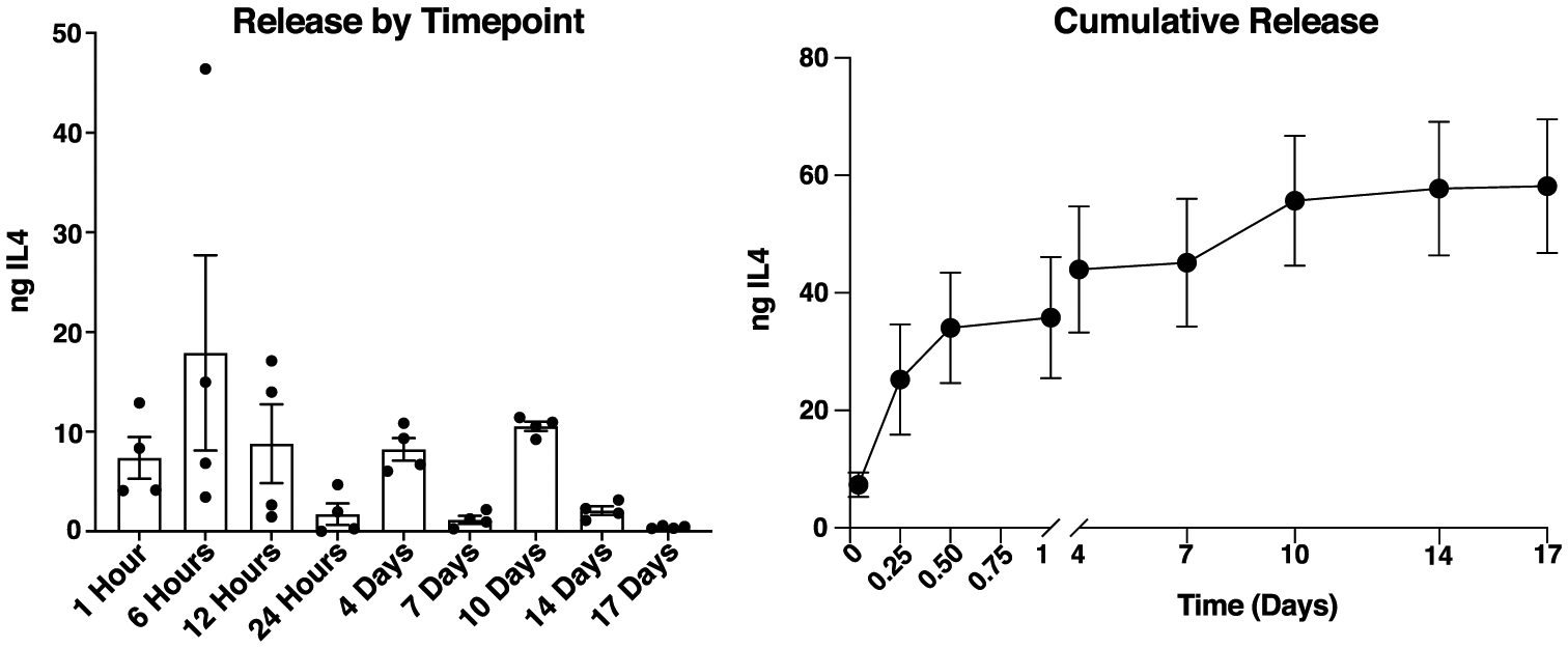
Methods: Interleukin 4 was adsorbed onto ADM samples and drug elution curves were measured. Next, experimental groups of 8 C57BL/6 mice had 5-mm ADM discs surgically placed in a dorsal window chamber with a vascularized skin flap on one side and a plastic cover slip on the other in a model of implant-based breast reconstruction. Group 1 consisted of IL-4 (5 μg) adsorbed into the ADM preoperatively and group 2 consisted of an untreated ADM control. Serial gross examinations were performed with histology at day 21 for markers of vascularization, mesenchymal cell infiltration, and macrophage lineage.
Results: Drug elution curves showed sustained IL-4 release for 10 days after adsorption. Serial gross examination showed similar rates of superficial vascular investment of the ADM beginning at the periphery by day 14 and increasing through day 21. Interleukin-4 treatment led to significantly increased CD31 staining of vascular endothelial cells within the ADM over the control group (P < 0.05) at 21 days. Although vimentin staining did not indicate a significant increase in fibroblasts overall, IL-4 did result in a significant increase in expression of α-smooth muscle actin. The expression of macrophage phenotype markers Arginase1 and iNOS present within the ADM were not significantly affected by IL-4 treatment at the day 21 time point.
Conclusions: Acellular dermal matrix has the potential to be used for immunomodulatory cytokine delivery during the timeframe of healing. Using implanted ADM as a delivery vehicle to drive IL-4 mediated angiogenesis and vascular remodeling significantly enhanced vascularity within the ADM substrate.
Copyright © 2022 Wolters Kluwer Health, Inc. All rights reserved.
Conflict of interest statement
Conflicts of interest and sources of funding: none declared.
Figures


References
-
- American Cancer Society. Cancer Facts & Figures 2021. Atlanta, GA; 2021.
-
- Kummerow KL, Du L, Penson DF, et al. Nationwide trends in mastectomy for early-stage breast cancer. JAMA Surg. 2015;150:9–16. - PubMed
-
- American Society of Plastics Surgeons. 2020 Plastic Surgery Statistics Report. 2020. Accessed online September 15, 2021.
-
- Breuing KH, Warren SM. Immediate bilateral breast reconstruction with implants and inferolateral AlloDerm slings. Ann Plast Surg. 2005;55:232–239. - PubMed
-
- Alderman A, Gutowski K, Ahuja A, et al. ASPS clinical practice guideline summary on breast reconstruction with expanders and implants. Plast Reconstr Surg. 2014;134:648e–655e. - PubMed
MeSH terms
Substances
Grants and funding
LinkOut - more resources
Full Text Sources
