

Artificial Intelligence for Screening of Multiple Retinal and Optic Nerve Diseases
- PMID: 35503220
- PMCID: PMC9066285
- DOI: 10.1001/jamanetworkopen.2022.9960
Artificial Intelligence for Screening of Multiple Retinal and Optic Nerve Diseases
Abstract
Importance: The lack of experienced ophthalmologists limits the early diagnosis of retinal diseases. Artificial intelligence can be an efficient real-time way for screening retinal diseases.
Objective: To develop and prospectively validate a deep learning (DL) algorithm that, based on ocular fundus images, recognizes numerous retinal diseases simultaneously in clinical practice.
Design, setting, and participants: This multicenter, diagnostic study at 65 public medical screening centers and hospitals in 19 Chinese provinces included individuals attending annual routine medical examinations and participants of population-based and community-based studies.
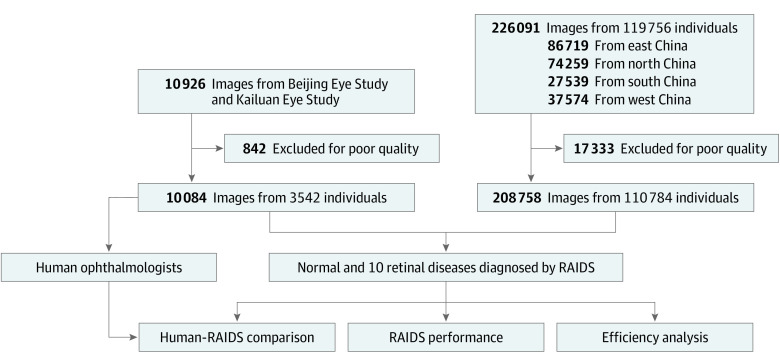
Exposures: Based on 120 002 ocular fundus photographs, the Retinal Artificial Intelligence Diagnosis System (RAIDS) was developed to identify 10 retinal diseases. RAIDS was validated in a prospective collected data set, and the performance between RAIDS and ophthalmologists was compared in the data sets of the population-based Beijing Eye Study and the community-based Kailuan Eye Study.
Main outcomes and measures: The performance of each classifier included sensitivity, specificity, accuracy, F1 score, and Cohen κ score.
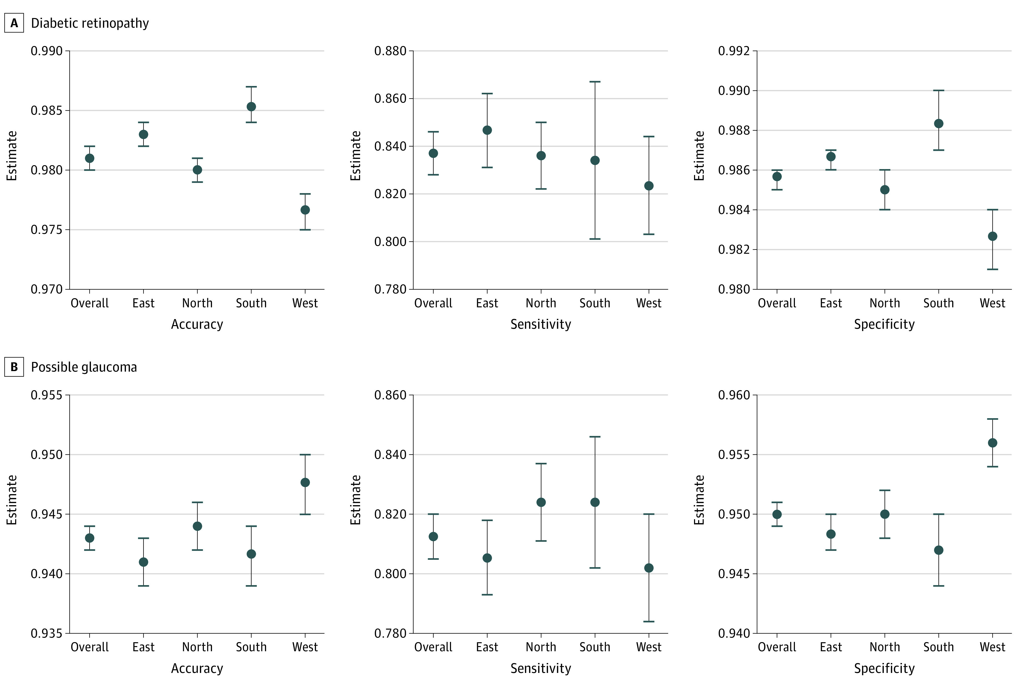
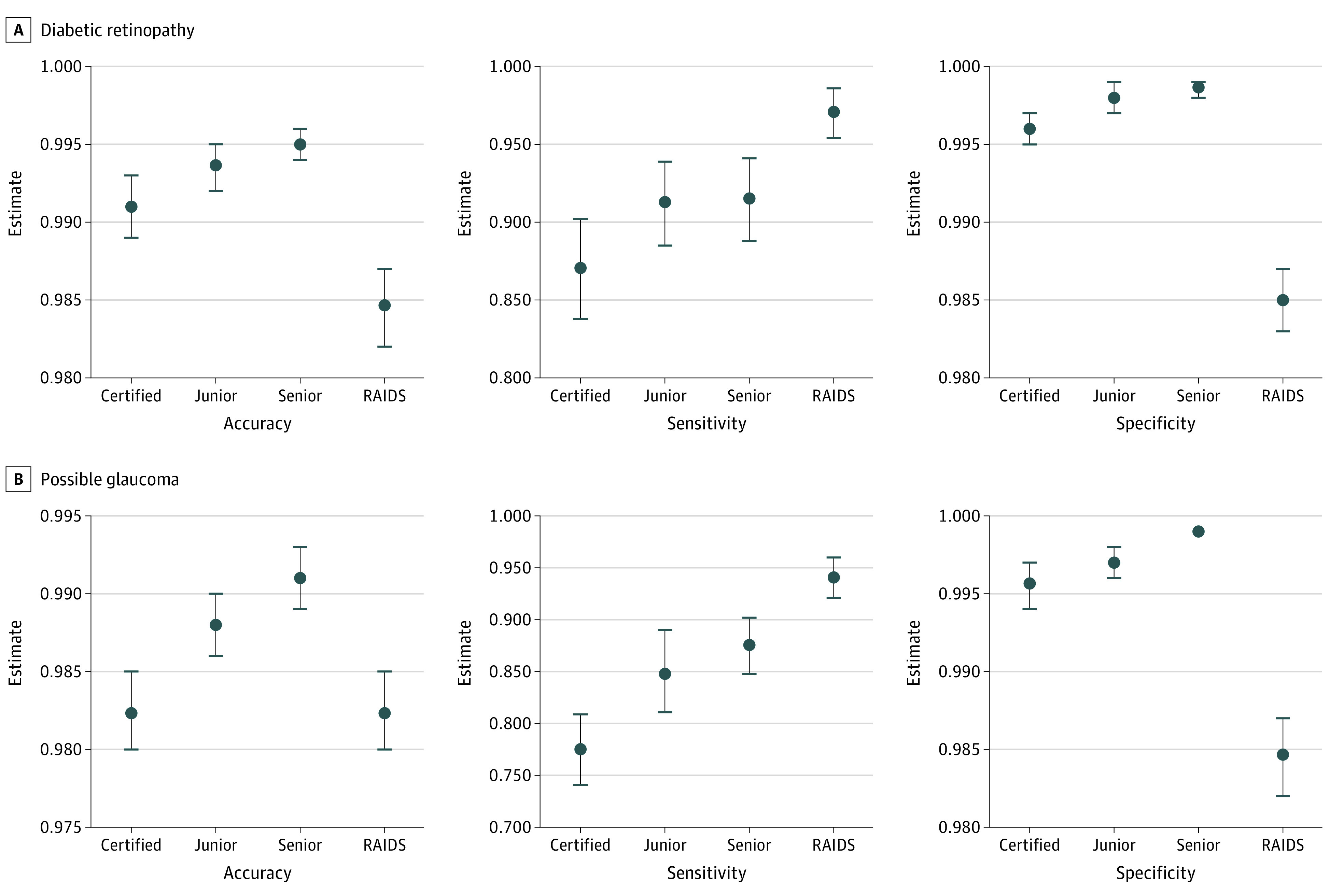
Results: In the prospective validation data set of 208 758 images collected from 110 784 individuals (median [range] age, 42 [8-87] years; 115 443 [55.3%] female), RAIDS achieved a sensitivity of 89.8% (95% CI, 89.5%-90.1%) to detect any of 10 retinal diseases. RAIDS differentiated 10 retinal diseases with accuracies ranging from 95.3% to 99.9%, without marked differences between medical screening centers and geographical regions in China. Compared with retinal specialists, RAIDS achieved a higher sensitivity for detection of any retinal abnormality (RAIDS, 91.7% [95% CI, 90.6%-92.8%]; certified ophthalmologists, 83.7% [95% CI, 82.1%-85.1%]; junior retinal specialists, 86.4% [95% CI, 84.9%-87.7%]; and senior retinal specialists, 88.5% [95% CI, 87.1%-89.8%]). RAIDS reached a superior or similar diagnostic sensitivity compared with senior retinal specialists in the detection of 7 of 10 retinal diseases (ie, referral diabetic retinopathy, referral possible glaucoma, macular hole, epiretinal macular membrane, hypertensive retinopathy, myelinated fibers, and retinitis pigmentosa). It achieved a performance comparable with the performance by certified ophthalmologists in 2 diseases (ie, age-related macular degeneration and retinal vein occlusion). Compared with ophthalmologists, RAIDS needed 96% to 97% less time for the image assessment.
Conclusions and relevance: In this diagnostic study, the DL system was associated with accurately distinguishing 10 retinal diseases in real time. This technology may help overcome the lack of experienced ophthalmologists in underdeveloped areas.
Conflict of interest statement
Figures
References
-
- GBD 2019 Blindness and Vision Impairment Collaborators; Vision Loss Expert Group of the Global Burden of Disease Study . Causes of blindness and vision impairment in 2020 and trends over 30 years, and prevalence of avoidable blindness in relation to VISION 2020: the Right to Sight: an analysis for the Global Burden of Disease Study. Lancet Glob Health. 2021;9(2):e144-e160. doi: 10.1016/S2214-109X(20)30489-7 - DOI - PMC - PubMed
Publication types
MeSH terms
LinkOut - more resources
Full Text Sources
Other Literature Sources
Medical
