. 2022 Jan-Dec:14:17590914221099112.
doi: 10.1177/17590914221099112.

Cathepsin B Relocalization in Late Membrane Disrupted Neurons Following Diffuse Brain Injury in Rats

Affiliations

Cathepsin B Relocalization in Late Membrane Disrupted Neurons Following Diffuse Brain Injury in Rats

Martina L Hernandez et al. ASN Neuro. 2022 Jan-Dec.

Abstract

Traumatic brain injury (TBI) has consequences that last for years following injury. While TBI can precipitate a variety of diffuse pathologies, the mechanisms involved in injury-induced neuronal membrane disruption remain elusive. The lysosomal cysteine protease, Cathepsin B (Cath B), and specifically its redistribution into the cytosol has been implicated in cell death. Little is known about Cath B or neuronal membrane disruption chronically following diffuse TBI. Therefore, the current study evaluated Cath B and diffuse neuronal membrane disruption over a more chronic post-injury window (6 h-4 w). We evaluated Cath B in adult male Sprague-Dawley rats following central fluid percussion injury (CFPI). Expression of Cath B, as well as Cath B-associated pro (Bak and AIF) and anti-apoptotic (Bcl-xl) proteins, were assessed using western blot analysis. Cath B activity was also assessed. Localization of Cath B was evaluated in the membrane disrupted and non-disrupted population following CFPI using immunohistochemistry paired with quantitative image analysis and ultrastructural verification. There was no difference in expression or activity of Cath B or any of the associated proteins between sham and CFPI at any time post-injury. Immunohistological studies, however, showed a sub-cellular re-localization of Cath B at 2 w and 4 w post-injury in the membrane disrupted neuronal population as compared to the time-point matched non-disrupted neurons. Both membrane disruption and Cath B relocalization appear linked to neuronal atrophy. These observations are indicative of a late secondary pathology that represents an opportunity for therapeutic treatment of these neurons following diffuse TBI. Summary Statement Lysosomal cathepsin B relocalizes to the cytosol in neurons with disrupted plasmalemmal membranes weeks following diffuse brain injury. Both the membrane disrupted and cathepsin B relocalized neuronal subpopulations displayed smaller soma and nucleus size compared to non-pathological neurons, indicating atrophy.

Keywords: AIF; Bak/Bcl-XL; cathepsin B; membrane disruption; neuronal atrophy; traumatic brain injury.


Conflict of interest statement

Declaration of conflicting interests: The author(s) declared no potential conflicts of interest with respect to the research, authorship, and/or publication of this article.

Figures

Figure 1.
Figure 1.
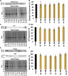
There were no differences in the expression of Cathepsin B (Cath-B) or Cath-B signaling proteins 6 h to 4w post-CFPI compared to sham. (A) representative chemiluminescent western blots depicting (A) Cath-B protein bands at 24/27 kDa, (C) AIF protein bands at ∼67 kDa, as well as (E) Bcl-XL and Bak protein bands at 30 and 25 kDa, respectively, from homogenized lateral cortices. All westerns were normalized to total protein as depicted below the corresponding western blot. Bar graphs depicting the averaged normalized protein expression of (B) Cath-B, (D) AIF, and (F) the expression ratio of Bcl-XL/Bak in sham animals (n = 6) [black bars] and at 6 h–4w post-injury in TBI animals [yellow bars] (n = 6/time point). There were no differences in the expression of any proteins assessed following CFPI compared to sham. Mean ± S.E.M.
Figure 2.
Figure 2.
Cathepsin B activity does not significantly change following injury. Normalized average fluorescence intensity of Cathepsin B activity within whole homogenates of the lateral neocortex is reported in the bar graph. Sham animal (n = 5) [black bars] (black bar) Cathepsin B activity was not found to be different from 6 h–4w post-CFPI (yellow bar; n = 4/ time point) reported as mean ± S.E.M.
Figure 3.
Figure 3.
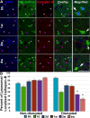
Representative confocal images at 6 h, 1w, 2w, and 4w of immunolabeled Cathepsin B (red) in the lateral neocortex following CFPI (A). Membrane disrupted neurons were identified as containing the cell-impermeable Alexa-488 conjugated dextran (green). The nuclei were labeled with DAPI (blue) to identify the location of non-membrane disrupted neurons. The fourth panel is an overlay, and the last panel is a higher magnification image from the overlay panel. White arrows indicate membrane disrupted neurons and purple arrow heads indicate non membrane disrupted neurons. Scale bar = 20 µm. Cathepsin B localization shifts in membrane disrupted neurons at 2 and 4w post-CFPI (B). Bar graph depicts the percentage of total analyzed neurons per timepoint with punctate lysosomal cathepsin B localization at 6 h (n = 290 neurons, 5 animals) [teal blue], 1d (n = 282 neurons, 5 animals) [green], 3d (n = 207 neurons, 4 animals) [dark blue], 1w (n = 237 neurons, 5 animals) [red], 2w (n = 300 neurons, 5 animals) [purple], and 4w (n = 235 neurons, 5 animals) [pink] in non-disrupted and membrane disrupted neuronal populations reported as mean ± S.E.M. * p < 0.05.
Figure 4.
Figure 4.
Qualitative ultrastructural confirmation of Cathepsin B redistribution outside of lysosomes following CFPI. Electron micrographs of neurons (“N” indicates the nucleus) immunolabeled for Cath-B (black granules) from a representative 2w and 4w TBI animal. (A) Some neurons in the injured brain displayed normally localized cathepsin B within lysosomes (black arrows). (B & C) Diffuse localization of Cathepsin B outside of the lysosomes (red arrow heads), however, was observed in many of the lateral neocortical neurons in these cases, substantiating the confocal quantitative assessments. Scale bar = 2 µm.
Figure 5.
Figure 5.
Membrane disruption and Cathepsin B mislocalization affect cell morphology. Bar graphs comparing the mean (A & C) somal area and (B & D) nucleus area 6 h (n = 738 neurons/5 animals), 1d (n = 527 neurons/5 animals), 3d (n = 388 neurons/4 animals), 1w (n = 426 neurons/5 animals), 2w (n = 577 neurons/ 5 animals), and 4w (n = 434 neurons/5 animals) following CFPI. These neurons were designated as (A & B) membrane disrupted (light purple bars) and non-disrupted (dark purple bars) or (C & D) based on Cathepsin B localization into puncta (dark blue bars) or diffusely distributed throughout the cytoplasm (light blue bars) in each cell. (E) Representative image indicating somal area (dashed circle) and nucleus area (dotted circle) in a membrane disrupted and non-disrupted neuron at 1w post-injury. Scale bar = 20 µm. The area of both the soma and nucleus was decreased in membrane disrupted neurons, and neurons with diffusely localized Cathepsin B. * p < 0.05 compared to timepoint-matched internal controls [(A & B) non-membrane disrupted or (C & D) punctate localized Cath B neurons].

References

    1. Amritraj A., Peake K., Kodam A., Salio C., Merighi A., Vance J. E., Kar S. (2009). Increased activity and altered subcellular distribution of lysosomal enzymes determine neuronal vulnerability in Niemann-Pick type C1-deficient mice. The American Journal of Pathology, 175(6), 2540–2556. 10.2353/ajpath.2009.081096 - DOI - PMC - PubMed
    1. Benchoua A., Braudeau J., Reis A., Couriaud C., Onténiente B. (2004). Activation of proinflammatory caspases by cathepsin B in focal cerebral ischemia. Journal of Cerebral Blood Flow & Metabolism, 24(11), 1272–1279. 10.1097/01.WCB.0000140272.54583.FB - DOI - PubMed
    1. Boutté A. M., Hook V., Thangavelu B., Sarkis G. A., Abbatiello B. N., Hook G., Jacobsen J. S., Gilsdorf J, Yang Z., Wang K. K. W., Shear D. A. (2020). Penetrating traumatic brain injury triggers dysregulation of Cathepsin B protein levels independent of cysteine protease activity in brain and cerebral spinal fluid. Journal of Neurotrauma, 37(13), 1574–1586. 10.1089/neu.2019.6537 - DOI - PMC - PubMed
    1. Boya P., Andreau K., Poncet D., Zamzami N., Perfettini J. L., Metivier D., Ojcius D. M., Jäättelä M., Kroemer G. (2003). Lysosomal membrane permeabilization induces cell death in a mitochondrion-dependent fashion. Journal of Experimental Medicine, 197(10), 1323–1334. 10.1084/jem.20021952 - DOI - PMC - PubMed
    1. Cavallo-Medved D., Moin K., Sloane B. (2011). Cathepsin B: basis sequence: mouse. AFCS-nature Molecule Pages, 2011, 1–17. PMCID: PMC5541861 - PMC - PubMed

Publication types