

The incremental value of myocardial viability, evaluated by 18F-fluorodeoxyglucose positron emission tomography, and cardiovascular magnetic resonance for mortality prediction in patients with previous myocardial infarction and symptomatic heart failure
- PMID: 35503304
- PMCID: PMC10466976
- DOI: 10.1177/02676591221100739
The incremental value of myocardial viability, evaluated by 18F-fluorodeoxyglucose positron emission tomography, and cardiovascular magnetic resonance for mortality prediction in patients with previous myocardial infarction and symptomatic heart failure
Abstract
Objectives: To find the imaging mortality predictors in patients with previous myocardial infarction (MI), symptomatic heart failure (HF), and reduced left ventricle (LV) ejection fraction (EF).
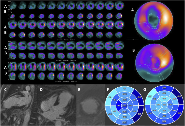
Methods: for the study 39 patients were selected prospectively with prior MI, symptomatic HF, and LVEF ≤40%. All patients underwent transthoracic echocardiography (TTE), single-photon emission computed tomography myocardial perfusion imaging (SPECT MPI), 18F-FDG positron emission tomography (FDG PET). 31 patients underwent cardiovascular magnetic resonance (CMR) with late gadolinium enhancement (LGE). Patients were divided into two groups: 1 group - cardiac death; 2 group - no cardiac death. Myocardial scars were assessed on a 5-point-scale. Follow-up data was obtained.
Results: Imaging features disclosed significant difference (p < 0.05) of defect score (CMR and SPECT-PET), LV end-diastolic diameter (EDD) (TTE), LVEDD index (CMR), LV global longitudinal strain (CMR) and LV global circumferential strain (CMR) between the groups. Predictors of cardiac death were: LVEDD index (TTE) and LV global longitudinal strain. The cut-off values to predict cardiac death were: defect score (CMR) 25 (AUC, 79.5%; OR 1.8, 95% CI 1.2-2.7), SPECT-PET defect score 22 (AUC, 73.9%; OR 0.5, 95% CI 0.3-0.7), LVEDD (TTE) 58 mm (AUC, 88.4%; OR 23.6, 95% CI 2.6-217.7), LVEDDi 30 mm/m2 (TTE) (AUC, 73.6%; OR 22.0, 95% CI 1.9-251.5), LVEDDi 33.6 mm/m2 (CMR) (AUC, 73.6%; OR 22.0, 95% CI 1.9-251.5), LV global longitudinal strain -13.4 (AUC, 87.8%; OR 2.1, 95% CI 1.2-3.7) and LV global circumferential strain -16.3 (AUC, 76.1%; OR 1.9, 95% CI 1.2-3.0).
Conclusions: Imaging features, such as defect score (CMR) >25, SPECT-PET defect score >22, LVEDD (TTE) >58 mm, LVEDDi (TTE) >30 mm/m2, LVEDDi (CMR) >33.6 mm/m2, LV global longitudinal strain -13.4 and LV global circumferential strain -16.3, may increase sensitivity and specificity of FDG PET and LGE CMR predicting of late mortality.
Keywords: 18F-fluorodeoxyglucose positron emission tomography; late gadolinium enhancement; myocardial perfusion imaging; myocardial viability; strain-encoded imaging.
Conflict of interest statement
The author(s) declared no potential conflicts of interest with respect to the research, authorship, and/or publication of this article.
Figures

Similar articles
-
Comparative Analysis of Myocardial Viability Multimodality Imaging in Patients with Previous Myocardial Infarction and Symptomatic Heart Failure.Medicina (Kaunas). 2022 Mar 1;58(3):368. doi: 10.3390/medicina58030368. Medicina (Kaunas). 2022. PMID: 35334543 Free PMC article.
-
Scar imaging in the dyssynchronous left ventricle: Accuracy of myocardial metabolism by positron emission tomography and function by echocardiographic strain.Int J Cardiol. 2023 Feb 1;372:122-129. doi: 10.1016/j.ijcard.2022.11.042. Epub 2022 Nov 29. Int J Cardiol. 2023. PMID: 36460211
-
Positron emission tomography for the assessment of myocardial viability: an evidence-based analysis.Ont Health Technol Assess Ser. 2005;5(16):1-167. Epub 2005 Oct 1. Ont Health Technol Assess Ser. 2005. PMID: 23074467 Free PMC article.
-
Recent advances in cardiac imaging for patients with heart failure.Curr Opin Cardiol. 2011 Mar;26(2):132-43. doi: 10.1097/HCO.0b013e32834380e7. Curr Opin Cardiol. 2011. PMID: 21297464 Review.
-
Imaging heart failure: current and future applications.Can J Cardiol. 2013 Mar;29(3):317-28. doi: 10.1016/j.cjca.2013.01.006. Can J Cardiol. 2013. PMID: 23439018 Review.
Cited by
-
Impact of CT attenuation correction on viable myocardium detection in combined SPECT and PET/CT: A retrospective cohort study.Medicine (Baltimore). 2024 Oct 25;103(43):e40175. doi: 10.1097/MD.0000000000040175. Medicine (Baltimore). 2024. PMID: 39470532 Free PMC article.
-
Increased [18F]FDG uptake in the infarcted myocardial area displayed by combined PET/CMR correlates with snRNA-seq-detected inflammatory cell invasion.Basic Res Cardiol. 2024 Oct;119(5):807-829. doi: 10.1007/s00395-024-01064-y. Epub 2024 Jun 26. Basic Res Cardiol. 2024. PMID: 38922408 Free PMC article.
References
-
- Rahimtoola SH. The hibernating myocardium. Am Heart J 1989; 117(1): 211–221. - PubMed
-
- Braunwald E, Rutherford JD. Reversible ischemic left ventricular dysfunction: evidence for the "hibernating myocardium. J Am Coll Cardiol 1986; 8(6): 1467–1470. - PubMed
-
- Wijns W, Vatner SF, Camici PG. Hibernating myocardium. N Engl J Med 1998; 339(3): 173–181. - PubMed
-
- Beanlands RS, Nichol G, Huszti E, et al. F-18-fluorodeoxyglucose positron emission tomography imaging-assisted management of patients with severe left ventricular dysfunction and suspected coronary disease: a randomized, controlled trial (PARR-2). J Am Coll Cardiol 2007; 50(20): 2002–2012. - PubMed
-
- McDiarmid AK, Loh H, Nikitin N, et al. Predictive power of late gadolinium enhancement for myocardial recovery in chronic ischaemic heart failure: a HEART sub-study. ESC Heart Fail 2014; 1(2): 146–153. - PubMed
MeSH terms
Substances
LinkOut - more resources
Full Text Sources
Medical
Research Materials
Miscellaneous
