Review
. 2022 May 3;117(1):26.
doi: 10.1007/s00395-022-00931-w.

Neonatal injury models: integral tools to decipher the molecular basis of cardiac regeneration

Affiliations
Review

Neonatal injury models: integral tools to decipher the molecular basis of cardiac regeneration

Alessia Costa et al. Basic Res Cardiol. .

Abstract

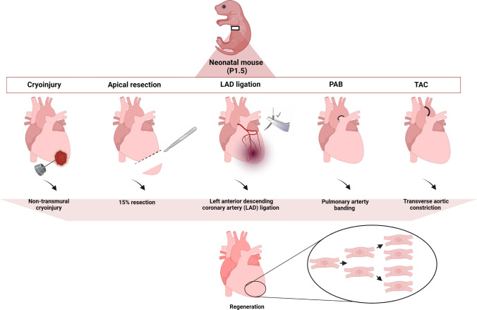
Myocardial injury often leads to heart failure due to the loss and insufficient regeneration of resident cardiomyocytes. The low regenerative potential of the mammalian heart is one of the main drivers of heart failure progression, especially after myocardial infarction accompanied by large contractile muscle loss. Preclinical therapies for cardiac regeneration are promising, but clinically still missing. Mammalian models represent an excellent translational in vivo platform to test drugs and treatments for the promotion of cardiac regeneration. Particularly, short-lived mice offer the possibility to monitor the outcome of such treatments throughout the life span. Importantly, there is a short period of time in newborn mice in which the heart retains full regenerative capacity after cardiac injury, which potentially also holds true for the neonatal human heart. Thus, in vivo neonatal mouse models of cardiac injury are crucial to gain insights into the molecular mechanisms underlying the cardiac regenerative processes and to devise novel therapeutic strategies for the treatment of diseased adult hearts. Here, we provide an overview of the established injury models to study cardiac regeneration. We summarize pioneering studies that demonstrate the potential of using neonatal cardiac injury models to identify factors that may stimulate heart regeneration by inducing endogenous cardiomyocyte proliferation in the adult heart. To conclude, we briefly summarize studies in large animal models and the insights gained in humans, which may pave the way toward the development of novel approaches in regenerative medicine.

Keywords: Cardiac regeneration; Cardiomyocyte proliferation; Myocardial infarction; Neonatal heart injury; Regenerative medicine; microRNA.


Conflict of interest statement

The authors declare no competing interest in relation to this work.

Figures

Fig. 1
Fig. 1
Neonatal cardiac injury models. Cryoinjury is performed through a cryoprobe, resulting in severe and immediate damage to the heart. A part of the left ventricle apex is removed for the apical resection procedure. MI is induced by surgical ligation of the LAD. Non-transmural cryoinjury, apical resection (in the amount 15%) and LAD ligation can trigger full heart regeneration in neonatal mice. Pulmonary artery banding (PAB) surgery allows for cardiac remodeling in a pressure overload system in the right ventricle and proliferation in neonates. TAC is performed by placing a suture under the transverse aorta causing constriction and hypertrophy and left ventricle pressure overload. Common to all models is that the regeneration observed is mainly attributed to the proliferation of pre-existing cardiomyocytes rather than to the differentiation of progenitor cells or transdifferentiation of non-cardiomyocytes. “Created with BioRender.com”
Fig. 2
Fig. 2
MicroRNA modes of actions. A MiRNAs bind mRNA targets at the 3` UTR by base pairing. Targets are inhibited through translational repression or by mediating degradation, leading to specific regulation of gene expression levels. B miR-199a as an example of a possible mode of action for a miRNA. miR-199a downregulates the mRNA levels of TAOK1 (TAO kinase 1) and beta-TrCP (beta-transducing repeat containing protein). TAOK1 targets MST1 and LATS1/2, while beta-TrCP promotes the dephosphorylation of YAP and the subsequential degradation. By inhibiting TAOK1 and beta. TrCP, miR-199a promotes the nuclear translocation of YAP, its binding with TEAD (transcriptional enhanced associated domain) and the regulation of target genes involved in the cell cycle, growth and proliferation. “Created with BioRender.com”
Fig. 3
Fig. 3
Cardiac regenerative potential in animal models. In response to injury, zebrafish can regenerate the heart without scarring within 2 months as well as in adulthood. Embryonic and neonatal mice retain the capacity to regenerate the heart after injury, but this ability is lost at 7 days postnatal, and in adult mice, thereafter the heart undergoes adverse and pathological remodeling after injury with fibrotic scar formation. Recently, important insights are gained in large mammalian animals. The hearts of neonatal pigs are capable of regeneration during the first 2 days of life. The human heart may have a similar capacity to other mammalians to regenerate, but the heart regenerative potential in humans needs still to be explored. “Created with BioRender.com”

References

    1. Aix E, Gutiérrez-Gutiérrez Ó, Sánchez-Ferrer C, Aguado T, Flores I. Postnatal telomere dysfunction induces cardiomyocyte cell-cycle arrest through p21 activation. J Cell Biol. 2016 doi: 10.1083/jcb.201510091. - DOI - PMC - PubMed
    1. Aly S, Aguet J, Dragulescu A, Grosse-Wortmann L. Neonatal myocardial infarction in association with gestational diabetes. Can J Cardiol. 2021 doi: 10.1016/j.cjca.2021.04.005. - DOI - PubMed
    1. Andersen DC, Ganesalingam S, Jensen CH, Sheikh SP. Do neonatal mouse hearts regenerate following heart apex resection? Stem Cell Rep. 2014;2:406–413. doi: 10.1016/j.stemcr.2014.02.008. - DOI - PMC - PubMed
    1. Andersen DC, Jensen CH, Baun C, Hvidsten S, Zebrowski DC, Engel FB, Sheikh SP. Persistent scarring and dilated cardiomyopathy suggest incomplete regeneration of the apex resected neonatal mouse myocardium—a 180 days follow up study. J Mol Cell Cardiol. 2016;90:47–52. doi: 10.1016/j.yjmcc.2015.11.031. - DOI - PubMed
    1. Anversa P, Rota M, Urbanek K, Hosoda T, Sonnenblick EH, Leri A, Kajstura J, Bolli R. Myocardial aging: a stem cell problem. Basic Res Cardiol. 2005;100:482–493. doi: 10.1007/s00395-005-0554-3. - DOI - PubMed

Publication types