. 2022 May 3;18(5):e1010488.
doi: 10.1371/journal.ppat.1010488. eCollection 2022 May.

A neutralizing antibody target in early HIV-1 infection was recapitulated in rhesus macaques immunized with the transmitted/founder envelope sequence

Affiliations

A neutralizing antibody target in early HIV-1 infection was recapitulated in rhesus macaques immunized with the transmitted/founder envelope sequence

Sarah Welbourn et al. PLoS Pathog. .

Abstract

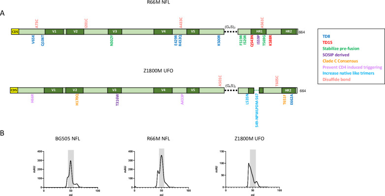
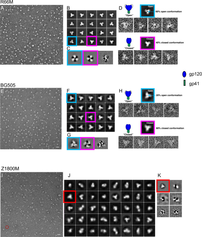
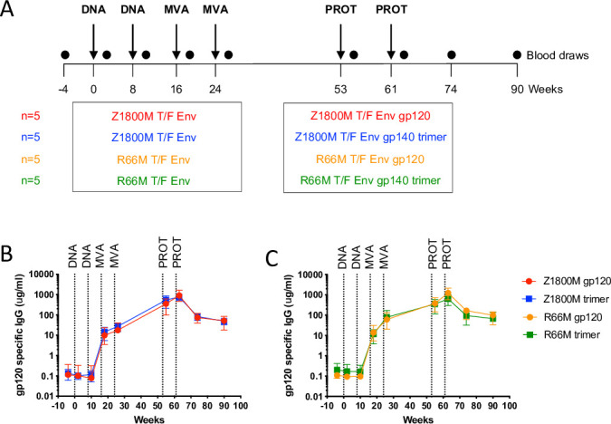
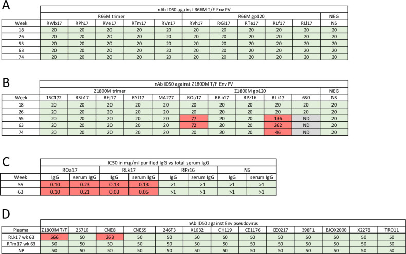
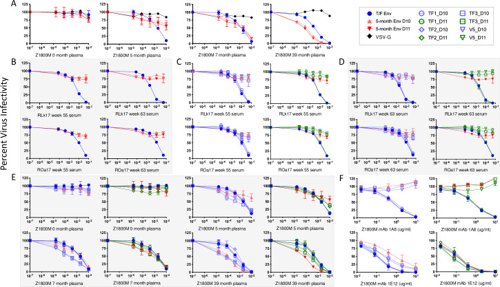
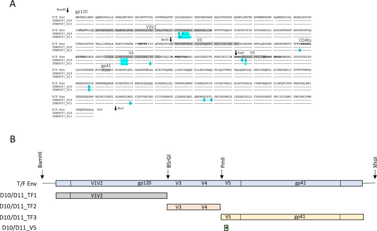
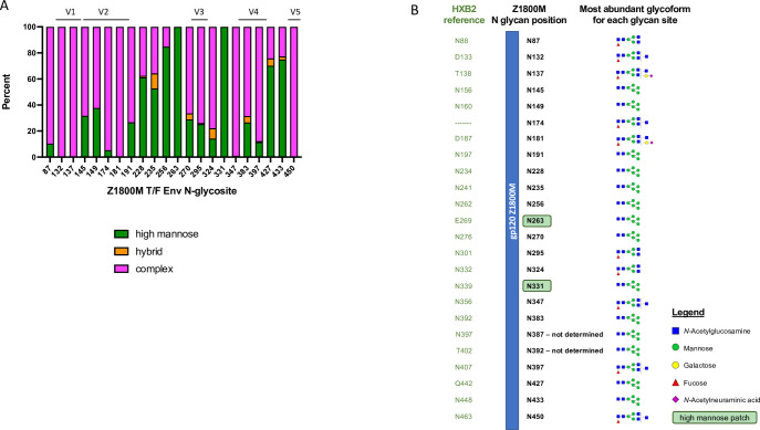
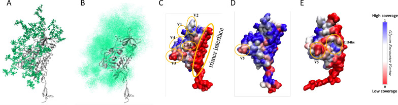
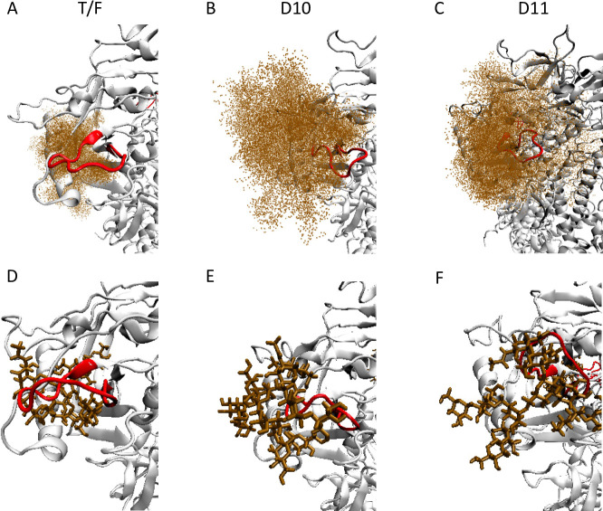
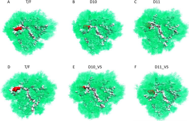
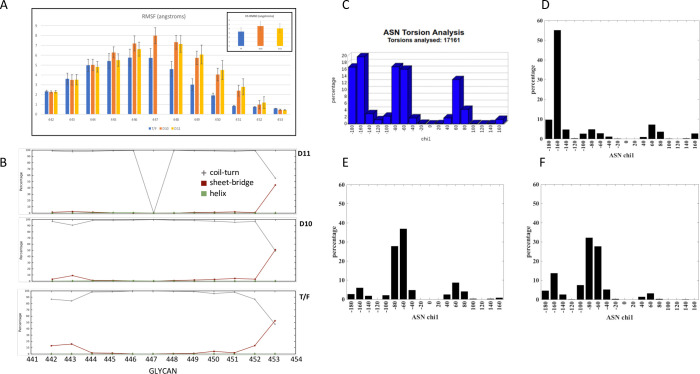
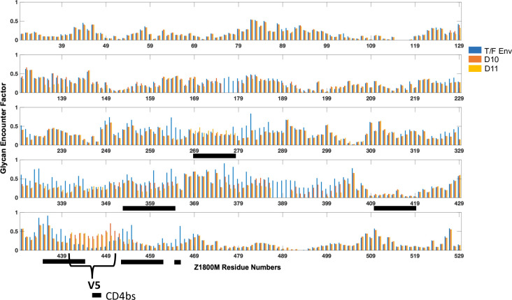
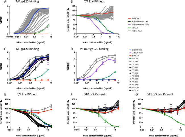
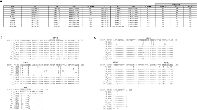
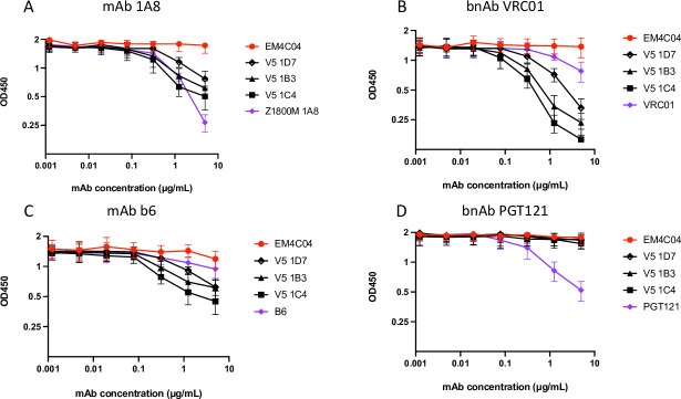
Transmitted/founder (T/F) HIV-1 envelope proteins (Envs) from infected individuals that developed neutralization breadth are likely to possess inherent features desirable for vaccine immunogen design. To explore this premise, we conducted an immunization study in rhesus macaques (RM) using T/F Env sequences from two human subjects, one of whom developed potent and broad neutralizing antibodies (Z1800M) while the other developed little to no neutralizing antibody responses (R66M) during HIV-1 infection. Using a DNA/MVA/protein immunization protocol, 10 RM were immunized with each T/F Env. Within each T/F Env group, the protein boosts were administered as either monomeric gp120 or stabilized trimeric gp140 protein. All vaccination regimens elicited high titers of antigen-specific IgG, and two animals that received monomeric Z1800M Env gp120 developed autologous neutralizing activity. Using early Env escape variants isolated from subject Z1800M as guides, the serum neutralizing activity of the two immunized RM was found to be dependent on the gp120 V5 region. Interestingly, the exact same residues of V5 were also targeted by a neutralizing monoclonal antibody (nmAb) isolated from the subject Z1800M early in infection. Glycan profiling and computational modeling of the Z1800M Env gp120 immunogen provided further evidence that the V5 loop is exposed in this T/F Env and was a dominant feature that drove neutralizing antibody targeting during infection and immunization. An expanded B cell clonotype was isolated from one of the neutralization-positive RM and nmAbs corresponding to this group demonstrated V5-dependent neutralization similar to both the RM serum and the human Z1800M nmAb. The results demonstrate that neutralizing antibody responses elicited by the Z1800M T/F Env in RM converged with those in the HIV-1 infected human subject, illustrating the potential of using immunogens based on this or other T/F Envs with well-defined immunogenicity as a starting point to drive breadth.


Conflict of interest statement

The authors have declared that no competing interests exist.

Figures

Fig 1
Fig 1. Z1800M and R66M trimers.
(A) Linear representations of the R66M NFL and the Z1800M UFO Env trimers are shown. Cysteine residues are indicated along the top; the (G4S)2 linker is indicated by a black dashed line; the optimized gp41 region in the UFO is indicated by a green dashed line; key amino acid substitutions with HXB2 numbering are indicated along the Env open reading frame and are color-coded to indicate their putative contribution to stabilization. The CD5 leader sequence is also shown. (B) Size exclusion chromatography profiles are shown for the BG505 NFL trimer as a process control, the R66M NFL trimer, and the Z1800M UFO trimer. The fractions corresponding to the trimeric proteins are shaded.
Fig 2
Fig 2. Negative stain electron microscopy analysis of Z1800M and R66M trimers.
Negative stain transmission electron microscopy images (A, E, I) and reference-free 2D class averages (B, F, J) are shown for R66M, BG505, and Z1800M trimers. Representative open and closed states for the R66M and BG505 trimers are indicated by blue and magenta boxes, respectively, in (B) and (F). Eigen vector classes of aligned particles of R66M (C) and BG505 (G) also demonstrate open and closed states. Reference-free 2D class averages of open (blue boxed) and closed (magenta boxed) representative particles of R66M (D) and BG505 (H) are shown. Z1800M trimers show a more heterogenous population (I). The red circle in (I) and red boxes in (J) and in the Eigen vector classes in (K) highlight stable trimers. For R66M, approximately 60% open and 40% closed out of 14,000 included particles for 2D classification were observed. For BG505, approximately 20% open and 80% closed out of 12,000 included particles for 2D classification were observed. Scale bar = 20 nm.
Fig 3
Fig 3. Immunization regimen and antigen-specific serum IgG responses.
(A) A representation of the immunizations, time scale, and vaccination groups are shown. The four vaccine groups consisted of 5 RM immunized intramuscularly with DNA expressing SIVmac239 Gag and the indicated HIV-1 Env, R66M or Z1800M T/F Env, at weeks 0 and 8; MVA expressing SIVmac239 Gag and the R66M or Z1800M T/F Env at weeks 16 and 24; and the R66M or Z1800M T/F Env as a stabilized gp140 trimer or gp120 protein in Adjuplex at weeks 53 and 61. Blood was collected at baseline (week -4) and weeks 2, 10, 18, 26, 55, 63, 74, and 90. Serum IgG with specificity for the autologous T/F Env gp120, Z1800M (B) and R66M (C), was quantified by ELISA. Serial dilutions of serum from RM in each of the four vaccine groups were tested at weeks -4, 2, 10, 18, 26, 55, 63, 74, and 90. The group medians with standard error of the mean are shown in μg/ml, based on a RM IgG standard curve. For samples where both replicates fell below the limit of detection, a value of 0.078 was used. If only one replicate fell below the limit of detection, the other value was used. All samples were measured in duplicate wells and in two independent experiments.
Fig 4
Fig 4. Characterization of immunized RM serum neutralization.
(A-B) Neutralization activity was measured against the autologous T/F Env PV, Z1800M or R66M, with heat-inactivated, serially diluted serum using the TZM-bl assay. Neutralization was evaluated at weeks 18, 26, 55, 63, and 74, corresponding to two weeks post MVA immunizations, two weeks post protein immunizations, and 13 weeks after the final protein immunization. Green boxes indicate that an ID50 titer could not be calculated using 1:20 as the highest dilution. Red boxes show a positive ID50 titer. Gray boxes indicate that the serum was not evaluated (ND). (C) For neutralization positive week 55 and 63 post-protein serum samples from ROa17 and RLk17, and neutralization negative week 55 and 63 post-protein samples from RPz16, IgG was purified from the serum and tested for neutralization. The results shown are IC50 titers calculated using the quantified amounts of purified IgG and total IgG present in serum as measured by ELISA. The infectivity curves corresponding to purified and total serum IgG IC50 values from panel C are shown in S6 Fig. NS represents serum or IgG from a naïve RM in (A-C). (D) Week 63 plasma from neutralization positive Z1800M gp120-immunized RLk17 was tested for neutralization activity against a global panel of envelope PV [48]. Plasma serial dilutions began at 1:50 and ID50 values are shown. A value of 50 indicates the highest concentration of plasma tested without 50% inhibition. Naïve RM plasma (NP) and week 63 plasma from a non-neutralizing animal (R66M trimer immunized RTm17) were used as negative controls. All experiments had duplicate wells and were repeated at least twice independently.
Fig 5
Fig 5. Neutralization mapping of immunized RM sera, and autologous plasma and mAbs from HIV+ subject Z1800M.
(A) Plasma from HIV+ subject Z1800M collected close to the time of infection (0-month) and longitudinally at 5, 7, and 39 months and (B) week 55 and 63 serum from neutralization positive RMs RLk17 and ROa17 were tested against PV expressing the Z1800M T/F Env and 5-month Envs D10 and D11. RLk17 and ROa17 week 55 and 63 serum (C and D), Z1800M longitudinal patient plasma (E), and Z1800M derived mAbs 1A8 and 1E12 (F) were tested against Env chimeras and mutants generated in the T/F Env. The TF1, TF2, and TF3 constructs correspond to fragments from D10 (lavender) or D11 (green) that were introduced into the T/F Env background (Fig 6B). The V5 constructs correspond to the changes in D10 and D11 V5 loops (Fig 6A) that were introduced into the T/F Env. Each graph represents percent viral infectivity on the y axis and plasma/serum/mAb dilution or concentration on the x axis on a log10 scale. All experiments had duplicate wells and were repeated at least twice independently, and error bars indicate the standard deviation.
Fig 6
Fig 6. Sequence differences and chimeras based on T/F Env and escape Envs.
(A) Amino acid sequence alignment of the Z1800M T/F Env and the 5-month neutralization escape Envs D10 and D11. Dashes indicate conserved residues relative to the T/F Env. Dots indicate deleted residues and amino acid differences from the T/F Env are indicated by cyan highlighted. Key Env regions are shaded and labeled. The positions of restriction sites used to generate chimeras shown in panel B are indicated with arrows. (B) The scheme for construction of the chimeras is shown. The restriction sites and fragments that were replaced in the T/F Env using D10 and D11 sequences are shown. The V5 constructs contained only the changes present in V5, which were two amino acid differences in D10 and one amino acid deletion in D11.
Fig 7
Fig 7. Site specific N-linked glycan profiling of the Z1800M T/F Env gp120 protein.
Glycoproteomic analysis was performed using nano-LC-MS/MS and site-specific glycosylation profiling was performed for 22 of 24 putative N-linked glycan sites (N387 and N392 could not be separated and therefore not assigned with confidence). (A) The bar graph depicts the relative percentage of classified glycoforms on each N-glycan site (numbered according to the Z1800M T/F Env), indicated as complex (pink), hybrid (orange), or high mannose (green) types. The hyper-variable domains are indicated above the graph. (B) For each glycan position, the equivalent HXB2 reference position is indicated as well as the most abundant glycoform observed at that site that was subsequently used for computational modeling. For N-linked glycosylation sites N387 (HXB2 N397) and N392 (HXB N402) no glycopeptides were detected that only contained the N-glycosylation site of interest. Therefore, both sites were represented by Man5.
Fig 8
Fig 8. Structural model of the glycan shield topology over Z1800M gp120 glycoprotein.
(A) A single protomer of Z1800M gp120 glycoprotein in a trimeric fold structure. Static representation of native glycans (green sticks) over Env protein (grey ribbon), with each glycan occupying only one of many possible positions. (B) Cumulative shielding effect due to the flexibility of glycans represented by a density of points over the monomer surface. (C) Z1800M gp120 surface colored by the normalized Glycan Encounter Factor (GEF) resulting from all the glycans over the monomer. Regions having high GEF or shielding are colored in blue, exposed regions are red. (D) Z1800M gp120 structure as part of a Man9-glycoform SOSIP trimer, colored by normalized GEF. This model includes gp120 and gp41 protein and glycans from the complete trimeric SOSIP. (E) Z1800M gp120 glycoprotein with monomeric conformation different from the trimer, colored by normalized GEF. This monomer was modeled using previously observed X-ray diffraction structures of HIV-1 Env monomers. The V5 loop remains exposed in all three different models.
Fig 9
Fig 9. Glycan N450 reorientation leads to higher coverage of V5 in Z1800M D10 and D11.
Ensemble model using the wildtype T/F, D10, and D11 Env gp120 sequences shows the glycan at N450 (HXB2 N463) on the top (brown points) and a single conformation of the glycan on the bottom (brown sticks). (A, D) Z1800M T/F Env where the glycan is inserted below the loop, exposing V5 and (B, E) D10 and (C, F) D11 generations where the glycan is oriented on top of the protein surface, shielding V5. The V5 hypervariable loop is shown in red.
Fig 10
Fig 10. Complete glycan shield over Z1800M SOSIP structure.
The glycan structural distribution is represented by green density of points. The underlying protein surface is represented in gray and the V5 loop in red. SOSIP-based models using the (A) T/F Env, (B) D10 Env and (C) D11 Env sequences illustrate that the V5 region remains exposed in the T/F but is shielded in the escape Envs. Because D10 and D11 contained other sequence and glycan differences outside of V5 compared to the T/F Env, the model was repeated with just the D10 and D11 V5 changes. The reorientation of the V5 glycan at N450 held true for this model suggesting that the V5 changes are sufficient: (D) another depiction of the T/F Env, (E) T/F Env with D10 V5 modifications and (F) T/F Env with D11 V5 loop modifications.
Fig 11
Fig 11. Structural variations of V5 and torsional distribution of N450 between Z1800M Envs.
(A) Residue-wise Root Mean Squared Fluctuations (RMSF) comparison between T/F, D10 and D11, with the overall V5-loop RMS Deviation shown in inset. (B) Secondary structural analysis of the V5 loop indicates higher beta-strand propensity at the two terminal ends of the loop for the T/F (3rd row) as compared to D10 and D11 (2nd and 1st rows respectively). (C) Chi1 distribution of glycosylated asparagines obtained from 17161 pdb structures, calculated using GlyTorsion [106]. Chi1 distribution of N450 in the T/F Env (D) peaks at -160°, while in D10 (E) and D11 (F) the peak is around -60°. This difference in chi1 dihedral influences the overall orientation of the glycan. The numbering in panels A and B correspond to HXB2 residues 455–465 and 456–466, respectively.
Fig 12
Fig 12. Comparison of residue-wise glycan shielding by GEF, between the T/F, D10 and D11 Envs.
Amino acid positions are shown below each bar graph panel. The GEF for the T/F, D10, and D11 Envs at each position are shown on the y axis. The V5 loop region is indicated, and residues that comprise the CD4bs are marked by black horizontal bars. The bars correspond to HXB2 residues 275–286, 362–374, 425–434, 450–459, 466–475, 478–479,
Fig 13
Fig 13. Monoclonal antibodies isolated from neutralization positive RM RLk17.
54 RLk17 mAbs recovered using the T/F Env gp120-specific B cell sort were tested in ELISA for binding to the Z1800M T/F Env gp120 protein starting at 5 μg/ml in (A) and for neutralization of the Z1800M T/F Env PV in the TZM-bl assay starting at 25 μg/ml in (B). The bnAb VRC01; the anti-influenza HA mAb EM4C04, and the Z1800M neutralizing mAbs 1A and 1E12 are also shown. Two non-neutralizing RLk17 mAbs from the T/F Env gp120 B cell sort and nine mAbs from the expanded clonotype isolated during the T/F Env gp120 positive/V5 mutant negative B cell sort were tested for binding to the Z1800M T/F Env gp120 protein in (C) and to the T/F Env gp120 protein with the D11 mutated V5 loop in (D). The Z1800M mAbs 1A8 and 1E12, VRC01, and EM4C04 were also included. mAbs were also tested for neutralization of the Z1800M T/F Env PV (E), the D10_V5 (F) and D11_V5 (G) chimeras starting at 25 μg/ml. All assays contained duplicate wells and were repeated independently at least twice, with the means and standard deviation shown.
Fig 14
Fig 14. Characterization of the RLk17 neutralizing mAb clonotype.
The 9 neutralizing mAbs from the expanded clonotype are shown in (A) along with the germline assignments for VH, VJ, VL, and VJ. The CDRH3 and CDRL3 amino acid sequences are shown, as are the percent nucleotide identity to germline for VH and VL. The last row shows the same characteristics for the Z1800M human neutralizing mAb 1A8. The last 3 columns show the IC50 values for each mAb against the autologous T/F Env PV (from Fig 14E–14G). A value of >25 indicates that there was no neutralization at the highest mAb concentration tested. Amino acid alignments of the VH (B) and VL (C) sequences are shown along with the corresponding germlines. Dashes represent identical residues and substitutions are shown. The CDR regions are highlighted in the germline sequences.
Fig 15
Fig 15. Epitope mapping through competition ELISA.
The ability of representative RLk17 neutralizing mAbs V5 1D7, V5 1B3 and V5 1C4 to compete for binding of biotinylated reference antibodies Z1800M 1A8, VRC01, b6 and PGT121 is represented on each graph (A-D). Immobilized Z1800M T/F gp120 protein was first incubated with serial dilutions of test antibody (starting at 5 μg/ml) and subsequently probed with biotinylated reference mAb. Concentrations of biotinylated reference mAb were used that resulted in an OD450 between 1 and 2 in absence of competitor (3 μg/ml b6 and VRC01, 0.1 μg/ml Z1800M 1A8, and 0.3 μg/ml PGT121). Each assay was independently repeated twice and the means with standard deviation are shown.

Similar articles

Cited by

References

    1. Wei X, Decker JM, Wang S, Hui H, Kappes JC, Wu X, et al.. Antibody neutralization and escape by HIV-1. Nature. 2003;422(6929):307–12. Epub 2003/03/21. doi: 10.1038/nature01470 . - DOI - PubMed
    1. Wagh K, Kreider EF, Li Y, Barbian HJ, Learn GH, Giorgi E, et al.. Completeness of HIV-1 Envelope Glycan Shield at Transmission Determines Neutralization Breadth. Cell reports. 2018;25(4):893–908 e7. Epub 2018/10/26. doi: 10.1016/j.celrep.2018.09.087 ; PubMed Central PMCID: PMC6426304. - DOI - PMC - PubMed
    1. van den Kerkhof TL, Feenstra KA, Euler Z, van Gils MJ, Rijsdijk LW, Boeser-Nunnink BD, et al.. HIV-1 envelope glycoprotein signatures that correlate with the development of cross-reactive neutralizing activity. Retrovirology. 2013;10(1):102. Epub 2013/09/26. doi: 10.1186/1742-4690-10-102 ; PubMed Central PMCID: PMC3849187. - DOI - PMC - PubMed
    1. Smith SA, Burton SL, Kilembe W, Lakhi S, Karita E, Price M, et al.. Diversification in the HIV-1 Envelope Hyper-variable Domains V2, V4, and V5 and Higher Probability of Transmitted/Founder Envelope Glycosylation Favor the Development of Heterologous Neutralization Breadth. PLoS pathogens. 2016;12(11):e1005989. Epub 2016/11/17. doi: 10.1371/journal.ppat.1005989 ; PubMed Central PMCID: PMC5112890. - DOI - PMC - PubMed
    1. Townsley SM, Donofrio GC, Jian N, Leggat DJ, Dussupt V, Mendez-Rivera L, et al.. B cell engagement with HIV-1 founder virus envelope predicts development of broadly neutralizing antibodies. Cell host & microbe. 2021;29(4):564–78 e9. Epub 2021/03/05. doi: 10.1016/j.chom.2021.01.016 . - DOI - PMC - PubMed

Publication types

Substances