. 2022 Aug 1;44(8):575-580.
doi: 10.1097/DAD.0000000000002208. Epub 2022 Apr 27.

PRAME Expression Correlates With Genomic Aberration and Malignant Diagnosis of Spitzoid Melanocytic Neoplasms

Affiliations

PRAME Expression Correlates With Genomic Aberration and Malignant Diagnosis of Spitzoid Melanocytic Neoplasms

Pedram Gerami et al. Am J Dermatopathol. .

Abstract

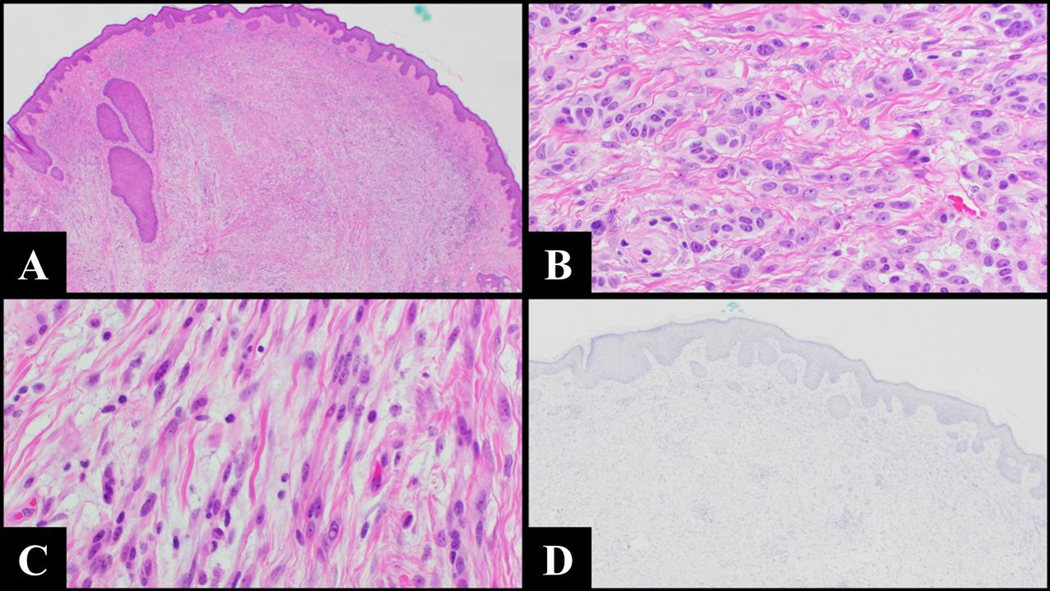
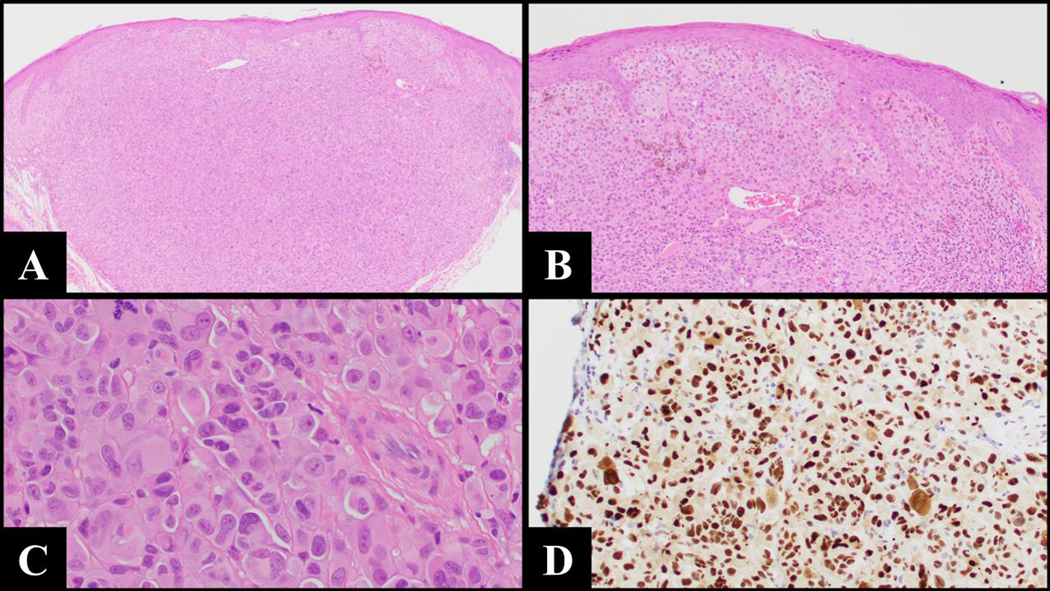
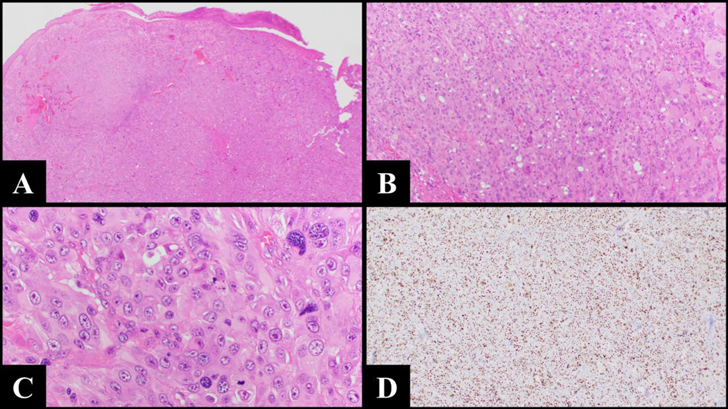
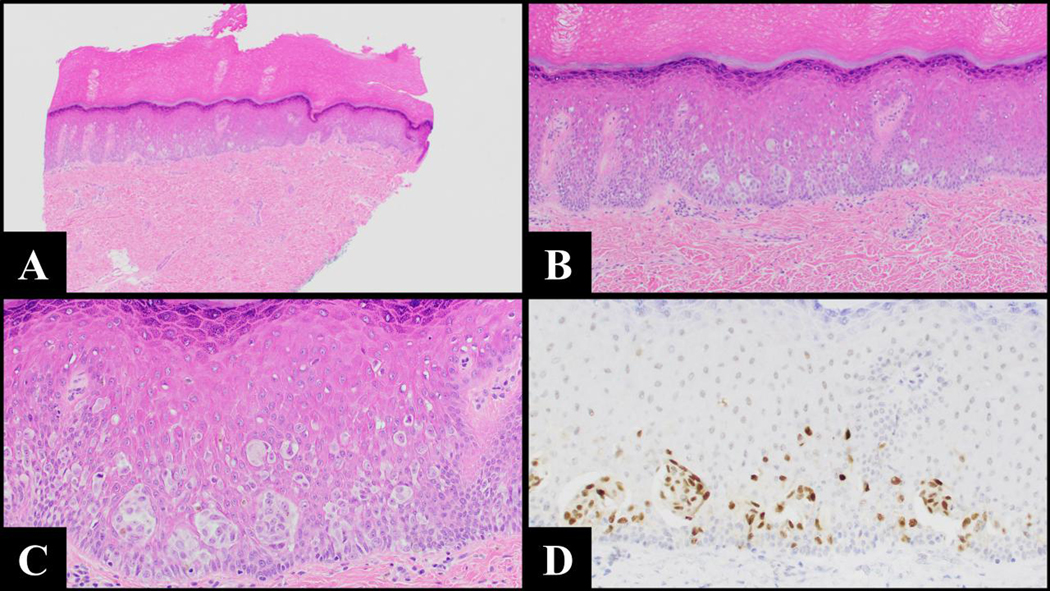
Spitzoid melanocytic neoplasms are a diagnostically challenging class of lesions in dermatopathology. Recently, molecular assays and immunohistochemical markers have been explored as ancillary methods to assist in the diagnostic workup. Specifically, preferentially expressed antigen in melanoma (PRAME) immunohistochemistry is a nuclear stain commonly positive in melanomas, but not in nevi. This study investigates PRAME immunoreactivity (≥75% positive nuclear staining in tumor cells) in a set of 59 spitzoid melanocytic neoplasms with known clinical outcomes. We compared PRAME status with (1) the clinical outcomes, (2) the morphologic diagnoses, and (3) the status of TERT promoter mutation. Regarding clinical outcomes, 3 cases developed metastatic disease, of which 2 expressed diffusely positive PRAME staining. Of the 56 cases that did not show evidence of metastasis, 6 expressed diffusely positive PRAME staining. Morphologically, diffusely positive PRAME staining was seen in 7 of 21 cases (33.3%) diagnosed as melanoma and only 1 benign tumor 1 of 38 (2.6%). There were 4 of 8 cases with a TERT promoter mutation which were diffusely PRAME-positive compared with 4 of 51 cases without TERT promoter mutation ( P = 0.001). Our results show a statistically significant correlation between PRAME expression and the diagnosis, outcome, and TERT promoter mutation status of atypical spitzoid melanocytic neoplasms, suggesting immunohistochemistry for PRAME can help support a suspected diagnosis. However, because of occasional false-positive and negative test results, correlation with the clinical and histologic findings as well as results from other tests is needed for the interpretation of diagnostically challenging spitzoid melanocytic neoplasms.


Conflict of interest statement

Conflict of Interest

The authors have disclosed that they have no significant relationships with, or financial interest in, any commercial companies pertaining to this article.

Figures

Figure 1.
Figure 1.
A) 2x magnification shows the surface of a bulky wedge-shaped melanocytic proliferation with mixed epithelioid and spindle components (2x, H&E). B) At 40x magnification of the more superficial aspect of the tumor, we can appreciate an epithelioid component in which the cells have some spitzoid morphology. This includes vesicular nuclei with prominent nucleoli and moderate amounts of eosinophilic cytoplasm (40x, H&E). C) The deeper component of the tumor shows fascicles of spindle shaped melanocytes also with some spitzoid cytomorphologic features but with severe nuclear atypia with irregular nuclear contours and course nuclear chromatin (40x, H&E). D) PRAME IHC with a normal positive control shows a complete absence of nuclear staining (4x, PRAME IHC). This case resulted in widespread metastatic distant disease.
Figure 2.
Figure 2.
A) and B) 4x and 10x magnifications, respectively, show a bulky, nodular proliferation of atypical melanocytes with expansile and coalescing nests (4x, H&E; 10x, H&E). C) High power magnification shows melanocytes with some spitzoid cytomorphologic features including moderate amounts of pale eosinophilic cytoplasm but with notable nuclear atypia and brisk mitotic activity (40x, H&E). D) PRAME IHC shows strong expression throughout the lesion (20x, PRAME IHC). This lesion resulted in bulky metastatic disease to the parotic gland.
Figure 3.
Figure 3.
A) At 2x, one can appreciate an ulcerated melanocytic neoplasm with sheet-like proliferation of spitzoid melanocytes (2x, H&E). B) and C) Higher magnification shows sheet-like proliferation of severely atypical melanocytes with spitzoid morphology and mitotic activity (10x, H&E; 40x, H&E). D) PRAME IHC shows strong expression throughout the lesion (4x, PRAME IHC). This tumor resulted in widely metastatic disease.
Figure 4.
Figure 4.
A) Acral Spitz nevus from the sole of the right foot of a 4-year old child with a well circumscribed proliferation of nested and single pagetoid melanocytes with overlying epidermal hyperplasia (4x, H&E). B) At 10x, one can appreciate uniform upward migration of melanocytes (10x, H&E). C) The melanocytes have spitzoid cytomorphology with abundant eosinophilic cytoplasm and vesicular nuclei with open chromatin (20x, H&E). D) The melanocytic cells show uniform strong nuclear expression of PRAME (20x, PRAME). This lesion showed no evidence of recurrence after complete excision.

Similar articles

Cited by

References

    1. Menezes FD, Mooi WJ. Spitz Tumors of the Skin. Surg Pathol Clin. 2017. Jun;10(2):281–298. doi: 10.1016/j.path.2017.01.004. Epub 2017 Mar 31. PMID: 28477881. - DOI - PubMed
    1. Barnhill RL. The Spitzoid lesion: rethinking Spitz tumors, atypical variants, ‘Spitzoid melanoma’ and risk assessment. Mod Pathol. 2006. Feb;19 Suppl 2:S21–33. doi: 10.1038/modpathol.3800519. PMID: 16446713. - DOI - PubMed
    1. Cerroni L, Barnhill R, Elder D, Gottlieb G, Heenan P, Kutzner H, et al. Melanocytic tumors of uncertain malignant potential: results of a tutorial held at the XXIX Symposium of the International Society of Dermatopathology in Graz, October 2008. Am J Surg Pathol. 2010. Mar;34(3):314–26. doi: 10.1097/PAS.0b013e3181cf7fa0. PMID: 20118771. - DOI - PubMed
    1. LeBoit PE. “Safe” Spitz and its alternatives. Pediatr Dermatol. 2002. Mar-Apr;19(2):163–5. doi: 10.1046/j.1525-1470.2002.00032.x. PMID: 11994185. - DOI - PubMed
    1. Mones JM, Ackerman AB. “Atypical” Spitz’s nevus, “malignant” Spitz’s nevus, and “metastasizing” Spitz’s nevus: a critique in historical perspective of three concepts flawed fatally. Am J Dermatopathol. 2004. Aug;26(4):310–33. doi: 10.1097/00000372-200408000-00008. PMID: 15249862. - DOI - PubMed