. 2022 Aug 1;137(2):212-231.
doi: 10.1097/ALN.0000000000004264.

Isoflurane Disrupts Postsynaptic Density-95 Protein Interactions Causing Neuronal Synapse Loss and Cognitive Impairment in Juvenile Mice via Canonical NO-mediated Protein Kinase-G Signaling

Affiliations

Isoflurane Disrupts Postsynaptic Density-95 Protein Interactions Causing Neuronal Synapse Loss and Cognitive Impairment in Juvenile Mice via Canonical NO-mediated Protein Kinase-G Signaling

Swati Agarwal et al. Anesthesiology. .

Abstract

Background: Inhalational anesthetics are known to disrupt PDZ2 domain-mediated protein-protein interactions of the postsynaptic density (PSD)-95 protein. The aim of this study is to investigate the underlying mechanisms in response to early isoflurane exposure on synaptic PSD-95 PDZ2 domain disruption that altered spine densities and cognitive function. The authors hypothesized that activation of protein kinase-G by the components of nitric oxide (NO) signaling pathway constitutes a mechanism that prevents loss of early dendritic spines and synapse in neurons and cognitive impairment in mice in response to disruption of PDZ2 domain of the PSD-95 protein.

Methods: Postnatal day 7 mice were exposed to 1.5% isoflurane for 4 h or injected with 8 mg/kg active PSD-95 wild-type PDZ2 peptide or soluble guanylyl cyclase activator YC-1 along with their respective controls. Primary neurons at 7 days in vitro were exposed to isoflurane or PSD-95 wild-type PDZ2 peptide for 4 h. Coimmunoprecipitation, spine density, synapses, cyclic guanosine monophosphate-dependent protein kinase activity, and novel object recognition memory were assessed.

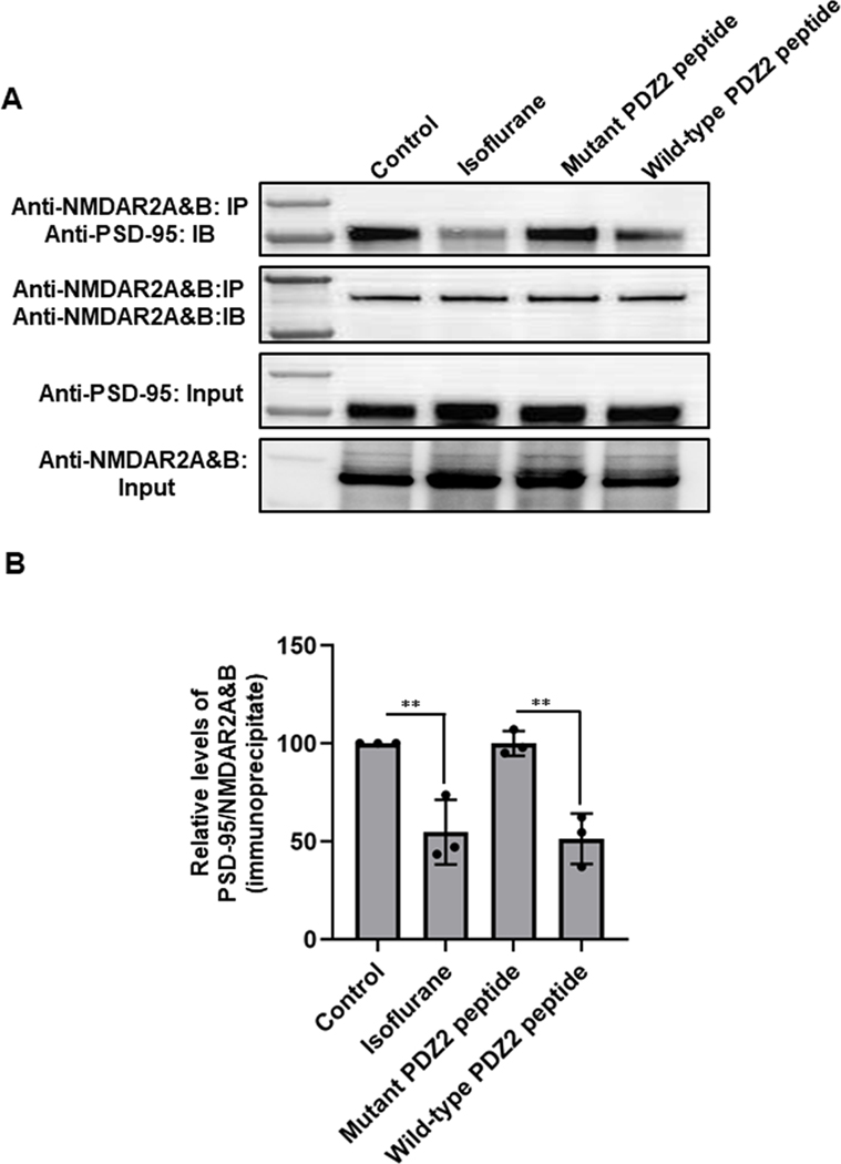
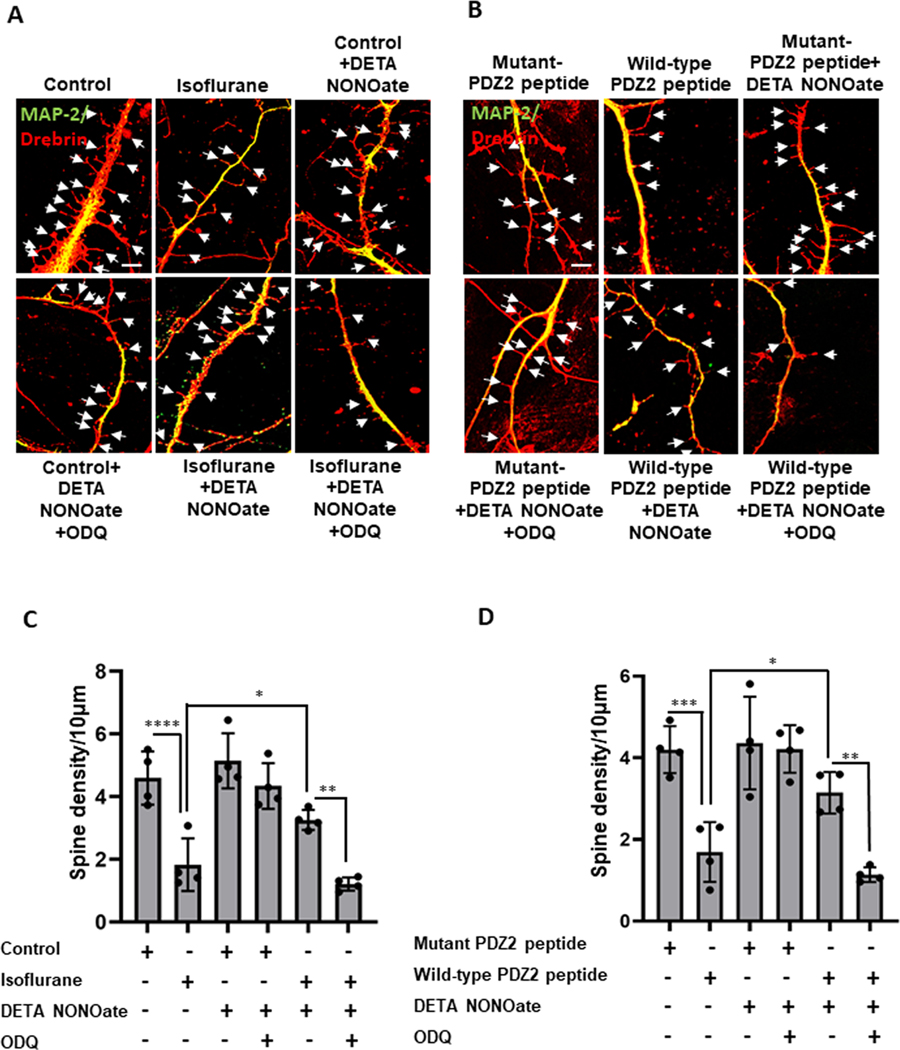
Results: Exposure of isoflurane or PSD-95 wild-type PDZ2 peptide relative to controls causes the following. First, there is a decrease in PSD-95 coimmunoprecipitate relative to N-methyl-d-aspartate receptor subunits NR2A and NR2B precipitate (mean ± SD [in percentage of control]: isoflurane, 54.73 ± 16.52, P = 0.001; and PSD-95 wild-type PDZ2 peptide, 51.32 ± 12.93, P = 0.001). Second, there is a loss in spine density (mean ± SD [spine density per 10 µm]: control, 5.28 ± 0.56 vs. isoflurane, 2.23 ± 0.67, P < 0.0001; and PSD-95 mutant PDZ2 peptide, 4.74 ± 0.94 vs. PSD-95 wild-type PDZ2 peptide, 1.47 ± 0.87, P < 0.001) and a decrease in synaptic puncta (mean ± SD [in percentage of control]: isoflurane, 41.1 ± 14.38, P = 0.001; and PSD-95 wild-type PDZ2 peptide, 50.49 ± 14.31, P < 0.001). NO donor or cyclic guanosine monophosphate analog prevents the spines and synapse loss and decline in the cyclic guanosine monophosphate-dependent protein kinase activity, but this prevention was blocked by soluble guanylyl cyclase or protein kinase-G inhibitors in primary neurons. Third, there were deficits in object recognition at 5 weeks (mean ± SD [recognition index]: male, control, 64.08 ± 10.57 vs. isoflurane, 48.49 ± 13.41, P = 0.001, n = 60; and female, control, 67.13 ± 11.17 vs. isoflurane, 53.76 ± 6.64, P = 0.003, n = 58). Isoflurane-induced impairment in recognition memory was preventable by the introduction of YC-1.

Conclusions: Activation of soluble guanylyl cyclase or protein kinase-G prevents isoflurane or PSD-95 wild-type PDZ2 peptide-induced loss of dendritic spines and synapse. Prevention of recognition memory with YC-1, a NO-independent activator of guanylyl cyclase, supports a role for the soluble guanylyl cyclase mediated protein kinase-G signaling in countering the effects of isoflurane-induced cognitive impairment.


Conflict of interest statement

Conflicts of Interest: The authors declare no competing interests.

Figures

Fig.1.
Fig.1.
Disruption of NMDA receptor NR2A/2B and PSD-95PDZ2 protein interactions by isoflurane and PSD-95 wild-type PDZ2 peptide in the hippocampus of neonatal mouse brain. At postnatal day 7, mouse brains were harvested immediately after exposure to control conditions (50% oxygen: control), 1.5% isoflurane in 50% O2 (isoflurane), 8 mg/kg inactive PSD-95 mutant PDZ2 peptide referred as (mutant PDZ2 peptide) or 8 mg/kg active PSD-95 wild-type PDZ2 peptide referred as (wild-type PDZ2 peptide), in figures. (A) Representative Western blot shows co-immunoprecipitation of PSD-95 and NMDAR2A&B subunits in control, isoflurane, PSD-95 mutant PDZ2 peptide and PSD-95 wild-type PDZ2 peptide treated groups. The panel represents equal amounts of the NMDAR2A&B immunoprecipitated from mouse brain hippocampus with NMDAR2A&B antibody. The amount of sample loaded for the input was 10% of that for the immunoprecipitation. (B) Representative densitometric analysis of PSD-95 co-immunoprecipitate relative to NMDAR2A&B precipitate. Data represent mean ± SD, n=3 independent experiments. **P < 0.01 compared to respective control. Data were analyzed using one-way ANOVA with Bonferroni’s multiple comparisons test. IP, immunoprecipitate; IB, immunoblot.
Fig.2.
Fig.2.
Effect of isoflurane and PSD-95 wild-type PDZ2 peptide on neuronal cell viability and dendritic spines. Primary hippocampal neurons were cultured until 7 days in vitro. (A) Representative fluorescent photomicrograph (40X) with neuronal-specific marker MAP-2. DAPI counterstain was used to identify nuclei. Scale bar =5 µm. (B, C) Neuronal cells were exposed for 4 h to control conditions of 25% O2 and 5% CO2 that was balanced with N2 or to 1.5% isoflurane in a gas of 25% O2 and 5% CO2 that was balanced with N2. Other hippocampal neurons were exposed to 1 µM inactive PSD-95 mutant PDZ2 peptide or 1 µM active PSD-95 wild-type PDZ2 peptide for 4 h. Cell viability was assessed with the MTT assay. Viability was not significantly different in the experimental, control and untreated groups. Data represent mean ± SD, n=3 independent culture experiments and were analyzed by one-way ANOVA with Tukey’s multiple comparisons test. (D, E) Neuronal cells were co-immunostained with MAP-2 and drebrin. Representative confocal photomicrographs (63x) show dendritic segments and spines after Isoflurane (D) or PSD-95 wild-type PDZ2 peptide (E) exposures. Lower panels show (2x) zoomed image of the boxed areas in the upper images. Arrows indicate the dendritic spines. Scale bar=20 µm upper panel and 5=µm lower panel. (F, G) Summary plots represent total spine density per 10 µm dendrite length after isoflurane (F) or PSD-95 wild-type PDZ2 peptide (G) exposure in comparison with respective controls. (H, I) Summary plots represent mean spine length after isoflurane (H) or PSD-95 wild-type PDZ2 peptide (I) exposure in comparison with respective controls. Data were analyzed by independent two-tailed unpaired t-test. Data represent mean ± SD, n=5 independent experiments for spine density and spine length. *P < 0.05, *** P < 0.001, **** P <0.0001, compared with respective control.
Fig.3.
Fig.3.
Soluble guanylate cyclase inhibitor ODQ blocked the ability of NO donor DETA NONOate to prevent isoflurane and PSD-95 wild-type PDZ2 peptide induced dendritic spine loss. (A, B) Primary neuronal cells were pre-treated with the DETA NONOate or the sGC inhibitor ODQ for 4 h, neuronal cells were exposed to the conditions shown and were co-immunostained for MAP-2/drebrin and total spine density was calculated. Representative photomicrographs (63x) show dendritic spines in the hippocampal neurons after respective treatments, scale bar = 10 µm. Arrows indicate the dendritic spines. (C, D) Summary plots represent spine density per 10 µm dendrite length. Data were analyzed using one-way ANOVA with Bonferroni’s multiple comparisons test. Data represent mean ± SD, n=4 independent experiments. *P < 0.05, **P < 0.01, ***P < 0.001, ****P < 0.0001 compared to respective control or as indicated.
Fig.4.
Fig.4.
Protein kinase-G inhibitor KT5823 blocked the ability of 8-Br-cGMP to prevent isoflurane- and PSD-95 wild-type PDZ2 peptide induced dendritic spine loss and decline in cyclic GMP-dependent protein kinase activity. (A, B) Primary neuronal cells were pre-treated with 8-Br-cGMP or KT5823 at the time of gas or peptides exposure and then co-immunostained for MAP-2/drebrin and total spine density was calculated. Representative photomicrographs (63x) show dendritic spines in the hippocampal neurons after respective treatments, scale bar=10 µm. Arrows indicate the dendritic spines. (C, D) Summary plots represent spine density per 10 µm dendrite length. Data were analyzed using one-way ANOVA with Bonferroni’s multiple comparisons test. Data represent mean ± SD, n=4 independent experiments. *P < 0.05, ***P < 0.001, ****P < 0.0001 compared with respective control or as indicated. Hippocampal neurons were exposed to isoflurane or PSD-95 wild-type PDZ2 peptide in the presence/absence of 8-Br-cGMP or KT5823, and cyclic GMP-dependent kinase activity was assayed. (E, F) Graphs represent the relative cyclic GMP-dependent protein kinase activity levels after respective treatments. Data were analyzed using one-way ANOVA with Bonferroni’s multiple comparisons test. Data represent mean ± SD, n=3 independent experiments. *P < 0.05, **P < 0.01, ***P < 0.001 compared with respective controls or as indicated.
Fig.5
Fig.5
-Br-cGMP and YC-1 prevent isoflurane induced loss in synapse, blocked in presence of KT5823. Primary neuronal cells were pre-treated with 8-Br-cGMP or YC-1 or KT5823 at the time of gas exposure. (A) Representative fluorescent images of primary neurons co-immunostained at 14 days in vitro for pre- and post-synaptic markers synaptophysin (green), and PSD-95 (red), respectively, and MAP-2 (magenta). Arrows in the inset indicate the co-localized puncta of synaptophysin and PSD-95. Scale bar = 20μm (B) Co-localization between synaptophysin and PSD-95 was analyzed for quantification of synapses in neuronal cells. Graph represents mean ± SD [in percentage], n = 4 independent experiments. Data were analyzed using one-way ANOVA with Bonferroni’s multiple comparisons test. *p < 0.05 and **p < 0.01 compared with respective controls or as indicated.
Fig.6.
Fig.6.
PSD-95 wild-type PDZ2 peptide exposure causes synapse loss which is mitigated in the presence of 8-Br-cGMP and YC-1. Primary neuronal cells were pre-treated with 8-Br-cGMP or YC-1 or KT5823 at the time of peptides exposure. (A) Representative fluorescent images of primary neurons co-immunostained at 14 days in vitro for pre- and post-synaptic markers synaptophysin (green) and PSD-95 (red), respectively, and MAP2 (magenta). Arrows in the inset indicate the co-localized puncta of synaptophysin and PSD-95. Scale bars = 20μm (B) Co-localization between synaptophysin and PSD-95 was analyzed for quantification of synapses in neuronal cells. Graph represents mean ± SD [in percentage], n = 4 independent experiments. Data were analyzed using one-way ANOVA with Bonferroni’s multiple comparisons test. *p < 0.05 and ***p < 0.001 compared with respective controls or as indicated.
Fig.7.
Fig.7.
Neonatal exposure to isoflurane impairs recognition memory at 5 weeks in both male and female mice. At 1 week, mice were exposed to oxygen (control) or isoflurane and were injected with YC-1 or Vehicle (A, C) Plots representing time mice spent investigating novel or known objects among experimental groups. Data are plotted as; mean [seconds] ± SD for novel versus known ; male ; oxygen control +vehicle (N=15), P=0.001; oxygen control +YC-1 (N=15), P=0.001; isoflurane +YC-1 (N=15) , P=0.014; isoflurane + vehicle (N=15), P = 0.940, Female; oxygen control +vehicle (N=14), P<0.0001; oxygen control +YC-1(N=15), P=0.007; isoflurane +YC-1(N=15), P=0.009 ; isoflurane + vehicle (N=14), P = 0.091 Data were analyzed with paired-t-test. (B, D) Plot showing recognition index as % time investigating novel object/total time investigating both objects × 100; Data were analyzed using one-way ANOVA with Bonferroni’s multiple comparisons test. Mean recognition index ± SD, in male; oxygen control+vehicle versus isoflurane+vehicle, P=0.001, oxygen control+vehicle versus oxygen control+YC-1, P>0.999, oxygen control+vehicle versus isoflurane+YC-1, P=0.486 and in female; oxygen control+vehicle versus isoflurane+vehicle P=0.003, oxygen control+vehicle versus oxygen control+YC-1, P=0.667, oxygen control+vehicle versus isoflurane+YC-1, P=0.100 *P < 0.050, **P < 0.01, ****P < 0.0001 compared with respective controls or as indicated.
Fig.8.
Fig.8.
Schematic representation illustrates the dissociation of NMDAR-PSD95-nNOS interaction induced by isoflurane or PSD-95 wild-type PDZ2 peptide. # To avoid complexity, not all PSD-95 family members are shown. Basal conditions: The NMDA receptor associates with neuronal nitric oxide synthase (nNOS) a downstream signaling molecule through PSD-95, linked through its first and second PDZ domain, PSD-95 forms a ternary complex by binding to NMDAR NR2 subunit and PDZ domain in nNOS. NMDAR-PSD-95/93-nNOS complexes helps Ca2+ influx through NMDAR on the postsynaptic membrane to activate nNOS. The NO generated is required for neuronal plasticity. It acts predominantly via cGMP and protein kinase G (PKG) for activation of kinase pathways. NO-independent pathways may also be activated by NMDAR. NO exerts significant control over gene expression.

Similar articles

Cited by

References

    1. Liu X, Ji J, Zhao GQ: General anesthesia affecting on developing brain: evidence from animal to clinical research. J Anesth 2020; 34: 765–772 - PMC - PubMed
    1. Davidson AJ, Sun LS: Clinical Evidence for Any Effect of Anesthesia on the Developing Brain. Anesthesiology 2018; 128: 840–853 - PubMed
    1. Walters JL, Paule MG: Review of preclinical studies on pediatric general anesthesia-induced developmental neurotoxicity. Neurotoxicol Teratol 2017; 60: 2–23 - PubMed
    1. Lee JR, Loepke AW: Does pediatric anesthesia cause brain damage? - Addressing parental and provider concerns in light of compelling animal studies and seemingly ambivalent human data. Korean J Anesthesiol 2018; 71: 255–273 - PMC - PubMed
    1. Disma N, O’Leary JD, Loepke AW, Brambrink AM, Becke K, Clausen NG, De Graaff JC, Liu F, Hansen TG, McCann ME, Salorio CF, Soriano S, Sun LS, Szmuk P, Warner DO, Vutskits L, Davidson AJ: Anesthesia and the developing brain: A way forward for laboratory and clinical research. Paediatr Anaesth 2018; 28: 758–763 - PubMed

Publication types