. 2022 Jun 2;30(6):886-899.e4.
doi: 10.1016/j.str.2022.04.004. Epub 2022 May 2.

The GTP responsiveness of PI5P4Kβ evolved from a compromised trade-off between activity and specificity

Affiliations

The GTP responsiveness of PI5P4Kβ evolved from a compromised trade-off between activity and specificity

Koh Takeuchi et al. Structure. .

Abstract

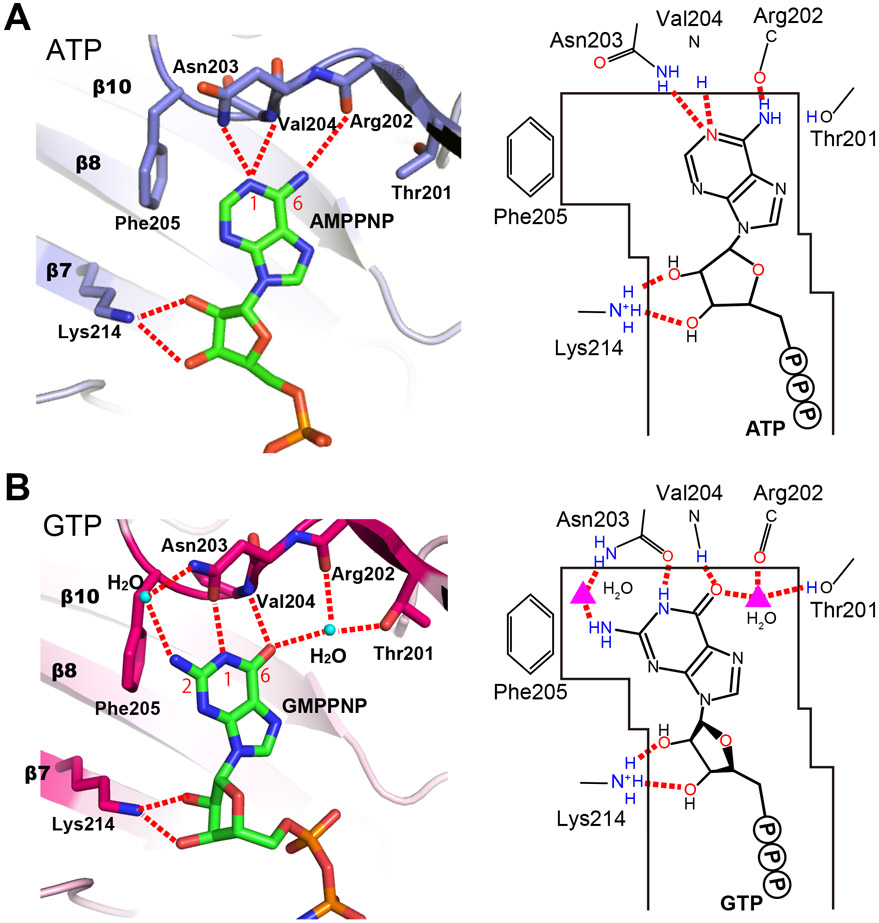
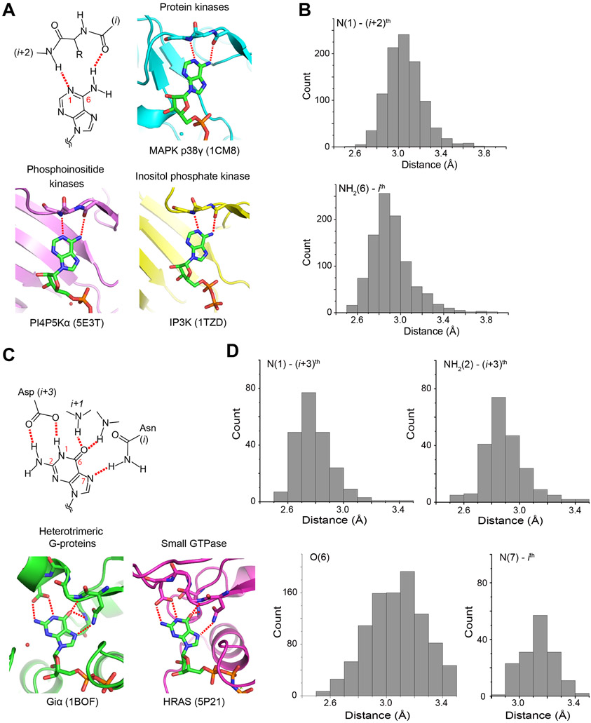
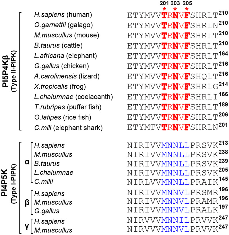
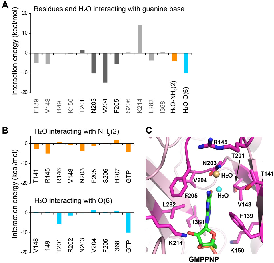
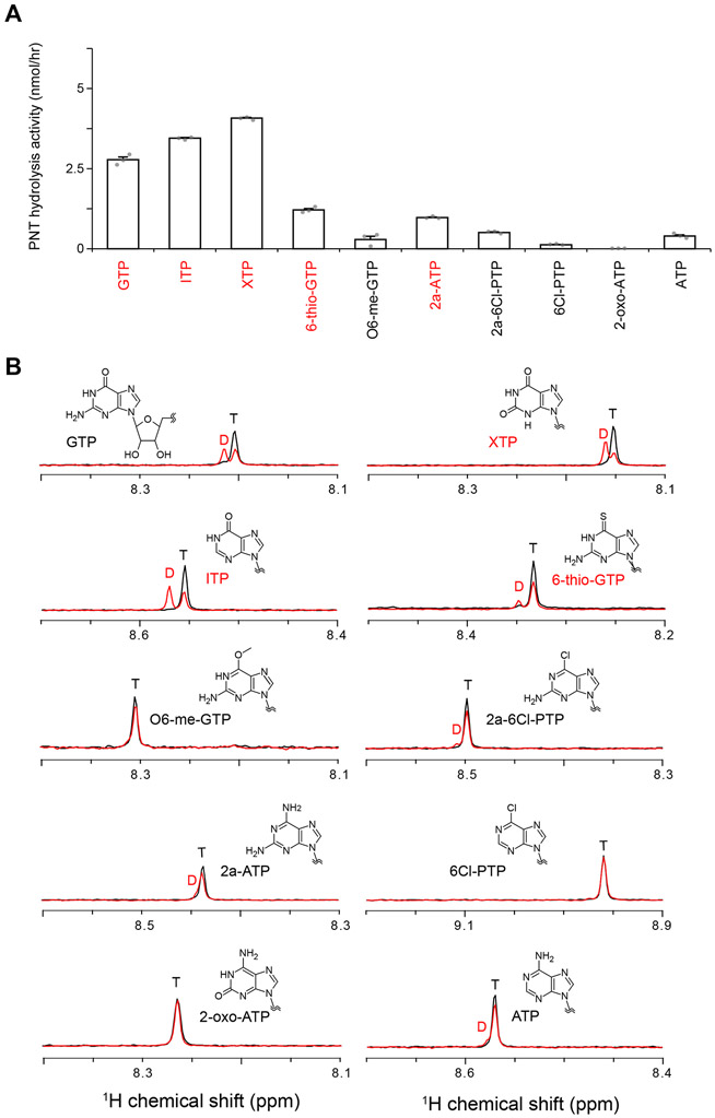
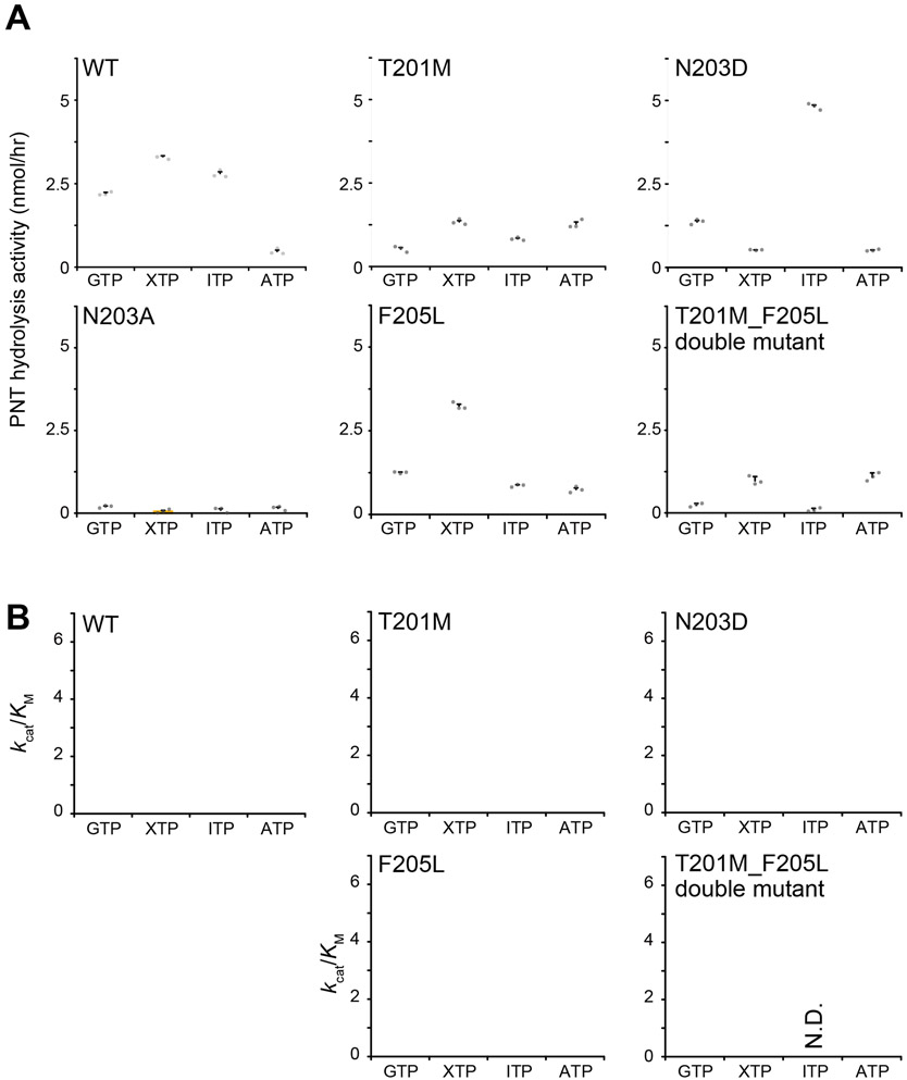
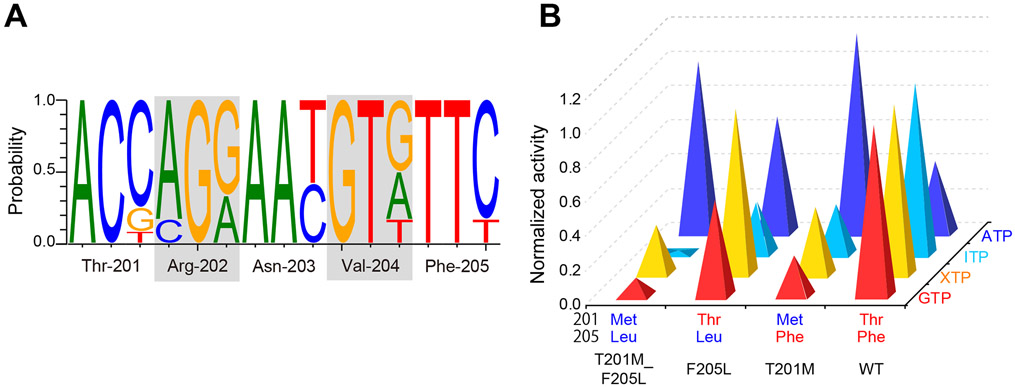
Unlike most kinases, phosphatidylinositol 5-phosphate 4-kinase β (PI5P4Kβ) utilizes GTP as a physiological phosphate donor and regulates cell growth under stress (i.e., GTP-dependent stress resilience). However, the genesis and evolution of its GTP responsiveness remain unknown. Here, we reveal that PI5P4Kβ has acquired GTP preference by generating a short dual-nucleotide-recognizing motif called the guanine efficient association (GEA) motif. Comparison of nucleobase recognition with 660 kinases and 128 G proteins has uncovered that most kinases and PI5P4Kβ use their main-chain atoms for adenine recognition, while the side-chain atoms are required for guanine recognition. Mutational analysis of the GEA motif revealed that the acquisition of GTP reactivity is accompanied by an extended activity toward inosine triphosphate (ITP) and xanthosine triphosphate (XTP). Along with the evolutionary analysis data that point to strong negative selection of the GEA motif, these results suggest that the GTP responsiveness of PI5P4Kβ has evolved from a compromised trade-off between activity and specificity, underpinning the development of the GTP-dependent stress resilience.

Keywords: GTP; GTP sensor; PI5P4K; activity; evolution; kinase; specificity; stress resilience.


Conflict of interest statement

Declaration of interests The authors declare no competing interests.

Figures

Figure 1.
Figure 1.. Chemical structures of PNTs.
Chemical structures of ATP and GTP, along with other PNTs used in this study.
Figure 2.
Figure 2.. Distinct ATP- and GTP-binding modes of PI5P4Kβ.
(A) ATP- and (B) GTP-binding modes of PI5P4Kβ determined in our previous study (Sumita et al., 2016). The hydrogen bonds between PNTs and PI5P4Kβ are shown in red dotted lines. PDB ID for the ATP- and GTP-bound structures are 6K4H and 6K4G, respectively. See also Table S6.
Figure 3.
Figure 3.. Nucleotide-base binding by kinases and G-proteins.
(A and C) Schematic representations of typical hydrogen-bond interactions between nucleotide bases and proteins are shown for (A) kinases and (C) G-proteins. Representative nucleotide-complex structures from major protein families are also shown with the PDB IDs. The hydrogen bonds are shown in red dotted lines. (B and D) Histogram of hydrogen bond distances between (B) kinases and adenine bases, and between (D) G-proteins and guanine bases. The interactions are listed in supplemental materials for kinases (Data S1) and G-proteins (Data S2). See also Fig. S1 for structural figures of other kinases and G-proteins, and Table S1 and S2 for the list of PDB ID, respectively.
Figure 4.
Figure 4.. Sequence alignment of PI5P4K and PI4P5K family proteins.
The GTP-recognizing TRNVF sequences and the ATP-recognizing MNNψL sequences are colored in red and blue, respectively. Asterisks indicate the positions Thr-201, Asn-203, and Phe-205 in human PI5P4Kβ.
Figure 5.
Figure 5.. FMO calculation of the PI5P4Kβ-GTP complex.
(A) The energetic contributions of each residue to PI5P4Kβ-guanine base interaction are indicated. The energetic contributions of two water molecules bound to the NH2(2) and O(6) positions of the guanine base moieties are also shown. (B) The energetic contributions of each residue of PI5P4Kβ and GTP to the interaction with water molecules that are bound to the (top) NH2(2) and (bottom) O(6) positions, respectively, of guanine base moieties are indicated. (C) Interacting residues are shown (stick model in magenta) in the PI5P4Kβ-GMPPNP complex structure determined in our previous study (15) (PDB ID 6K4G). The water molecules that interact with NH2(2) and O(6) positions are shown in orange and cyan, respectively.
Figure 6.
Figure 6.. PNT hydrolysis activity of PI5P4Kβ.
(A) PNT hydrolysis activity of PI5P4Kβ. The PNT hydrolysis activity (nmol/hr) of PI5P4Kβ was calculated from the signal intensities of diphosphorylated and triphosphorylated nucleotides NMR signals after the reaction (see Star Methods and panel B). In the PNT-hydrolysis assay, 250 μM PNTs were mixed with 2 μM PI5P4Kβ. The average values from three experiments are shown with error bars (standard deviations; S.D.). The values from each experiment are shown as gray dots. Highly hydrolyzed nucleotides (> 0.1 of the ratios) are indicated by red. (B) Representative spectra in the NMR-based PNT hydrolysis assay. The intensities of H8 protons of diphosphorylated (D)/triphosphorylated (T) nucleotide signals are quantified from the spectra. See also Fig. S3.
Figure 7.
Figure 7.. 2a-ATP, ITP, and XTP binding modes of PI5P4Kβ.
(A) Interaction of 2a-ATP to PI5P4Kβ (PDB ID 7EM3). (B) Interactions of the ITP to PI5P4Kβ(PDB ID 7EM1). (C and D) Interactions of XTP to PI5P4Kβ (C) in the mode similar to GTP (GTP-binding mode) and (D) in the mode unique to XTP (XTP-binding mode) (PDB ID 7EM2). Red dotted lines represent hydrogen bonds between the PNTs and PI5P4Kβ. As for the ITP complex, the water position visible with a lower σ value is also indicated with a gray circle. See also Fig. S4 for overlay of the 2a-ATP, ITP, and XTP binding modes with the ATP or GTP binding modes. See also Table S6 for structural statistics.
Figure 8.
Figure 8.. PNT hydrolysis activities of PI5P4Kβ mutants.
(A) PNT-hydrolysis activities of the WT PI5P4Kβ and T201M, N203D, N203A, F205L, and T201M_F205L double mutants. The PNT hydrolysis activity (nmol/hr) of PI5P4Kβ was calculated from the signal intensity of diphosphorylated nucleotides after the reaction (see Materials and Methods section for details). For the PNT-hydrolysis assay, 250 μM PNTs were mixed with 2 μM PI5P4Kβ. The average values from the three experiments are shown with error bars (S.D.). The values from each experiment are shown as gray dots. (B) The kcat/KM values of PNT hydrolysis activities of the WT PI5P4Kβ and T201M, N203D, F205L, and T201M_F205L double mutants. The values are calculated from KM and kcat values shown in Table S3 and S4, respectively. N.D.: not determined. For the determination of KM and kcat values, the reactions were carried out for different concentrations of PNTs from 63.5 μM, 125 μM, 250 μM, 500 μM, 1 mM, and 2 mM.
Figure 9.
Figure 9.. A trade-off between activity and specificity, and evolution of PI5P4Kβ
(A) Normalized GTP, XTP, ITP, and ATP hydrolysis activity of each mutant compared to WT (for GTP, XTP, and ITP) and PI5P4KβT201M_F205L. The average values taken from Figure 8A were used. (B) Nucleotide fraction of each site that encodes a GEA motif of PI5P4Kβ. The species used are shown in Figure 4 and are listed in Star Methods.

References

    1. Abascal F, Zardoya R, and Telford MJ (2010). TranslatorX: multiple alignment of nucleotide sequences guided by amino acid translations. Nucleic Acids Res 38, W7–13. - PMC - PubMed
    1. Bazenet CE, Ruano AR, Brockman JL, and Anderson RA (1990). The human erythrocyte contains two forms of phosphatidylinositol-4-phosphate 5-kinase which are differentially active toward membranes. Journal of Biological Chemistry 265, 18012–18022. - PubMed
    1. Berman H, Henrick K, and Nakamura H (2003). Announcing the worldwide Protein Data Bank. Nature Structural & Molecular Biology 10, 980–980. - PubMed
    1. Bossemeyer D (1995). Protein kinases--structure and function. FEBS Lett 369, 57–61. - PubMed
    1. Burke JE (2018). Structural Basis for Regulation of Phosphoinositide Kinases and Their Involvement in Human Disease. Mol Cell 71, 653–673. - PubMed

Publication types

MeSH terms

LinkOut - more resources