

Pathogenesis of neural tube defects: The regulation and disruption of cellular processes underlying neural tube closure
- PMID: 35504597
- PMCID: PMC9605354
- DOI: 10.1002/wsbm.1559
Pathogenesis of neural tube defects: The regulation and disruption of cellular processes underlying neural tube closure
Abstract
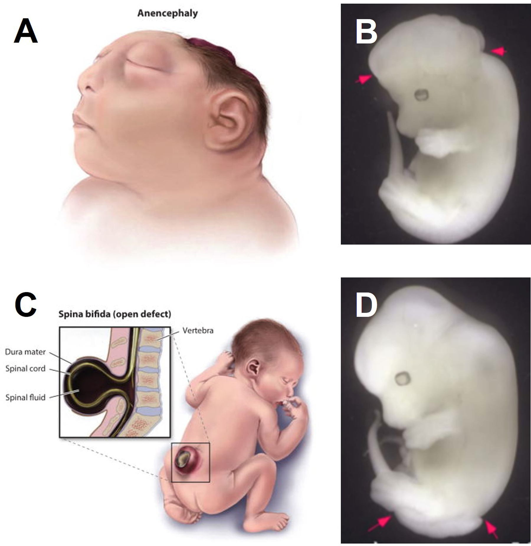
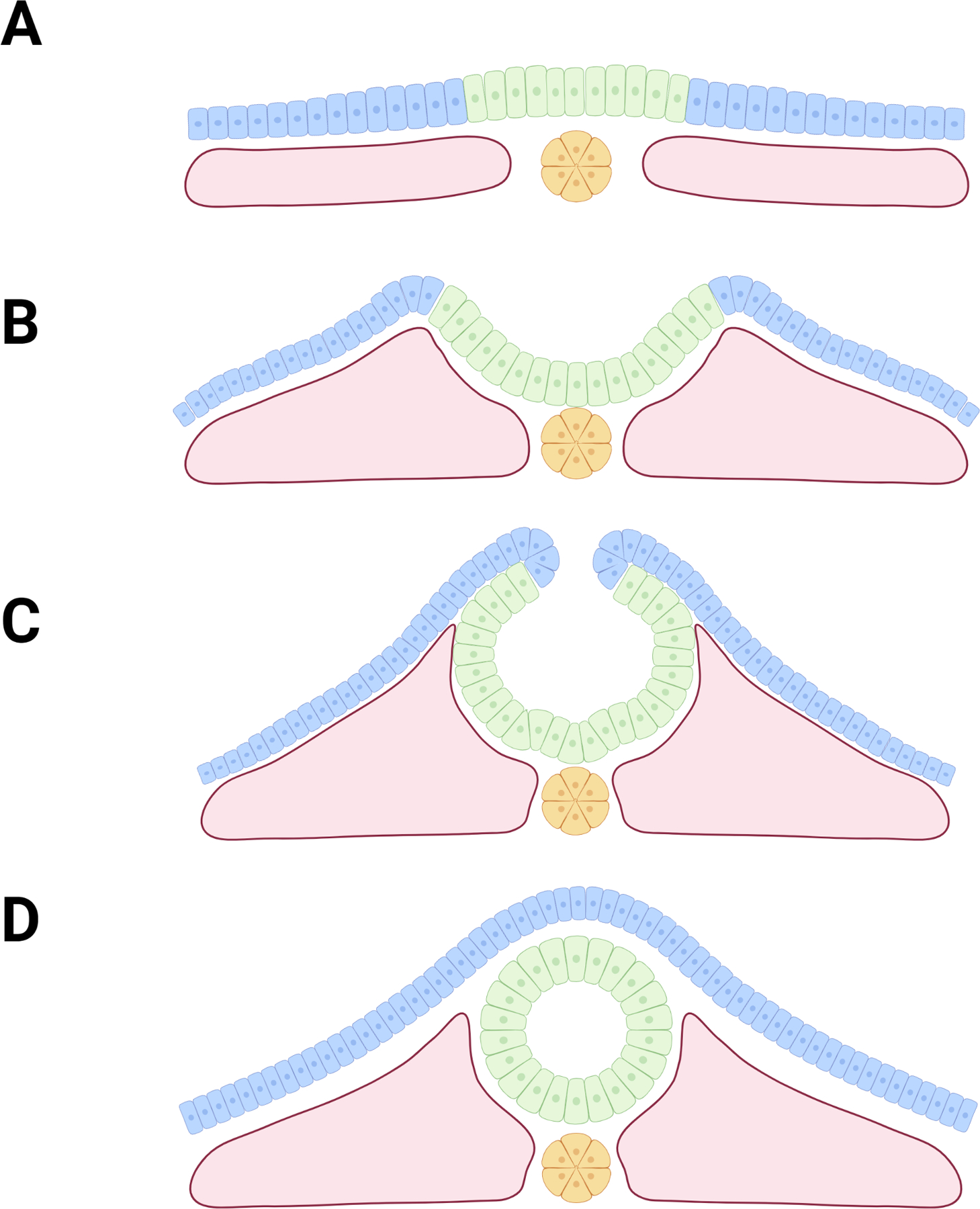
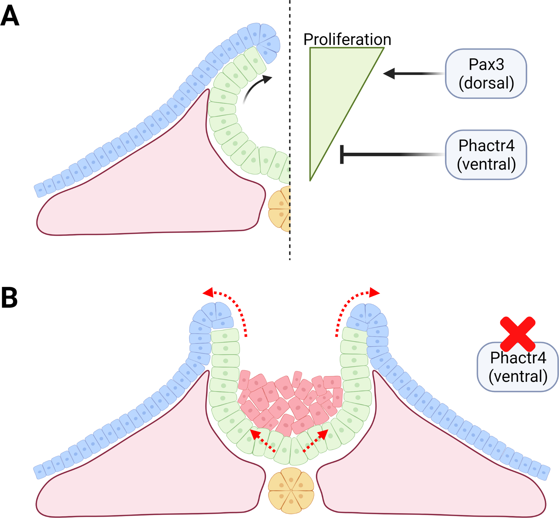
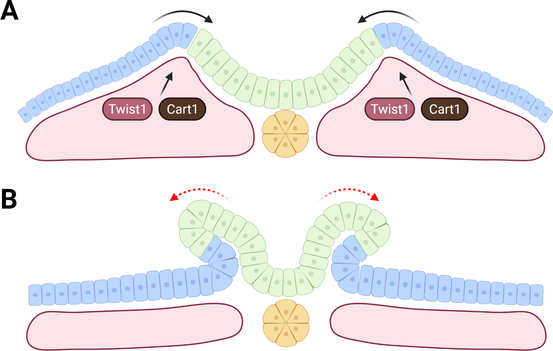
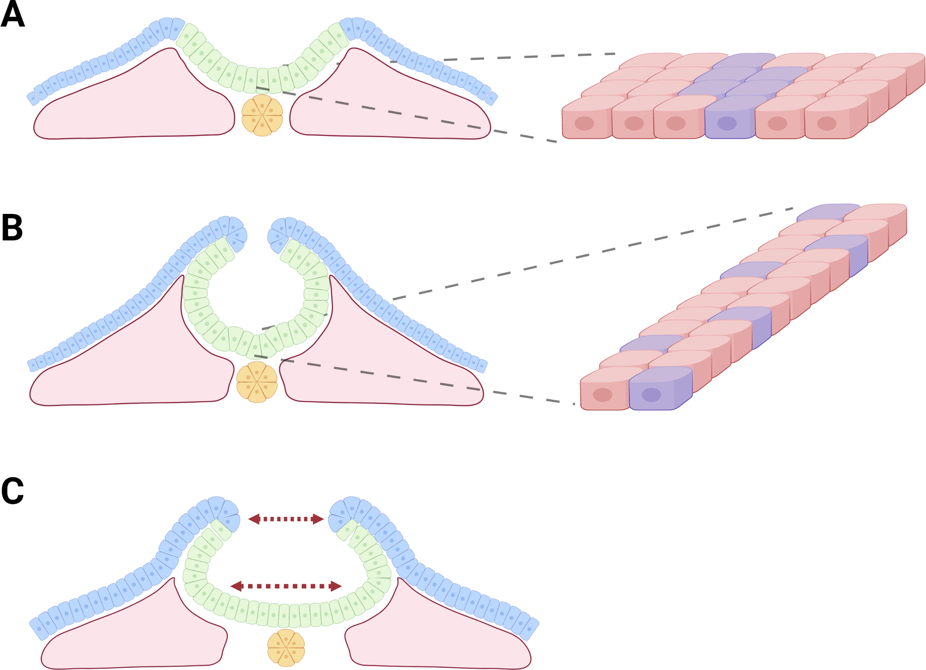
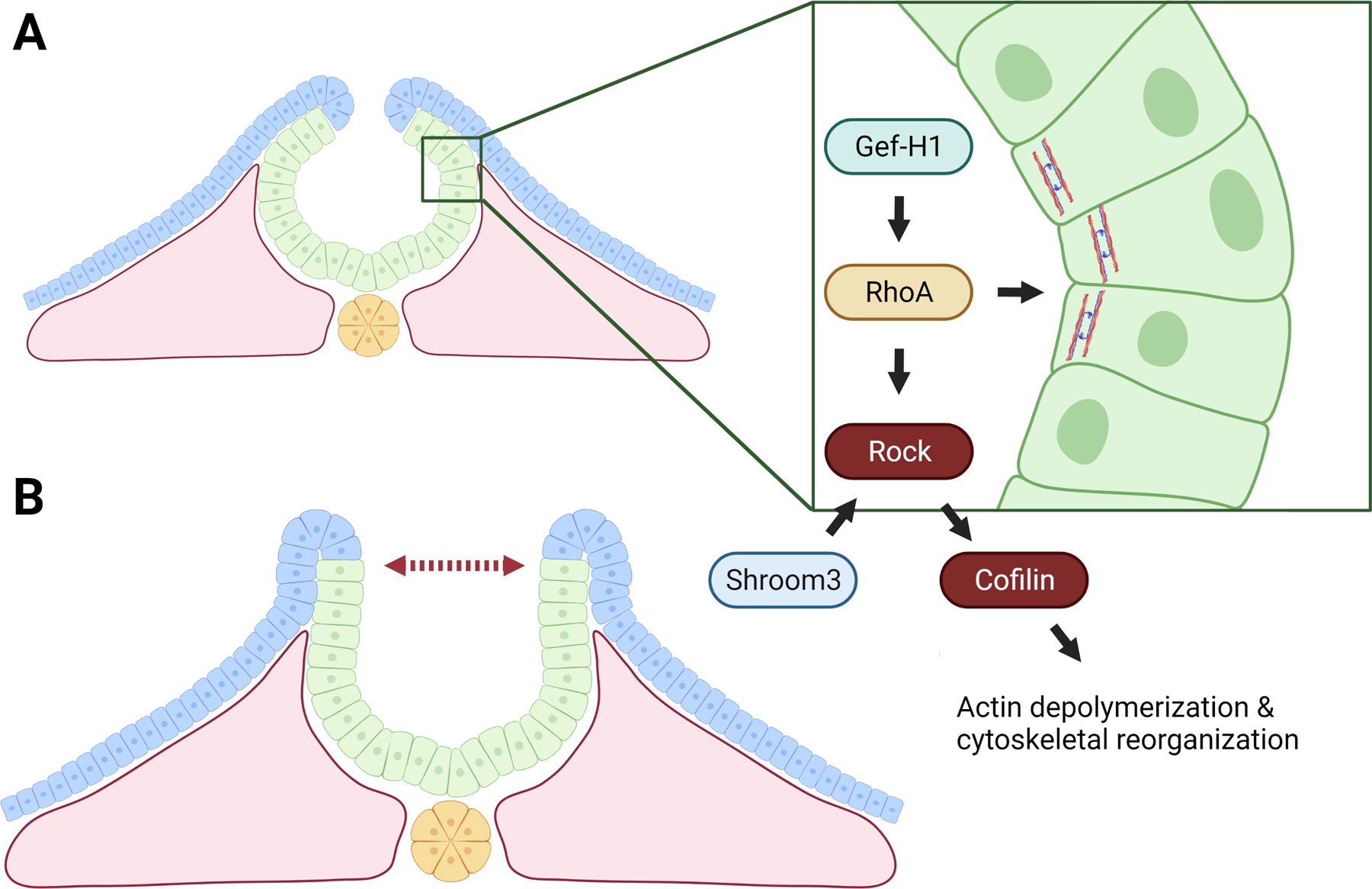
Neural tube closure (NTC) is crucial for proper development of the brain and spinal cord and requires precise morphogenesis from a sheet of cells to an intact three-dimensional structure. NTC is dependent on successful regulation of hundreds of genes, a myriad of signaling pathways, concentration gradients, and is influenced by epigenetic and environmental cues. Failure of NTC is termed a neural tube defect (NTD) and is a leading class of congenital defects in the United States and worldwide. Though NTDs are all defined as incomplete closure of the neural tube, the pathogenesis of an NTD determines the type, severity, positioning, and accompanying phenotypes. In this review, we survey pathogenesis of NTDs relating to disruption of cellular processes arising from genetic mutations, altered epigenetic regulation, and environmental influences by micronutrients and maternal condition. This article is categorized under: Congenital Diseases > Genetics/Genomics/Epigenetics Neurological Diseases > Genetics/Genomics/Epigenetics Neurological Diseases > Stem Cells and Development.
Keywords: environmental factors; epigenetics; folic acid; neural tube defect; neurulation; pathogenesis.
© 2022 Wiley Periodicals LLC.
Conflict of interest statement
Conflict of Interest The Authors declare that there is no conflict of interest.
Figures
References
-
- Abu-Abed S, Dollé P, Metzger D, Beckett B, Chambon P, & Petkovich M (2001). The retinoic acid-metabolizing enzyme, CYP26A1, is essential for normal hindbrain patterning, vertebral identity, and development of posterior structures. Genes & Development, 15, 226–240. 10.1101/gad.855001 - DOI - PMC - PubMed
-
- Araya García CA (2017). Formation of Neural Tube. In Reference Module in Biomedical Sciences. Elsevier. 10.1016/B978-0-12-801238-3.11055-4 - DOI
-
- Arnold C, Demirel E, Feldner A, Genové G, Zhang H, Sticht C, Wieland T, Hecker M, Heximer S, & Korff T (2018). Hypertension-evoked RhoA activity in vascular smooth muscle cells requires RGS5. FASEB Journal: Official Publication of the Federation of American Societies for Experimental Biology, 32(4), 2021–2035. 10.1096/fj.201700384RR - DOI - PubMed
Publication types
MeSH terms
Substances
Grants and funding
LinkOut - more resources
Full Text Sources
Medical
