. 2022 May 3;12(1):7188.
doi: 10.1038/s41598-022-11085-z.

A human pancreatic ECM hydrogel optimized for 3-D modeling of the islet microenvironment

Affiliations

A human pancreatic ECM hydrogel optimized for 3-D modeling of the islet microenvironment

Daniel M Tremmel et al. Sci Rep. .

Abstract

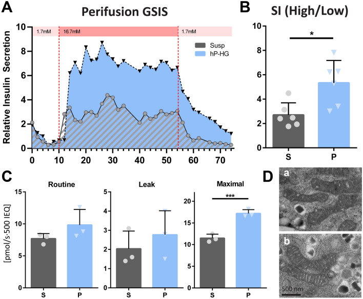
Extracellular matrix (ECM) plays a multitude of roles, including supporting cells through structural and biochemical interactions. ECM is damaged in the process of isolating human islets for clinical transplantation and basic research. A platform in which islets can be cultured in contact with natural pancreatic ECM is desirable to better understand and support islet health, and to recapitulate the native islet environment. Our study demonstrates the derivation of a practical and durable hydrogel from decellularized human pancreas that supports human islet survival and function. Islets embedded in this hydrogel show increased glucose- and KCl-stimulated insulin secretion, and improved mitochondrial function compared to islets cultured without pancreatic matrix. In extended culture, hydrogel co-culture significantly reduced levels of apoptosis compared to suspension culture and preserved controlled glucose-responsive function. Isolated islets displayed altered endocrine and non-endocrine cell arrangement compared to in situ islets; hydrogel preserved an islet architecture more similar to that observed in situ. RNA sequencing confirmed that gene expression differences between islets cultured in suspension and hydrogel largely fell within gene ontology terms related to extracellular signaling and adhesion. Natural pancreatic ECM improves the survival and physiology of isolated human islets.


Conflict of interest statement

The authors declare the following competing financial interest(s): JSO is scientific co-founder of Regenerative Medical Solutions, Inc. and has stock equity.

Figures

Figure 1
Figure 1
Protocol for the decellularization and gelation of human pancreas ECM. A schematic representation for the protocol to decell, digest and form a hydrogel from human pancreas ECM (left side). Images of the native tissue (A), homogenized tissue (B), decellularized and delipidized ECM (C) and multiple 5 μL hydrogel droplets in a 6 cm dish (D). Human islets can be embedded in the hydrogel prior to gelation to form stable droplets for in vitro culture (E); the droplets are durable enough to maintain shape and consistency throughout the transplantation process (F).
Figure 2
Figure 2
Optimized decell protocol removes lipids and DNA, resulting in an improved hydrogel. (A,B) Total lipid content by dry weight of the native and decellularized hP-ECM from the Homog and Optimized protocols, determined using a modified Folch method (A), also displayed on a donor-by-donor basis, in which donors with higher lipid content retain significantly more lipids in the Homog protocol (B). (C) Total DNA content of the native and decellularized hP-ECM from the Homog and Optimized protocols. (D–F) The storage (G’) and loss (G’’) moduli of the optimized hydrogels were less variable than the Homog hP-HG hydrogels, and compared to Col1 controls; temperature is plotted in green (F). (G) The complex viscosity curves of the Optimized protocol are less variable than the Homog protocol, and all hP-HG gels were less firm than Col1 hydrogel of the same concentration. (*p < 0.05, **p < 0.01, ***p < 0.001, ****p < 0.0001).
Figure 3
Figure 3
hP-HG co-culture improves islet function after 2 days of culture. (A) Representative phase contrast images of islets cultured in suspension (“S”) (a), hP-HG (“P”) (b), alginate (“A”) (c), Col1 (“C”) (d), or hK-HG (“K”) (e) were assessed for function on Day 2 of culture (scale = 200 microns). (B) A static glucose stimulated insulin secretion (GSIS) assay was performed using sequential low glucose (2.8 mM, “Low”), high glucose (28 mM, “High”), a return to low glucose, followed by low glucose + KCl (30 mM KCl, “KCl”); basal and stimulated C-peptide (C-pep) secreted during the static GSIS with human islets are shown as a percentage of total C-pep content. Statistics indicated above each bar are relative to the “S” control for the same treatment. (C) Total C-pep content of islets undergoing the indicated treatments. Stimulation index (D, high/low glucose) (E, KCl/low glucose) of human islets cultured in all five conditions, determined by static GSIS. Statistical comparisons indicated in black are relative to “S”, and indicated in blue are relative to “P”. (S,P: n = 9; A,C,K: n = 5 islet donors) (ns not significant, *p < 0.05, **p < 0.01, ***p < 0.001, ****p < 0.0001).
Figure 4
Figure 4
hP-HG culture enables proper dynamic function and enhances maximum respiration. (A) Perifusion GSIS was performed on human islets after 2 days of culture in either suspension (S) or embedded in hP-HG (P), from low glucose (1.7 mM) to high glucose (16.7 mM) and back to low glucose; representative plots shown were generated from 2 technical replicates per treatment, from one islet donor. Insulin secretion is normalized to first low glucose response (unitless). One representative donor is shown, data from additional donors can be found in Supplemental Fig. 4D,E. (B) Average stimulation index (SI, high/low glucose) is reported, N = 3 islet donors. (C) Islets were assessed for mitochondrial respiration after 2 days of culture in either suspension (S) or hP-HG (P); Routine (normal media), Leak (Complex V inhibition), and Maximal (uncoupled) respiration were measured. (D) β cell mitochondrial pathology in suspension (a) and hP-HG (b) culture was assessed with transmission electron microscopy. N = 3 islet donors. Scale = 500 nm. (ns not significant, *p < 0.05, ***p < 0.001).
Figure 5
Figure 5
hP-HG co-culture improves islet survival and function in extended culture. (A,B) Metabolic activity of islets cultured in suspension (S) or hP-HG (P) over a 7 day period was assessed by MTS assay, reflecting islet survival over time (presented as a percentage of the Day 1 value). No significant difference in survival was apparent on day 3 or 5 (A), but a significant difference between S and P was found on day 7 (B). N = 4 islet donors per treatment. (C–E) Islet function changed over the 7 day period, as assessed through static GSIS. Islets maintained a significantly higher stimulation index (ratio of C-pep secreted under high/low glucose) at day 7 (C). The percentage of total C-pep content secreted under low and high glucose is plotted for day 2 and day 7 under both suspension and hP-HG culture conditions (D). Total C-pep content was not significantly different among the two treatments at the two time points (E). N = 7 islet donors. (ns not significant, *p < 0.05, **p < 0.01).
Figure 6
Figure 6
Apoptosis rates and islet architecture are altered in suspension culture and partially preserved in hP-HG. (A) Immunofluorescent staining images for native (in situ) islets, and islets cultured in suspension or hP-HG for 7 days. Islets are stained for TUNEL/Insulin (a–c), Ki67/Insulin (d–f), insulin/glucagon/somatostatin (g–i), and insulin/Tie2/αSMA (j–l). White arrowheads indicate projections of Tie2+ and αSMA+ cells into the hydrogel. All scale bars = 50 microns. (B) Quantification of cells positive for Ki67 (blue bars, significance indicated by blue stars) and TUNEL (red bars, significance indicated by red stars) in native islets, and on day (d) 0, 2 and 7 in suspension (S) and hP-HG (P) culture. N = 4 islet donors. (C) Assessment of islet architecture by counting α (Gcg+), β (Ins+), and δ (Sst+) cells in the islet mantle (checkered) and core (solid). Data is represented as a percentage of total endocrine cells. N = 5 islet donors. (Statistical analyses presented in Supplemental Fig. 7D,E.) (D) Localization of positive staining for Tie2 (light gray) and αSMA (dark gray) in the islet mantle (checkered) and core (solid). N = 5 islet donors. p-values depicted in italics and with the † symbol are relative to the native tissue. (ns not significant, */p < 0.05, **/††p < 0.01, ****/††††p < 0.0001).

References

    1. Prince E, Kumacheva E. Design and applications of man-made biomimetic fibrillar hydrogels. Nat. Rev. Mater. 2019;4:99–115. doi: 10.1038/s41578-018-0077-9. - DOI
    1. Amin J, et al. A simple, reliable method for high-throughput screening for diabetes drugs using 3D beta-cell spheroids. J. Pharmacol. Toxicol. Methods. 2016;82:83–89. doi: 10.1016/j.vascn.2016.08.005. - DOI - PubMed
    1. Walpita D, et al. A human islet cell culture system for high-throughput screening. J. Biomol. Screen. 2012;17:509–518. doi: 10.1177/1087057111430253. - DOI - PMC - PubMed
    1. Foster ED, et al. Improved health-related quality of life in a phase 3 islet transplantation trial in type 1 diabetes complicated by severe hypoglycemia. Diabetes Care. 2018;41:1001–1008. doi: 10.2337/dc17-1779. - DOI - PMC - PubMed
    1. Shapiro AM, et al. Islet transplantation in seven patients with type 1 diabetes mellitus using a glucocorticoid-free immunosuppressive regimen. N. Engl. J. Med. 2000;343:230–238. doi: 10.1056/NEJM200007273430401. - DOI - PubMed

Publication types