. 2022 May 3;12(1):7193.
doi: 10.1038/s41598-022-11212-w.

Nanoscale copper and silver thin film systems display differences in antiviral and antibacterial properties

Affiliations

Nanoscale copper and silver thin film systems display differences in antiviral and antibacterial properties

Toni Luise Meister et al. Sci Rep. .

Abstract

The current Coronavirus Disease 19 (COVID-19) pandemic has exemplified the need for simple and efficient prevention strategies that can be rapidly implemented to mitigate infection risks. Various surfaces have a long history of antimicrobial properties and are well described for the prevention of bacterial infections. However, their effect on many viruses has not been studied in depth. In the context of COVID-19, several surfaces, including copper (Cu) and silver (Ag) coatings have been described as efficient antiviral measures that can easily be implemented to slow viral transmission. In this study, we detected antiviral properties against Severe Acute Respiratory Syndrome Coronavirus-2 (SARS-CoV-2) on surfaces, which were coated with Cu by magnetron sputtering as thin Cu films or as Cu/Ag ultrathin bimetallic nanopatches. However, no effect of Ag on viral titers was observed, in clear contrast to its well-known antibacterial properties. Further enhancement of Ag ion release kinetics based on an electrochemical sacrificial anode mechanism did not increase antiviral activity. These results clearly demonstrate that Cu and Ag thin film systems display significant differences in antiviral and antibacterial properties which need to be considered upon implementation.


Conflict of interest statement

The authors declare no competing interests.

Figures

Figure 1
Figure 1
Schematic illustration (not to scale) of the fabrication of Cu and Ag thin film nanostructures by sputter deposition. (A) Elemental Cu sputtered homogeneously as a dense and continuous film with 50 nm thickness; (B) Cu- and Ag-nanopatches co-sputtered for 120 s; (C) Cu- and Ag-nanopatches sputtered sequentially for each 120 s. Nanopatches are island-like nanostructures with nominal thickness < 5 nm.
Figure 2
Figure 2
Exemplary electron microscopy images of continuous nanoscale films (A, B) and nanopatches (CF). (A) SEM top view of 50 nm Ag, (B) SEM top view of 50 nm Cu, (C) SEM top view of Ag nanopatches sputtered for 60 s, (D) Ag & Pt nanopatches co-sputtered for 60 s, (E) TEM image of Ag & Cu nanopatches co-sputtered for 60 s on a TEM grid, (F) Cu on Ag nanopatches sequentially sputtered for 60 s on a TEM grid. (E, F) taken from reference.
Figure 3
Figure 3
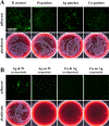
Antibacterial activity towards S. aureus (104 CFU/mL) of (A) a continuous Ti thin film (Ti control) as well as Pt, Ag, and Cu thin nanopatches sputtered on Ti thin film compared to (B) thin Ag/Pt and thin Ag/Cu nanopatches sputtered simultaneously (i.e., co-deposited) or sequentially (first Pt, second Ag or first Ag, second Cu). Sputter time for all samples 60 s. Upper figures: representative fluorescence images of adherent bacteria on sample surfaces after 24 h of incubation and staining with SYTO-9 (green fluorescence); lower images: representative blood agar plates of plated planktonic bacteria in the drop fluid after 24 h of incubation on the different samples (white bacterial colonies indicate viable cells).
Figure 4
Figure 4
Quantitative analysis of the antibacterial activity of silver acetate (AgAc, panel A) and copper sulfate (CuSO4, panel B) solutions towards S. aureus (different bacterial concentrations) performed by the AlamarBlue assay. Data are expressed as mean ± SD of at least three independent experiments and given as the percentage of untreated bacteria (no exposure). Asterisks (*) indicate significant differences (*p ≤ 0.05) compared to the untreated control; hash marks indicate significant differences (*p ≤ 0.05) between AgAc and CuSO4.
Figure 5
Figure 5
Results of antiviral activity for (A) Cu and Ag thin films sputtered on Si/SiO2 pieces or (BD) nanopatches sputtered on Si/SiO2 which were incubated with SARS-CoV-2 for indicated time periods. (EF) Silver acetate (AgAc) and copper sulfate (CuSO4) solutions, used as ionic controls, were spiked with SARS-CoV-2 an incubated for similar time periods. Residual infectious virus was quantified by TCID50 calculation. Dotted line indicates the lower limit of quantification. Data are expressed as mean ± SD of three independent experiments. Asterisks (*) indicate significant differences (*p < 0.05; **p < 0.01; and ***p < 0.001) compared to MOCK (untreated control) or Si/SiO2.

References

    1. Goodman SB, Yao Z, Keeney M, Yang F. The future of biologic coatings for orthopaedic implants. Biomaterials. 2013;34:3174–3183. doi: 10.1016/j.biomaterials.2013.01.074. - DOI - PMC - PubMed
    1. Chernousova S, Epple M. Silver as antibacterial agent: ion, nanoparticle, and metal. Angew. Chem. Int. Ed. 2013;52:1636–1653. doi: 10.1002/anie.201205923. - DOI - PubMed
    1. Grass G, Rensing C, Solioz M. Metallic copper as an antimicrobial surface. Appl. Environ. Microbiol. 2011;77:1541–1547. doi: 10.1128/AEM.02766-10. - DOI - PMC - PubMed
    1. Cameron SJ, Hosseinian F, Willmore WG. A current overview of the biological and cellular effects of nanosilver. Int. J. Mol. Sci. 2018 doi: 10.3390/ijms19072030. - DOI - PMC - PubMed
    1. Loza K, Diendorf J, Sengstock C, Ruiz-Gonzalez L, Gonzalez-Calbet JM, Vallet-Regi M, Köller M, Epple M. The dissolution and biological effects of silver nanoparticles in biological media. J. Mater. Chem. B. 2014;2:1634–1643. doi: 10.1039/c3tb21569e. - DOI - PubMed

Publication types