Review
. 2022 Jul;23(7):411-427.
doi: 10.1038/s41583-022-00588-3. Epub 2022 May 3.

Macroautophagy in CNS health and disease

Affiliations
Review

Macroautophagy in CNS health and disease

Christopher J Griffey et al. Nat Rev Neurosci. 2022 Jul.

Abstract

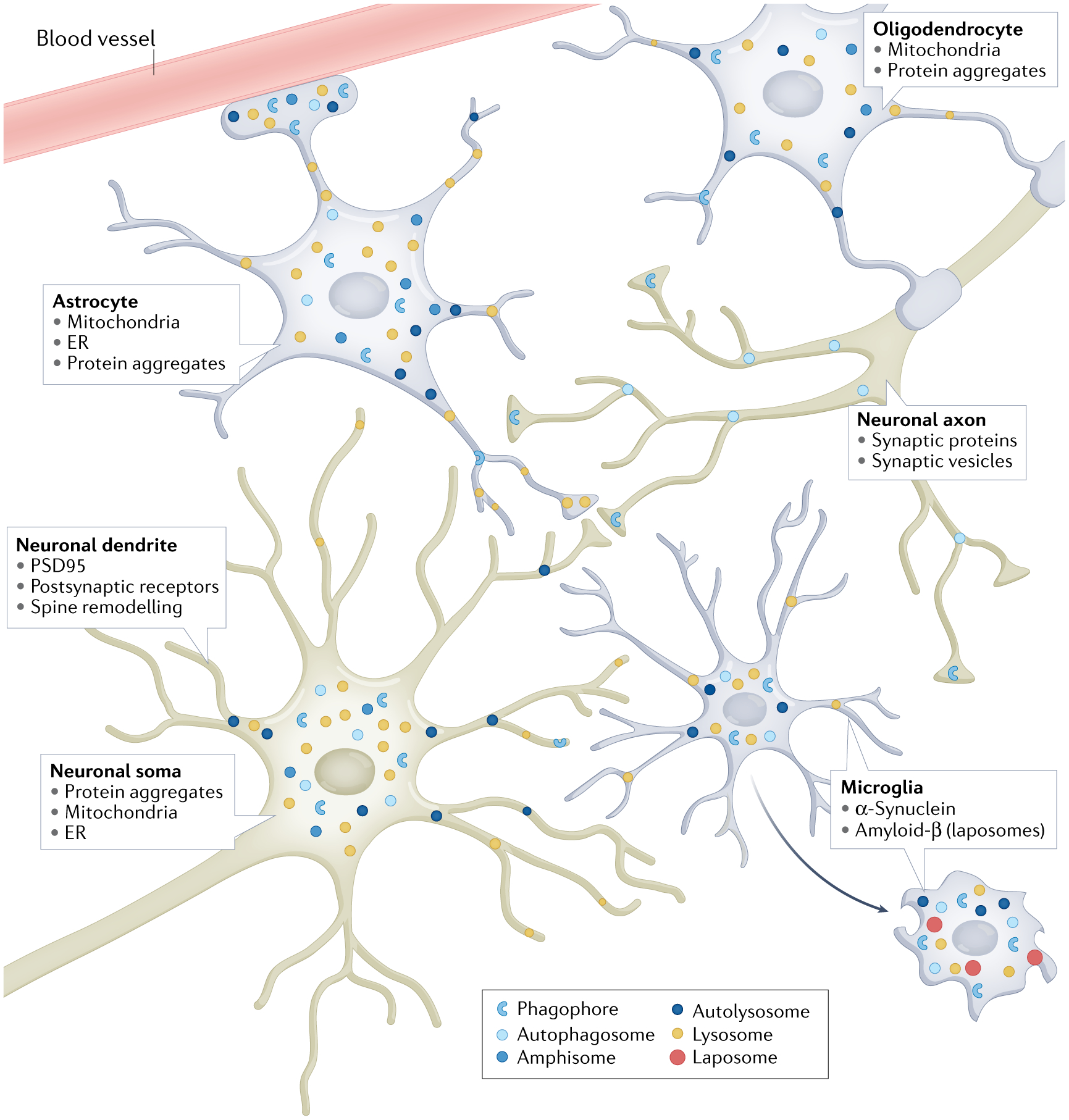
Macroautophagy is an evolutionarily conserved process that delivers diverse cellular contents to lysosomes for degradation. As our understanding of this pathway grows, so does our appreciation for its importance in disorders of the CNS. Once implicated primarily in neurodegenerative events owing to acute injury and ageing, macroautophagy is now also linked to disorders of neurodevelopment, indicating that it is essential for both the formation and maintenance of a healthy CNS. In parallel to understanding the significance of macroautophagy across contexts, we have gained a greater mechanistic insight into its physiological regulation and the breadth of cargoes it can degrade. Macroautophagy is a broadly used homeostatic process, giving rise to questions surrounding how defects in this single pathway could cause diseases with distinct clinical and pathological signatures. To address this complexity, we herein review macroautophagy in the mammalian CNS by examining three key features of the process and its relationship to disease: how it functions at a basal level in the discrete cell types of the brain and spinal cord; which cargoes are being degraded in physiological and pathological settings; and how the different stages of the macroautophagy pathway intersect with diseases of neurodevelopment and adult-onset neurodegeneration.


Conflict of interest statement

Competing interests

The authors declare no competing interests.

Figures

Fig. 1 |
Fig. 1 |. Molecular overview of autophagy and related membrane trafficking events.
Autophagy initiates with ULK complex-mediated autophagosome biogenesis, promoting formation of the isolation membrane, or phagophore, by a series of enzymatic reactions involving autophagy-related (ATG) proteins and incorporation of ATG8 homologues (for example, LC3) into the growing membrane. Autophagosomes form either non-selectively around bulk cytosol or coordinate with adaptor proteins or autophagy receptors (ARs) to selectively capture specific cargo. Nascent autophagosomes can fuse to endosomes and multivesicular bodies to form the intermediate structure, called the amphisome, or directly with lysosomes to form the autolysosome. Ultimately, lysosomal hydrolases degrade the cargo captured by the autophagosome. Autophagy exhibits crosstalk with other membrane trafficking events. Critical for this crosstalk is the regulation of phosphatidylinositol 3-monophosphate (PI3P) by the lipid kinase complex VPS34–VPS15–beclin 1. Depending on binding partners, PI3P can be shunted to autophagy, endosome–lysosome formation or non-canonical membrane fates, including ATG-dependent secretion and LC3-associated phagocytosis (LAP) or LC3-associated endocytosis (not shown).
Fig. 2 |
Fig. 2 |. Autophagy in cells of the CNS.
Schematic depicting spatial dynamics and example cargo captured by autophagosomes in neurons, astrocytes, oligodendrocytes and microglia. The boxes indicate the cell type and the most prevalently described cargoes. Blue c-shaped structures represent newly forming autophagosomes (phagophores) to indicate potential sites of autophagosome biogenesis; differently shaded blue circles are autophagosomes, amphisomes and autolysosomes; yellow circles indicate the localization of lysosomes. In microglia, red circles represent vesicles formed by LC3-associated phagocytosis and endocytosis, a feature noted in such cells. Only in neuronal axons, the newly formed autophagosomes must traffic in a retrograde manner to the soma to permit fusion to lysosomes, which are concentrated at the soma. Autophagosome biogenesis in the dendritic and somatic compartments likely represents capture of postsynaptic cargo, mitochondria and endoplasmic reticulum (ER). Compartmentalization does not seem to be as rigid for astrocytes and oligodendrocytes as autophagosome biogenesis and lysosomal fusion occurs throughout these cells.
Fig. 3 |
Fig. 3 |. Models of autophagy dysfunction in neurodegenerative diseases.
The neurodegenerative diseases amyotrophic lateral sclerosis (ALS), frontotemporal dementia (FTD), Parkinson disease (PD) and late-onset Alzheimer disease (LOAD) occur predominantly without known genetic causes. Even within the small proportion of cases for which monogenic aetiologies or risk loci have been identified, convergent pathophysiology remains elusive. To begin to understand the role of autophagy in these mechanisms of disease, disease-causative, disease-associated and disease-susceptibility genes for familial or known genetic forms of ALS/FTD, PD and LOAD are mapped onto their putative sites of function in the autophagy pathway. a | Schematic of autophagosome biogenesis and cargo capture, autophagosome maturation, and fusion of structures to lysosomes for degradation. b | In ALS/FTD, mutations in FUS (encoding fused in sarcoma (FUS)), TARDBP (encoding TAR DNA-binding protein 43 (TDP43)) and SOD1 (encoding superoxide dismutase 1 (SOD1)) can cause disease, and their protein products as well as UBQLN2 have been identified as autophagic cargo and can accumulate in tissue from idiopathic cases and cases with other genetic aetiologies. Other genetic aetiologies include hexanucleotide repeat expansion at the C9ORF72 locus, whose protein product forms a complex that is implicated in autophagosome biogenesis. Optineurin (OPTN) and p62 are known autophagy adaptors, which are phosphorylated by TANK-binding kinase 1 (TBK1), an event essential for targeting these adaptors and their associated cargoes for autophagic degradation. VCP, progranulin (GRN) and FIG4 are implicated in membrane trafficking and lysosome function. c | In PD, mutations in PINK1 and PRKN (encoding parkin) can cause autosomal recessive, early-onset PD, with cell biological studies connecting them to mitophagy. SNCA encodes α-synuclein (SNCA), a cargo for autophagy and a pathological component of Lewy bodies in brain tissue from patients with idiopathic PD and other synucleinopathies. SNCA and leucine-rich repeat kinase 2 (LRRK2) have been broadly associated with different steps of autophagy. Other known causes of familial PD are primarily associated with autophagosome maturation. d | The genetic determinants of AD are not well understood, but a number of risk loci for the development of LOAD have been identified. The risk-associated genes have roles in the regulation of endocytic transport or are localized to the lysosome. These include TREM2, APOE4, BIN1, CD2AP, PICALM and PLD3. Orange ovals depict protein cargoes that are degraded by autophagy and can be mutated in disease, and beige ovals represent all other types of molecular players. BIN1, bridging integrator 1; CD2AP, CD2-associated protein; CHMP2A, charged multivesicular body protein 2A; PINK1, PTEN-induced kinase 1; PLD3, phospholipase D3; SYNJ1, synaptojanin 1.

References

    1. Stavoe AKH & Holzbaur ELF Autophagy in neurons. Annu. Rev. Cell Dev. Biol 35, 477–500 (2019). - PMC - PubMed
    1. Dixon JS “Phagocytic” lysosomes in chromatolytic neurones. Nature 215, 657–658 (1967). - PubMed
    1. Holtzman E & Novikoff AB Lysosomes in the rat sciatic nerve following crush. J. Cell Biol 27, 651–669 (1965). - PMC - PubMed
    1. De Duve C & Wattiaux R Functions of lysosomes. Annu. Rev. Physiol 28, 435–492 (1966). - PubMed
    2. After discovering the lysosome, De Duve created the term ‘autophagy’ and, along with Novikoff and others, laid the foundation for the subsequent decades of study of this process in the CNS.

    1. Adhami F et al. Cerebral ischemia-hypoxia induces intravascular coagulation and autophagy. Am. J. Pathol 169, 566–583 (2006). - PMC - PubMed

Publication types