. 2022 Apr;52(2):141-154.
doi: 10.5051/jpis.2104360218.

A murine periodontitis model using coaggregation between human pathogens and a predominant mouse oral commensal bacterium

Affiliations

A murine periodontitis model using coaggregation between human pathogens and a predominant mouse oral commensal bacterium

Mengmeng Liu et al. J Periodontal Implant Sci. 2022 Apr.

Abstract

Purpose: C57BL/6 mice, which are among the most common backgrounds for genetically engineered mice, are resistant to the induction of periodontitis by oral infection with periodontal pathogens. This study aimed to develop a periodontitis model in C57BL/6 mice using coaggregation between human pathogens and the mouse oral commensal Streptococcus danieliae (Sd).

Methods: The abilities of Porphyromonas gingivalis ATCC 33277 (Pg33277), P. gingivalis ATCC 49417 (Pg49417), P. gingivalis KUMC-P4 (PgP4), Fusobacterium nucleatum subsp. nucleatum ATCC 25586 (Fnn), and F. nucleatum subsp. animalis KCOM 1280 (Fna) to coaggregate with Sd were tested by a sedimentation assay. The Sd-noncoaggregating Pg33277 and 2 Sd-coaggregating strains, PgP4 and Fna, were chosen for animal experiments. Eighty C57BL/6 mice received oral gavage with Sd once and subsequently received vehicle alone (sham), Fna, Pg33277, PgP4, or Fna+PgP4 6 times at 2-day intervals. Mice were evaluated at 5 or 8 weeks after the first gavage of human strains.

Results: Fnn, Fna, and PgP4 efficiently coaggregated with Sd, but Pg33277 and Pg49417 did not. Alveolar bone loss was significantly higher in the PgP4 group at both time points (weeks 5 and 8) and in all experimental groups at week 8 compared with the sham group. The PgP4 group presented greater alveolar bone loss than the other experimental groups at both time points. A higher degree of alveolar bone loss accompanied higher bacterial loads in the oral cavity, the invasion of not only PgP4 but also Sd and Fna, and the serum antibody responses to these bacteria.

Conclusions: Periodontitis was successfully induced in C57BL/6 mice by oral infection with a P. gingivalis strain that persists in the oral cavity through coaggregation with a mouse oral commensal bacterium. This new model will be useful for studying the role of human oral bacteria-host interactions in periodontitis using genetically engineered mice.

Keywords: Animal models; Mice; Periodontitis; Porphyromonas gingivalis; Streptococcus danieliae.


Conflict of interest statement

No potential conflict of interest relevant to this article was reported.

Figures

Figure 1
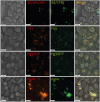
Figure 1. Autoaggregation and coaggregation of human and mouse oral bacteria. (A) The autoaggregation and coaggregation of bacterial cells were evaluated by sedimentation assays. Each column represents the mean and standard error of the mean for 5 experiments performed in triplicate. (B) Coaggregation of 2 species stained with different fluorescent dyes was examined by confocal microscopy. pHrodoRed-stained Sd (red) was mixed with CFSE-stained Fnn, Fna, Pg33277, Pg49417, or PgP4 (green). pHrodoRed-stained Fna (red) was mixed with CFSE-stained PgP4 (green). Representative images from 3 independent experiments are shown. Scale bars: 20 μm.
Sd: S. danieliae, Fnn: F. nucleatum. subsp. nucleatum ATCC 25586, Fna: F. nucleatum. subsp. animalis KCOM 1280, Pg33277: P. gingivalis ATCC 33277, Pg49417: P. gingivalis ATCC 49417, PgP4: P. gingivalis KUMC-P4. **P<0.005; ***P<0.0005 compared with the autoaggregation of Sd alone by the t-test.
Figure 2
Figure 2. Bacterial invasion of IMOK cells. IMOK cells were infected with CFSE/pHrodoRed double-stained Sd, Fna, Pg33277, or PgP4 at a multiplicity of infection of 1,000 for 4 hours. The IMOK cells were then live imaged with time-lapse on a 3D Cell Explorer microscope (×600). Representative results of 2 independent experiments with similar results are shown. Each panel represents an appropriately selected section from serial stacks taken on the z-axis to visualize intracellular and extracellular bacteria. The bacteria in yellow represent intracellular bacteria. Scale bars: 20 μm.
IMOK: immortalized murine oral keratinocyte, Sd: S. danieliae, Fnn: F. nucleatum. subsp. nucleatum ATCC 25586, Fna: F. nucleatum. subsp. animalis KCOM 1280, Pg33277: P. gingivalis ATCC 33277, Pg49417: P. gingivalis ATCC 49417, PgP4: P. gingivalis KUMC-P4.
Figure 3
Figure 3. Increased alveolar bone loss by PgP4 in C57BL/6 mice. (A) The overall outline of the animal experiments. (B) Representative sagittal images of alveolar bones at the hemimaxilla scanned with microcomputed tomography. (C) Distances from the CEJ to the ABC at 6 sites marked with white lines in B were measured. Red lines indicate mean values.
Fna: F. nucleatum. subsp. animalis KCOM 1280, Pg33277: P. gingivalis ATCC 33277, PgP4: P. gingivalis KUMC-P4, CEJ: cementoenamel junction, ABC: alveolar bone crest. #P<0.05, ###P<0.0005 compared with week 5 by the t-test. **P<0.005, ***P<0.0005 compared with the sham group by t-test. †††P<0.0005 compared with other experimental groups by analysis of variance with Bonferroni-adjusted post hoc tests.
Figure 4
Figure 4. Successful colonization of human strains in C57BL/6 mice. The copy numbers of Sd, Fna, or Pg in the total DNA of oral swabs were determined by qPCR. (G, H). The copy numbers of Pg33277 and PgP4 in the total DNA of oral swabs were determined by qPCR. The horizontal lines indicate the median values.
Sd: S. danieliae, Fna: F. nucleatum. subsp. animalis KCOM 1280, Pg33277: P. gingivalis ATCC 33277, PgP4: P. gingivalis KUMC-P4, qPCR: quantitative polymerase chain reaction. #P<0.05, ##P<0.005, ###P<0.005 compared with week 5 by the Mann-Whitney U test. *P<0.05, **P<0.005, ***P<0.005 compared with the sham group by the Mann-Whitney U test.
Figure 5
Figure 5. Gingival inflammation and bacterial invasion of gingival tissues. Sections of gingival tissues were subjected to hematoxylin and eosin staining and in situ hybridization using Sd-, Fna-, and P. gingivalis-specific probes. (A) Representative images of gingival tissues obtained at week 8 are shown. Scale bars: 50 μm. (B) The gingival inflammation index was scored by assigning 1 point each to inflammatory infiltration, vasodilation, and degradation of collagen fibers. (C-E) Bacterial signals in the epithelium and lamina propria were scored 0–3 (0, no signal; 1, signals visible only at high magnification; 2, signals visible at low magnification; 3, strong signals at low magnification) and presented as the sum. The horizontal lines indicate the median values.
H&E: hematoxylin and eosin, Fna: F. nucleatum. subsp. animalis KCOM 1280, Pg33277: P. gingivalis ATCC 33277, PgP4: P. gingivalis KUMC-P4, Sd: S. danieliae.
Figure 6
Figure 6. Antibody responses to commensal versus periodontopathic bacteria. The levels of sIgA, serum IgA, and serum IgG against Sd, Fna, and Pg were measured by enzyme-linked immunosorbent assays using the lysate of each species as antigens. The horizontal lines indicate the median values.
Sd: S. danieliae, sIgA: salivary immunoglobulin A, Fna: F. nucleatum. subsp. animalis KCOM 1280, Pg: P. gingivalis, Pg33277: P. gingivalis ATCC 33277, PgP4: P. gingivalis KUMC-P4, IgA: serum immunoglobulin A, IgG: immunoglobulin G. #P<0.05, ##P<0.005, ###P<0.005 compared with week 5 by Mann-Whitney U test. *P<0.05, **P<0.005, ***P<0.005 compared with the sham group by the Mann-Whitney U test.

Similar articles

Cited by

References

    1. Graves DT, Kang J, Andriankaja O, Wada K, Rossa C., Jr Animal models to study host-bacteria interactions involved in periodontitis. Front Oral Biol. 2012;15:117–132. - PMC - PubMed
    1. Gurumurthy CB, Lloyd KC. Generating mouse models for biomedical research: technological advances. Dis Model Mech. 2019;12:dmm029462. - PMC - PubMed
    1. Marchesan J, Girnary MS, Jing L, Miao MZ, Zhang S, Sun L, et al. An experimental murine model to study periodontitis. Nat Protoc. 2018;13:2247–2267. - PMC - PubMed
    1. Baker PJ, Dixon M, Roopenian DC. Genetic control of susceptibility to Porphyromonas gingivalis-induced alveolar bone loss in mice. Infect Immun. 2000;68:5864–5868. - PMC - PubMed
    1. Hajishengallis G, Diaz PI. Porphyromonas gingivalis: Immune subversion activities and role in periodontal dysbiosis. Curr Oral Health Rep. 2020;7:12–21. - PMC - PubMed

LinkOut - more resources