

Insulin growth factor binding protein-3 enhances dental implant osseointegration against methylglyoxal-induced bone deterioration in a rat model
- PMID: 35505576
- PMCID: PMC9064780
- DOI: 10.5051/jpis.2101200060
Insulin growth factor binding protein-3 enhances dental implant osseointegration against methylglyoxal-induced bone deterioration in a rat model
Abstract
Purpose: The aim of this study was to determine the effect of insulin growth factor binding protein-3 (IGFBP-3) on the inhibition of glucose oxidative stress and promotion of bone formation near the implant site in a rat model of methylglyoxal (MGO)-induced bone loss.
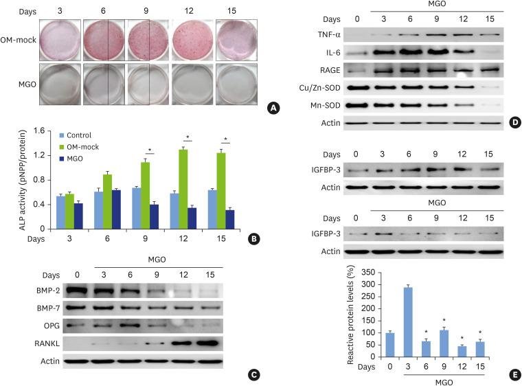
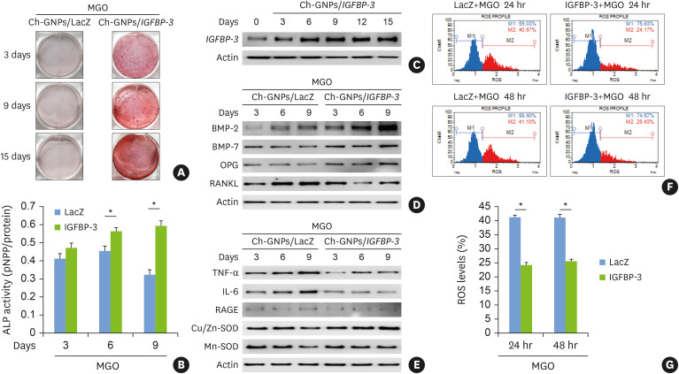
Methods: An in vitro study was performed in MC3T3 E1 cells treated with chitosan gold nanoparticles (Ch-GNPs) conjugated with IGFBP-3 cDNA followed by MGO. An in vivo study was conducted in a rat model induced by MGO administration after the insertion of a dental implant coated with IGFBP-3.
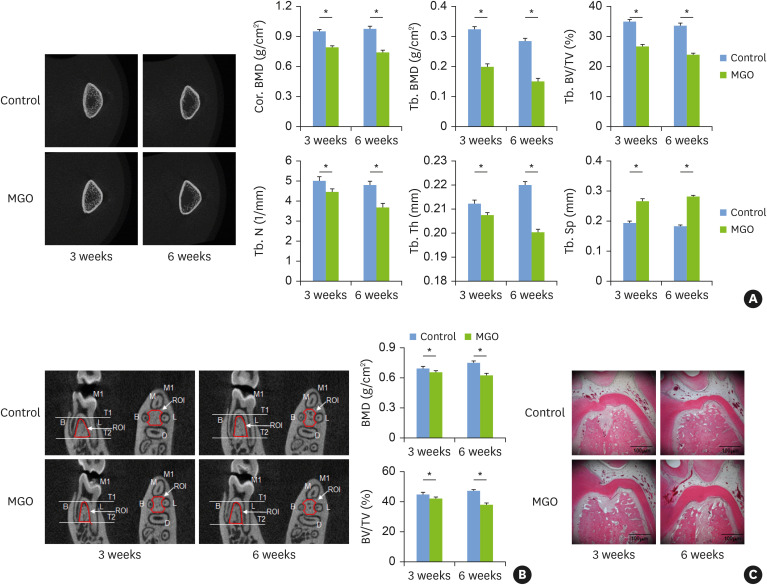
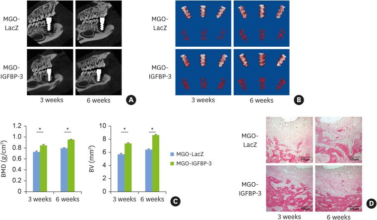
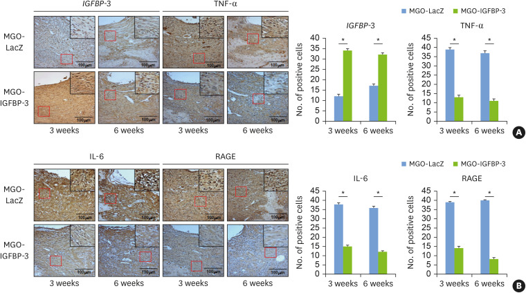
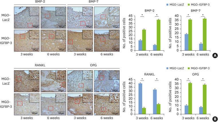
Results: MGO treatment downregulated molecules involved in osteogenic differentiation and bone formation in MC3T3 E1 cells and influenced the bone mineral density and bone volume of the femur and alveolar bone. In contrast, IGFBP-3 inhibited oxidative stress and inflammation and enhanced osteogenesis in MGO-treated MC3T3 E1 cells. In addition, IGFBP-3 promoted bone formation by reducing inflammatory proteins in MGO-administered rats. The application of Ch-GNPs conjugated with IGFBP-3 as a coating of titanium implants enhanced osteogenesis and the osseointegration of dental implants.
Conclusions: This study demonstrated that IGFBP-3 could be applied as a therapeutic component in dental implants to promote the osseointegration of dental implants in patients with diabetes, which affects MGO levels.
Keywords: Antioxidants; Bone formation; Diabetes mellitus; Inflammation; MC3T3 E1.
Copyright © 2022. Korean Academy of Periodontology.
Conflict of interest statement
No potential conflict of interest relevant to this article was reported.
Figures
Similar articles
-
Insulin-like growth factor binding protein-3 affects osteogenic efficacy on dental implants in rat mandible.Mater Sci Eng C Mater Biol Appl. 2015 Oct;55:490-6. doi: 10.1016/j.msec.2015.05.076. Epub 2015 May 31. Mater Sci Eng C Mater Biol Appl. 2015. PMID: 26117781
-
Chitosan-gold nanoparticles mediated gene delivery of c-myb facilitates osseointegration of dental implants in ovariectomized rat.Artif Cells Nanomed Biotechnol. 2018;46(sup3):S807-S817. doi: 10.1080/21691401.2018.1513940. Epub 2018 Oct 11. Artif Cells Nanomed Biotechnol. 2018. PMID: 30307328
-
Peroxisome proliferator activated receptor gamma loaded dental implant improves osteogenesis of rat mandible.J Biomed Mater Res B Appl Biomater. 2015 Apr;103(3):587-95. doi: 10.1002/jbm.b.33207. Epub 2014 Jun 25. J Biomed Mater Res B Appl Biomater. 2015. PMID: 24962969
-
Effects of magnesium-substituted nanohydroxyapatite coating on implant osseointegration.Clin Oral Implants Res. 2013 Aug;24 Suppl A100:34-41. doi: 10.1111/j.1600-0501.2011.02362.x. Epub 2011 Dec 6. Clin Oral Implants Res. 2013. PMID: 22145854
-
Osseointegration of titanium, titanium alloy and zirconia dental implants: current knowledge and open questions.Periodontol 2000. 2017 Feb;73(1):22-40. doi: 10.1111/prd.12179. Periodontol 2000. 2017. PMID: 28000277 Review.
Cited by
-
Progress in Surface Modification of Titanium Implants by Hydrogel Coatings.Gels. 2023 May 18;9(5):423. doi: 10.3390/gels9050423. Gels. 2023. PMID: 37233014 Free PMC article. Review.
-
The Integration of Gold Nanoparticles into Dental Biomaterials as a Novel Approach for Clinical Advancement: A Narrative Review.J Funct Biomater. 2024 Sep 30;15(10):291. doi: 10.3390/jfb15100291. J Funct Biomater. 2024. PMID: 39452589 Free PMC article. Review.
-
Comparison of the biocompatibility and osteogenesis potential of whitlockite and an activin A/BMP2 chimera using a rat calvarial defect model: a pilot study.J Periodontal Implant Sci. 2024 Dec;54(6):432-443. doi: 10.5051/jpis.2304280214. Epub 2024 Jun 21. J Periodontal Implant Sci. 2024. PMID: 39439104 Free PMC article.
-
Layer-by-Layer Coatings of Collagen-Hyaluronic acid Loaded with an Antibacterial Manuka Honey Bioactive Compound to Fight Metallic Implant Infections.ACS Appl Mater Interfaces. 2023 Dec 20;15(50):58119-58135. doi: 10.1021/acsami.3c11910. Epub 2023 Dec 6. ACS Appl Mater Interfaces. 2023. PMID: 38055248 Free PMC article.
-
The Impact of Implant Surface Modifications on the Osseointegration Process: An Overview.Cureus. 2025 Apr 1;17(4):e81576. doi: 10.7759/cureus.81576. eCollection 2025 Apr. Cureus. 2025. PMID: 40177230 Free PMC article. Review.
References
Grants and funding
LinkOut - more resources
Full Text Sources
Miscellaneous
