

A Tale of Two Colitides
- PMID: 35505712
- PMCID: PMC9055789
- DOI: 10.7759/cureus.23677
A Tale of Two Colitides
Abstract
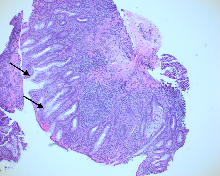
Inflammatory bowel disease (IBD) and microscopic colitis (MC) are two distinct subgroups within the larger group of colitides. MC could manifest as collagenous colitis (CC) or lymphocytic colitis (LC). The co-occurrence of MC in patients with IBD is rare, with few cases reported. No concurrent case of MC and ulcerative colitis (UC) each presenting with distinct clinical manifestations was found in the literature review. We report a case of a 76-year-old male presenting with concurrent CC and UC. The patient's initial flare of UC was characterized by episodes of bloody diarrhea while his flare of CC was evidenced by watery diarrhea.
Keywords: chronic colitis; crohn’s disease (cd); inflammatory bowel disease; microscopic colitis; ulcerative colitis (uc).
Copyright © 2022, Provenzano et al.
Conflict of interest statement
The authors have declared that no competing interests exist.
Figures


Similar articles
-
Microscopic colitis in patients with ulcerative colitis or Crohn's disease: a retrospective observational study and review of the literature.Scand J Gastroenterol. 2018 Apr;53(4):410-416. doi: 10.1080/00365521.2018.1430252. Epub 2018 Mar 16. Scand J Gastroenterol. 2018. PMID: 29546806
-
Clinicopathological significance of lymphocytic colitis/collagenous colitis in inflammatory bowel disease.Hum Pathol. 2020 Feb;96:87-95. doi: 10.1016/j.humpath.2019.09.014. Epub 2019 Nov 5. Hum Pathol. 2020. PMID: 31698005
-
Lysozyme expression in microscopic colitis.J Clin Pathol. 2011 Jun;64(6):510-5. doi: 10.1136/jcp.2010.086850. Epub 2011 Apr 2. J Clin Pathol. 2011. PMID: 21460390
-
[Evolution of ideas on microscopic colitis].Ter Arkh. 2015;87(4):69-76. doi: 10.17116/terarkh201587469-76. Ter Arkh. 2015. PMID: 26087638 Review. Russian.
-
Biomarkers and Microscopic Colitis: An Unmet Need in Clinical Practice.Front Med (Lausanne). 2017 May 10;4:54. doi: 10.3389/fmed.2017.00054. eCollection 2017. Front Med (Lausanne). 2017. PMID: 28540290 Free PMC article. Review.
References
-
- Inflammatory bowel disease. Podolsky DK. N Engl J Med. 1991;325:928–937. - PubMed
-
- Microscopic colitis: current status, present and future challenges: statements of the European Microscopic Colitis Group. Münch A, Aust D, Bohr J, et al. J Crohns Colitis. 2012;6:932–945. - PubMed
-
- [Lymphocytic colitis in a patient with ulcerative colitis: report of one case] Estay C, Simian D, Flores L, Piottante A, Quera R. Rev Med Chil. 2016;144:1088–1092. - PubMed
Publication types
LinkOut - more resources
Full Text Sources