. 2022 Jun;33(2):147-157.
doi: 10.1016/j.otot.2022.04.011. Epub 2022 Apr 28.

Imaging of the head and neck during the COVID19 pandemic

Affiliations

Imaging of the head and neck during the COVID19 pandemic

Joaquim Farinhas et al. Oper Tech Otolayngol Head Neck Surg. 2022 Jun.

Abstract

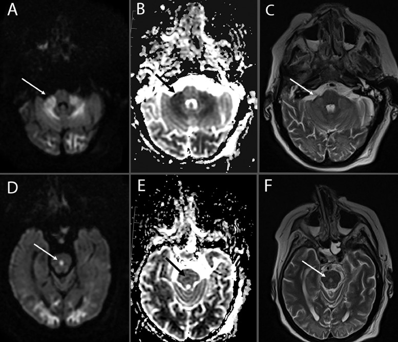
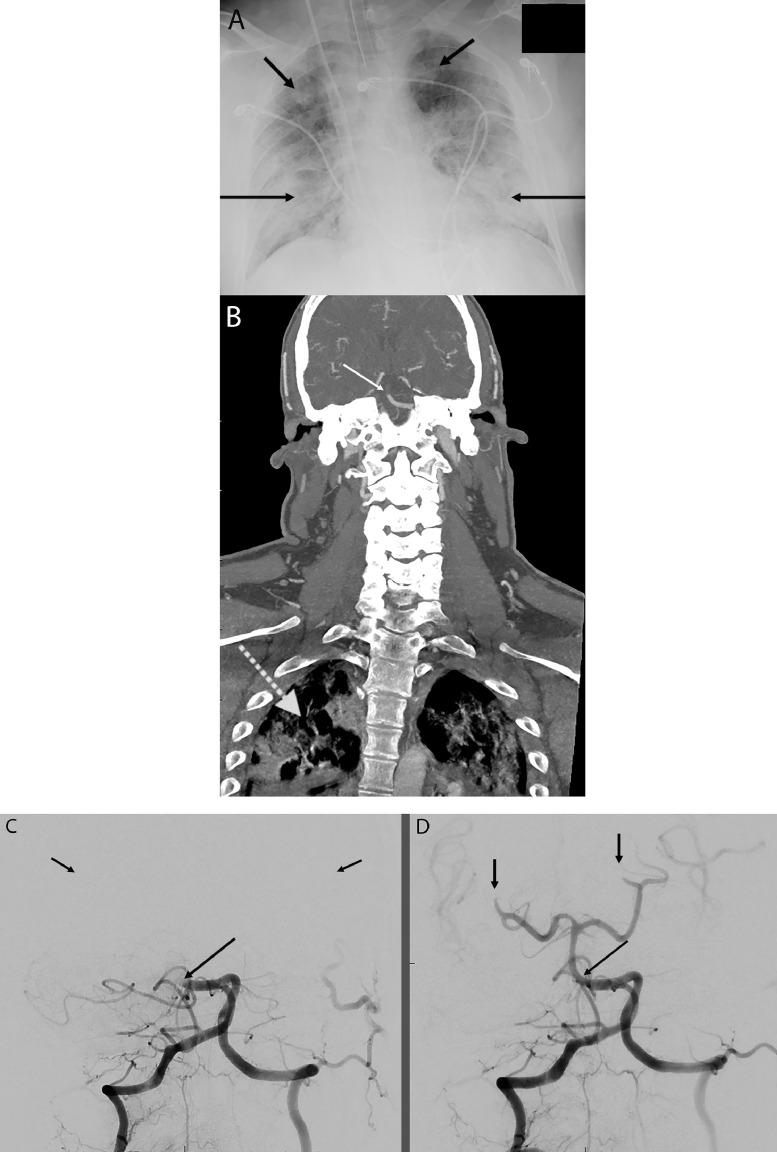
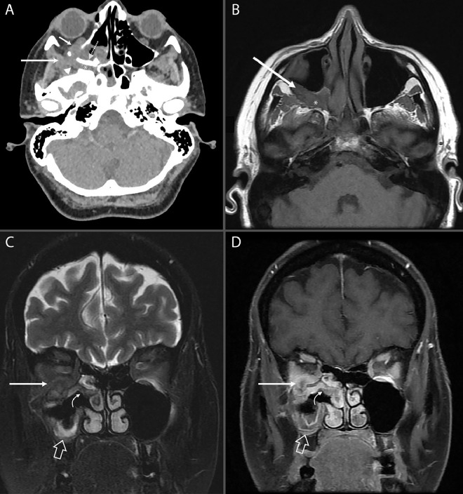
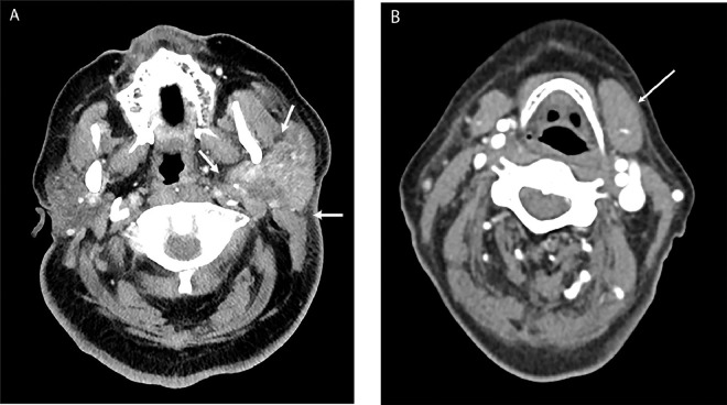
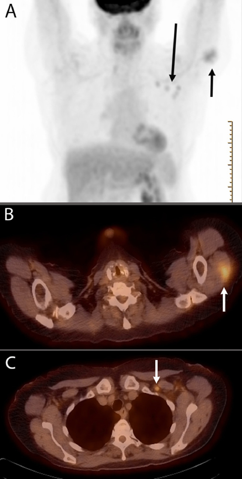
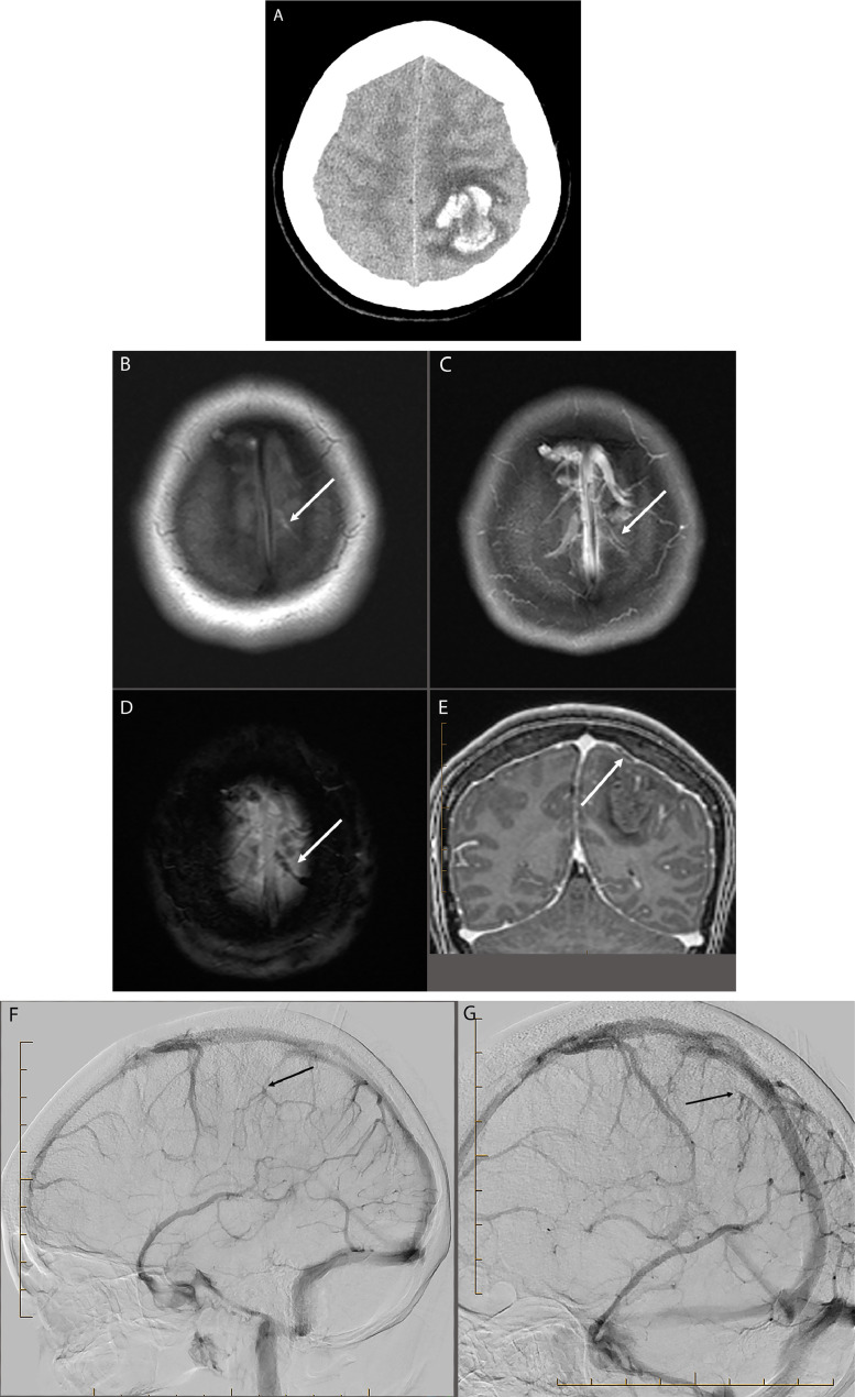
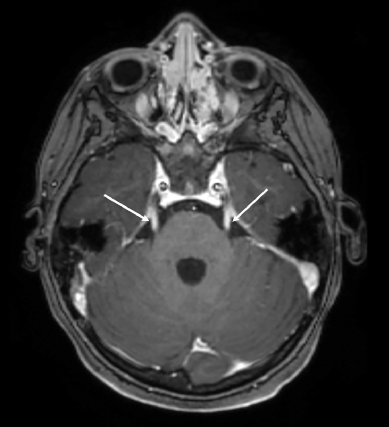
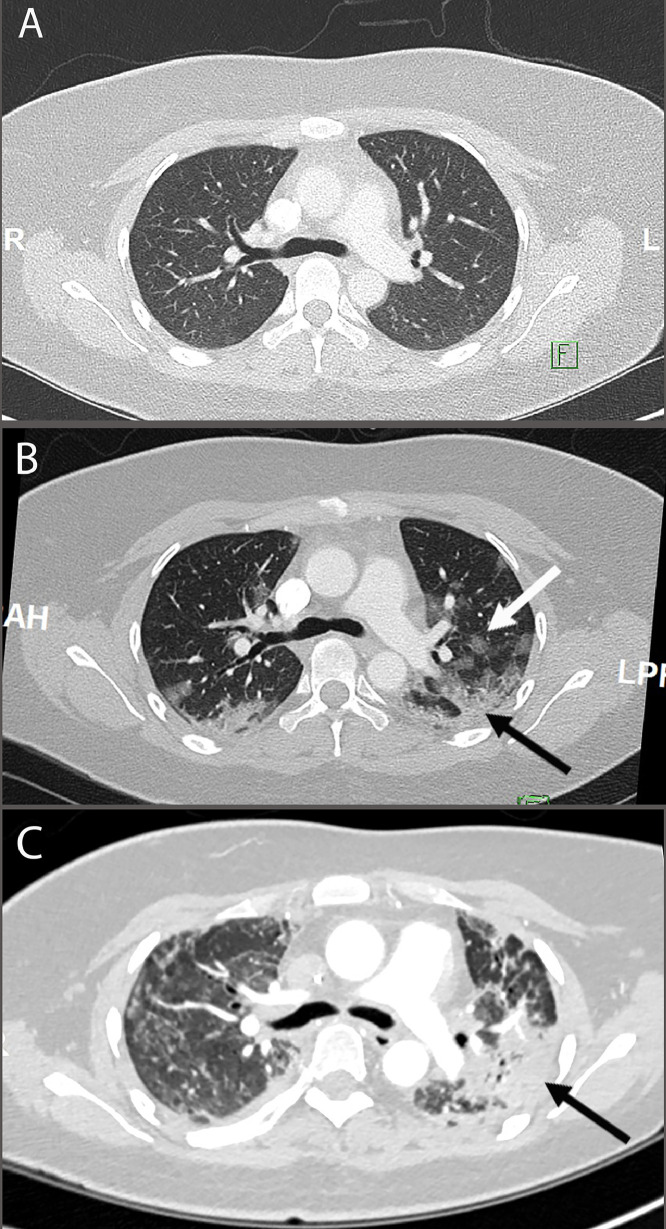
There is a wide spectrum of clinical manifestation of COVID-19 in the head and neck, but often these do not have an imaging correlate. This review will highlight the most common imaging features of COVID-19 in the head and neck that can be seen on routine head and neck CT and MRI. In addition, situations where a more dedicated imaging protocol is required will be highlighted. Finally, as mass vaccination efforts are underway worldwide, post vaccination imaging can often complicate cancer surveillance imaging. Post vaccination imaging features and recommendations will be discussed.

Keywords: Computed tomography (CT); Coronavirus disease 2019 (COVID-19); Head and Neck; Imaging; Magnetic resonance imaging (MRI).


Figures

Figure 1
Figure 1
Encephalitis: Acutely ill 60-year-old female with known COVID-19, presents with worsening headache, progressing weakness, seizure and mental status changes. Axial MRI images include (A) Diffusion weighted image (DWI); (B) Apparent diffusion coefficient (ADC); and (C) T2-weighted image through the level of the middle cerebellar peduncles (arrows) showing bilateral, symmetric edema and diffusion restriction consistent with acute inflammation expected with viral encephalitis. (D) DWI; (E) ADC and (F) T2-weighted MRI images through the level of the pons demonstrate both pontine (arrows) and bilateral occipital lobe signal abnormality as described in A-C above. This patient did not demonstrate hemorrhage, or enhancement, which can be present to varying degrees in viral encephalitides. Work up for other infectious agents was negative. Given known COVID diagnosis and eventual precipitous demise, the presumptive diagnosis of COVID-associated encephalitis was ascribed. *Images courtesy of Dr. David D. Pasquale, MD.
Figure 2
Figure 2
Thrombosis of the basilar artery: 49-year-old male with recent diagnosis of COVID-19 pneumonia, saturating at 50-60% oxygen carrying capacity, presents with headache, progressing weakness, seizure and AMS. Worsening respiratory failure and right body numbness and hemiparesis, progress to eventual coma. Final diagnosis was basilar artery occlusion syndrome. (A) Portable chest radiograph demonstrates bilateral lower lung air space opacities (long black arrows) with bilateral upper lung involvement (short black arrows). (B) CTA head and neck demonstrate the suspected basilar artery occlusion (solid white arrow). COVID-19 pneumonia is again seen in lung apices (dashed white arrow). (C) Thrombosis of the proximal basilar artery (long black arrow) is demonstrated on frontal radiographic projections of the posterior circulation during contrast injection in the right vertebral artery from mechanical thrombectomy procedure. The superior cerebellar and occipital circulation does not opacify (short black arrows) due to proximal occlusion. (D) After successful recanalization of the basilar occlusion, the basilar artery is now patent (black arrow), and the major vertebrobasilar vessel origins are seen. The distal posterior cerebellar and cerebral arteries do not fill, likely due to distal propagation of thrombus (short black arrows). *Images courtesy of Dr. David D. Pasquale, MD.
Figure 3
Figure 3
Rhino-orbital mucormycosis: 70-year-old man with history of follicular lymphoma status post chemotherapy, in remission, immunocompromised. Presents urgently with right orbital pain, proptosis and swelling, COVID positive and oxygen dependent. Failed first-line anti-fungal therapy. Subsequently underwent multiple surgeries to include: nasal endoscopy, right maxillectomy, sphenoidectomy, orbital decompression, resection of pterygopalatine fossa and pterygoid plates (postoperative images not shown). Lymphoma recurrence suspected on presentation; later pathology proven invasive fungal disease. (A) Axial, unenhanced CT of the head through the level of the orbits shows a mass centered in the right maxillary sinus invading the orbit (small white arrow), inducing mucoperiostial thickening of the adjacent maxillary sinus bone (dashed white arrow), with extension of soft issue into the right retroantral fat and masticator space soft tissues (long white arrow), and the pterygopalatine fossa (white asterisk). (B) T1-weighted non contrast axial image at the level of the pterygopalatine fossa shows similar soft tissue mass extending from right maxillary sinus into the retroantral fat and right masticator space (white arrow) in addition to the pterygopalatine fossa (white asterisk). (C) and (D) T2-weighted and T1-weighted post-contrast coronal images, respectively, through the orbits and middle meatus demonstrate the hypointense fungal elements invading the orbit from the maxillary sinus (white arrow). Non-enhancing mucosal soft tissue extending through the middle meatus toward the middle turbinate (curved white arrow) consistent with fungal invasion of the mucosa. Similar non-enhancing mucosal soft tissue in the inferior right maxillary sinus (white open arrow).
Figure 4
Figure 4
A 11-year-old girl COVID-19 positive 3 days prior presents with tinnitus and cranial neuropathy. This was followed by 4 days of fever, nausea, vomiting, ear pain, and diffuse abdominal pain with initial concern for multisystem inflammatory syndrome in children (MIS-C) –also known as pediatric inflammatory multisystem syndrome (PIMS)—in a patient with known COVID, elevated D-dimer of 936, lipase of 130, and whose course was complicated by ongoing fevers, worsening acute mental status changes. Lumbar puncture and clinical data suggesting possible encephalitis (infectious vs autoimmune). (A) Axial post contrast T1-weighted image shows bilateral IAC nerve root enhancement likely related to cochlear and/or vestibular cochlear nerves (white arrows) consistent with neuritis, likely contributing to tinnitus. (B) Axial post contrast T1-weighted image show bilateral trigeminal nerve, cisternal segment enhancement, consistent with trigeminal neuritis. *Images courtesy of Dr. David D. Pasquale, MD.
Figure 5
Figure 5
COVID-19 associated sialadenitis: Previous diagnosis of COVID-19, mild URI and headache. Admitted 5 days later with left sialoadenitis, bulging parotid and submandibular gland, facial pain, swelling and fever. (A) Enlarged left parotid gland with inflammation of the surrounding fatty soft tissue planes (white arrows). No salivary duct stones seen. (B) Ipsilateral, acalculous, left submandibular gland swelling (white arrow). *Images courtesy of Dr. David D. Pasquale, MD.
Figure 6
Figure 6
Post vaccination adenopathy: Mildly tender unilateral adenopathy incidentally found on routine PET/CT for bladder cancer staging 1 day post first COVID-19 vaccine. (A) PET/CT maximum intensity frontal projection demonstrates increased radiotracer uptake in the known left deltoid injection site (short black arrow) and left subpectoral lymphadenopathy (long black arrow). (B) Left deltoid injection site, and (C) Left subpectoral site confirmed on the fused, axial PET/CT images. *Images courtesy of Dr. David D. Pasquale, MD.
Figure 7
Figure 7
Cerebral venous thrombosis: 25-year-old female with headache, recently COVID positive. (A) Head CT showed left parietal lobe hemorrhage. (B) Unenhanced T1-weighted axial image at the vertex of the head shows a linear hyperintensity (white arrow) to the left of midline, corresponding to a superficial left anterior parietal cortical vein suggesting subacute thrombus. (C) Post-contrast T1-weighted axial image at the same level demonstrates the same vein containing a filling defect (white arrow) suspicious for presence of thrombus. (D) Axial susceptibility-weighted image (SWI) at the same level confirmed blood products in this cortical vein (white arrow). (E) Coronal T1-Weighted post-contrast image from reformatted volumetric acquisition demonstrates the filling defect (white arrow), supporting the diagnosis of CVT. Diagnostic catheter cerebral angiogram to evaluate the etiology of hemorrhage. (F) Lateral projection from the venous phase of catheter angiogram after injection of the left ICA shows an abrupt cut-off (black arrow) of the left parietal cortical draining vein. (G) Right anterior oblique projection of the same injection again shows the abrupt cut-off (black arrow) of the same left parietal cortical vein consistent with CVT. *Images courtesy of Dr. David D. Pasquale, MD.
Figure 8
Figure 8
A 4-year-old female with COVID-19, suspected Guillain-Barré: Axial, post-contrast, T1-weighted image at the level of the cisternal trigeminal nerve as it enters the pons shows bilateral cranial nerve V enhancement (white arrows), confirms clinical suspicion of Guillain-Barré syndrome.
Figure 9
Figure 9
COVID-19 pneumonia: (A) March 2021. Normal upper lung in patient undergoing surveillance for head and neck cancer. (B) May 2021. One week of dizziness, fatigue, productive cough, fevers, and abdominal pain prior to diagnosis of COVID-19 infection. Develops areas of ground-glass opacities (white arrow) in the posterior lung with peripheral nodularity, consolidation and atelectasis (black arrow). (C) October 2021. Diffuse ground-glass opacities, bronchiectasis compatible with post-COVID ARDS and developing pulmonary fibrosis. Consolidation in the left upper lobe suspected superimposed secondary infection (black arrow).

Similar articles

References

    1. Meppiel E, Peiffer-Smadja N, Maury A, et al. Neurologic manifestations associated with COVID-19: A multicentre registry. Clinical Microbiology and Infection. 2021;27:458–466. doi: 10.1016/j.cmi.2020.11.005. PMID - 33189873. - DOI - PMC - PubMed
    1. Ellul MA, Benjamin L, Singh B, et al. Neurological associations of COVID-19. The Lancet Neurology. 2020;19:767–783. doi: 10.1016/s1474-4422(20)30221-0. PMID - 32622375. - DOI - PMC - PubMed
    1. Samal P, Praharaj HN, Mishra B, et al. Acute Necrotizing Rhombencephalitis and Disemminated Thrombosis After SARS-CoV-2 Infection. Infectious Diseases in Clinical Practice (Baltimore, Md) 2021;29:e260–e261. doi: 10.1097/ipc.0000000000001029. PMID - 34276177. - DOI - PMC - PubMed
    1. Walter A, Kraemer M. A neurologist's rhombencephalitis after comirnaty vaccination. A change of perspective. Neurological Research and Practice. 2021;3:56. doi: 10.1186/s42466-021-00156-7. PMID - 34743758. - DOI - PMC - PubMed
    1. Vogrig A, Gigli GL, Bnà C, et al. Stroke in patients with COVID-19: Clinical and neuroimaging characteristics. Neuroscience Letters. 2021;743 doi: 10.1016/j.neulet.2020.135564. -135564PMID - 33352277. - DOI - PMC - PubMed

LinkOut - more resources