

CILP1 as a biomarker for right ventricular dysfunction in patients with ischemic cardiomyopathy
- PMID: 35506075
- PMCID: PMC9052998
- DOI: 10.1002/pul2.12062
CILP1 as a biomarker for right ventricular dysfunction in patients with ischemic cardiomyopathy
Abstract
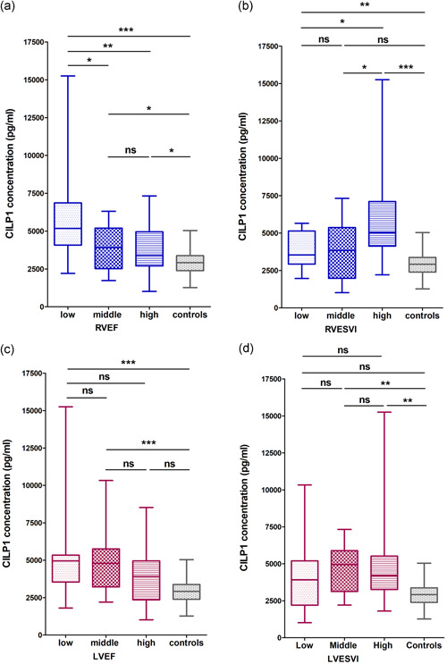
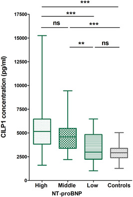
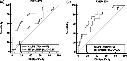
The aim of this study was to evaluate the cartilage intermediate layer protein 1 (CILP1) as a biomarker of right ventricular dysfunction in patients with ischemic cardiomyopathy (ICM). CILP1 plasma concentrations were measured in 98 patients with ICM and 30 controls without any cardiac abnormalities. All participants underwent cardiac magnetic resonance imaging. Median CILP1 concentrations were higher in ICM than in controls. In the tertile analysis, low right ventricular ejection fraction (RVEF) and high right ventricular end-systolic volume index and N-terminal pro-brain natriuretic peptide (NT-proBNP) were associated with higher CILP1 levels in ICM. However, there were no associations between CILP1 concentrations and left ventricular (LV) parameters in this group. In receiver-operating characteristic (ROC) analysis CILP1 was a good predictor of RVEF < 40% with an optimal cut-off value of 3545 pg/ml in ICM, whereas it was not predictive of LV ejection fraction (LVEF) < 40% (area under the curve [AUC] = 0.57) There was no significant difference between the ROC curves of CILP1 (AUC = 0.72) and NT-proBNP (AUC = 0.77) for RVEF < 40% (p = 0.42). In multivariable regression analysis, RVEF was the only independent predictor of elevated CILP1. CILP1 and LVEF were the only independent predictors of RVEF < 40% in ICM. Our analysis demonstrates the potential role of CILP1 as a novel cardiac biomarker of prognostically relevant RV dysfunction in patients with ICM.
Keywords: MRI; RVEF; fibrosis; myocardial remodeling.
© 2022 The Authors. Pulmonary Circulation published by Wiley Periodicals LLC on behalf of the Pulmonary Vascular Research Institute.
Conflict of interest statement
The authors declare no conflicts of interest.
Figures
Similar articles
-
CILP1 as a biomarker for right ventricular maladaptation in pulmonary hypertension.Eur Respir J. 2021 Apr 1;57(4):1901192. doi: 10.1183/13993003.01192-2019. Print 2021 Apr. Eur Respir J. 2021. PMID: 33184116
-
Can cartilage intermediate layer protein 1 (CILP1) use as a novel biomarker for canine myxomatous mitral valve degeneration levels or not?BMC Vet Res. 2023 Mar 7;19(1):59. doi: 10.1186/s12917-023-03583-7. BMC Vet Res. 2023. PMID: 36882760 Free PMC article.
-
NT-proBNP levels in the evaluation of right ventricular dysfunction in patients with coronary artery disease and abnormal left ventricular wall motion: a magnetic resonance imaging study.Coron Artery Dis. 2008 Nov;19(7):481-7. doi: 10.1097/MCA.0b013e32830b4d0e. Coron Artery Dis. 2008. PMID: 18923244
-
Right heart overload contributes to cardiac natriuretic hormone elevation in patients with heart failure.Int J Cardiol. 2005 Sep 15;104(1):39-45. doi: 10.1016/j.ijcard.2004.09.008. Int J Cardiol. 2005. PMID: 16137508
-
The prognostic value of cartilage intermediate layer protein 1 (CILP1) in patients with diabetic cardiomyopathy.BMC Cardiovasc Disord. 2024 Nov 14;24(1):646. doi: 10.1186/s12872-024-04331-x. BMC Cardiovasc Disord. 2024. PMID: 39543479 Free PMC article.
Cited by
-
CILP-1 Is a Biomarker for Backward Failure and Right Ventricular Dysfunction in HFrEF.Cells. 2023 Dec 13;12(24):2832. doi: 10.3390/cells12242832. Cells. 2023. PMID: 38132152 Free PMC article.
-
Assessment and diagnosis of right ventricular failure-retrospection and future directions.Front Cardiovasc Med. 2023 May 30;10:1030864. doi: 10.3389/fcvm.2023.1030864. eCollection 2023. Front Cardiovasc Med. 2023. PMID: 37324632 Free PMC article. Review.
-
Right Ventricular Hypertrophy in Spontaneously Hypertensive Rats (SHR/NHsd) Is Associated with Inter-Individual Variations of the Pulmonary Endothelin System.Biology (Basel). 2024 Sep 24;13(10):752. doi: 10.3390/biology13100752. Biology (Basel). 2024. PMID: 39452062 Free PMC article.
-
CILP1 interacting with YBX1 promotes hypertrophic scar formation by suppressing PPARs transcription.Cell Death Dis. 2025 May 9;16(1):371. doi: 10.1038/s41419-025-07554-8. Cell Death Dis. 2025. PMID: 40346063 Free PMC article.
References
-
- Wasemiller S, Earle T, Kashner M, Foster G, Silvet H. Right ventricular ejection fraction in ischemic versus nonischemic cardiomyopathy. Am J Cardiol. 2016;117(2):278–81. - PubMed
-
- Sabe MA, Sabe SA, Kusunose K, Flamm SD, Griffin BP, Kwon DH. Predictors and prognostic significance of right ventricular ejection fraction in patients with ischemic cardiomyopathy. Circulation. 2016;134(9):656–65. - PubMed
-
- Zornoff LA, Skali H, Pfeffer MA, St John Sutton M, Rouleau JL, Lamas GA, Plappert T, Rouleau JR, Moyé LA, Lewis SJ, Braunwald E, Solomon SD, SAVE I. Right ventricular dysfunction and risk of heart failure and mortality after myocardial infarction. J Am Coll Cardiol. 2002;39(9):1450–5. - PubMed
-
- van der Maas N, Braam RL, van der Zaag‐Loonen HJ, Meerman J, Cozijnsen L, Scholte AJ. Right ventricular ejection fraction measured by multigated planar equilibrium radionuclide ventriculography is an independent prognostic factor in patients with ischemic heart disease. J Nucl Cardiol. 2012;19(6):1162–9. - PubMed
-
- Larose E, Ganz P, Reynolds HG, Dorbala S, Di Carli MF, Brown KA, Kwong RY. Right ventricular dysfunction assessed by cardiovascular magnetic resonance imaging predicts poor prognosis late after myocardial infarction. J Am Coll Cardiol. 2007;49(8):855–62. - PubMed
LinkOut - more resources
Full Text Sources
Research Materials
