

Automatic Time-Resolved Cardiovascular Segmentation of 4D Flow MRI Using Deep Learning
- PMID: 35506525
- PMCID: PMC10946960
- DOI: 10.1002/jmri.28221
Automatic Time-Resolved Cardiovascular Segmentation of 4D Flow MRI Using Deep Learning
Abstract
Background: Segmenting the whole heart over the cardiac cycle in 4D flow MRI is a challenging and time-consuming process, as there is considerable motion and limited contrast between blood and tissue.
Purpose: To develop and evaluate a deep learning-based segmentation method to automatically segment the cardiac chambers and great thoracic vessels from 4D flow MRI.
Study type: Retrospective.
Subjects: A total of 205 subjects, including 40 healthy volunteers and 165 patients with a variety of cardiac disorders were included. Data were randomly divided into training (n = 144), validation (n = 20), and testing (n = 41) sets.
Field strength/sequence: A 3 T/time-resolved velocity encoded 3D gradient echo sequence (4D flow MRI).
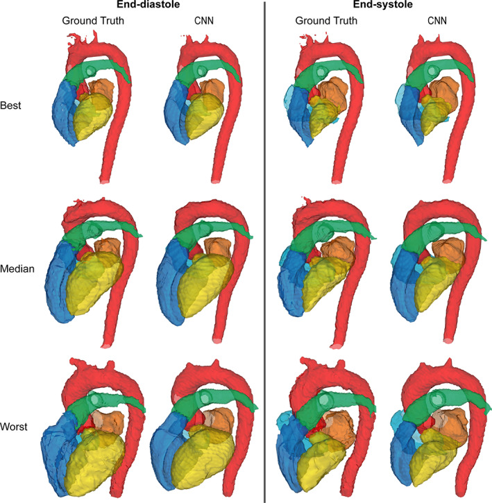
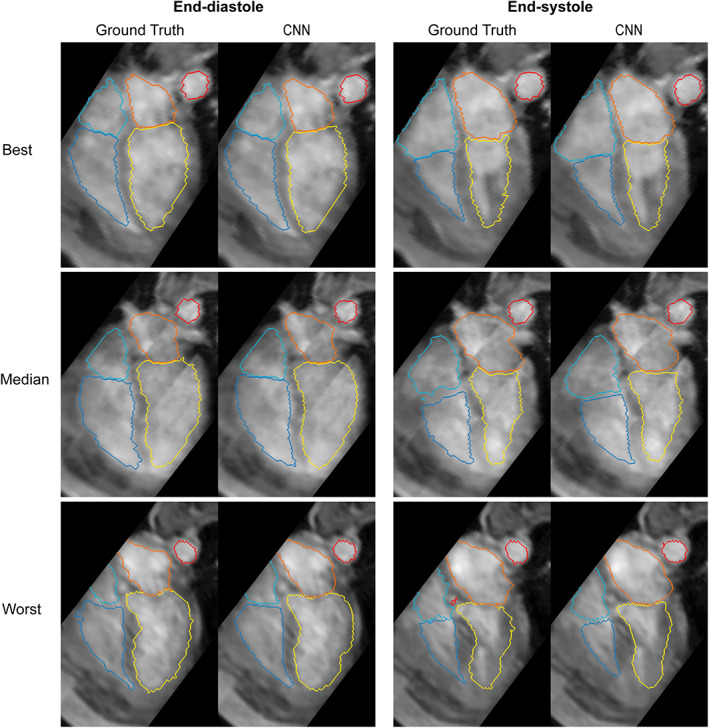
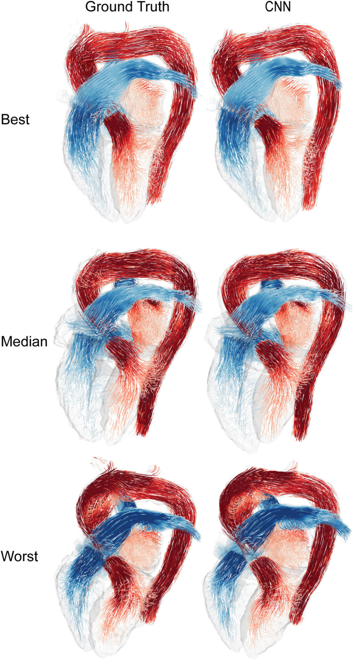
Assessment: A 3D neural network based on the U-net architecture was trained to segment the four cardiac chambers, aorta, and pulmonary artery. The segmentations generated were compared to manually corrected atlas-based segmentations. End-diastolic (ED) and end-systolic (ES) volumes of the four cardiac chambers were calculated for both segmentations.
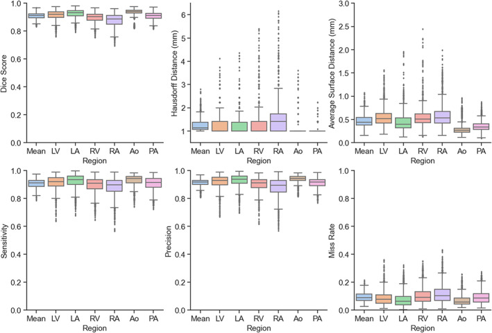
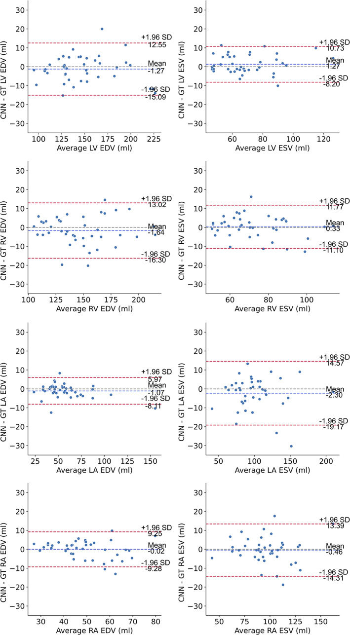
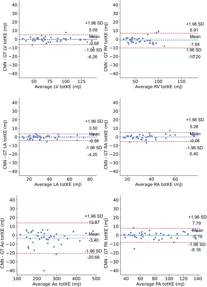
Statistical tests: Dice score, Hausdorff distance, average surface distance, sensitivity, precision, and miss rate were used to measure segmentation accuracy. Bland-Altman analysis was used to evaluate agreement between volumetric parameters.
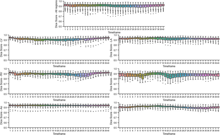
Results: The following evaluation metrics were computed: mean Dice score (0.908 ± 0.023) (mean ± SD), Hausdorff distance (1.253 ± 0.293 mm), average surface distance (0.466 ± 0.136 mm), sensitivity (0.907 ± 0.032), precision (0.913 ± 0.028), and miss rate (0.093 ± 0.032). Bland-Altman analyses showed good agreement between volumetric parameters for all chambers. Limits of agreement as percentage of mean chamber volume (LoA%), left ventricular: 9.3%, 13.5%, left atrial: 12.4%, 16.9%, right ventricular: 9.9%, 15.6%, and right atrial: 18.7%, 14.4%; for ED and ES, respectively.
Data conclusion: The addition of this technique to the 4D flow MRI assessment pipeline could expedite and improve the utility of this type of acquisition in the clinical setting.
Evidence level: 4 TECHNICAL EFFICACY: Stage 1.
Keywords: 4D flow MRI; cardiovascular MRI; convolutional neural networks; deep learning; segmentation.
© 2022 The Authors. Journal of Magnetic Resonance Imaging published by Wiley Periodicals LLC on behalf of International Society for Magnetic Resonance in Medicine.
Figures
Comment in
-
Editorial for "Automatic Time-Resolved Cardiovascular Segmentation of 4D Flow MRI Using Deep Learning".J Magn Reson Imaging. 2023 Jan;57(1):204-205. doi: 10.1002/jmri.28220. Epub 2022 May 5. J Magn Reson Imaging. 2023. PMID: 35510802 No abstract available.
References
-
- Markl M, Frydrychowicz A, Kozerke S, Hope M, Wieben O. 4D flow MRI. J Magn Reson Imaging 2012;36:1015‐1036. - PubMed
-
- Soulat G, McCarthy P, Markl M. 4D flow with MRI. Annu Rev Biomed Eng 2020;22:103‐126. - PubMed
-
- Köhler B, Born S, van Pelt RFP, Hennemuth A, Preim U, Preim B. A survey of cardiac 4D PC‐MRI data processing. Comput Graph Forum 2017;36:5‐35.
Publication types
MeSH terms
LinkOut - more resources
Full Text Sources
Other Literature Sources
Medical
