. 2022:2429:233-246.
doi: 10.1007/978-1-0716-1979-7_15.

Generation of Embryonic Origin-Specific Vascular Smooth Muscle Cells from Human Induced Pluripotent Stem Cells

Affiliations

Generation of Embryonic Origin-Specific Vascular Smooth Muscle Cells from Human Induced Pluripotent Stem Cells

Mengcheng Shen et al. Methods Mol Biol. 2022.

Abstract

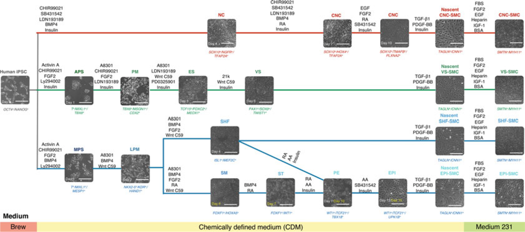
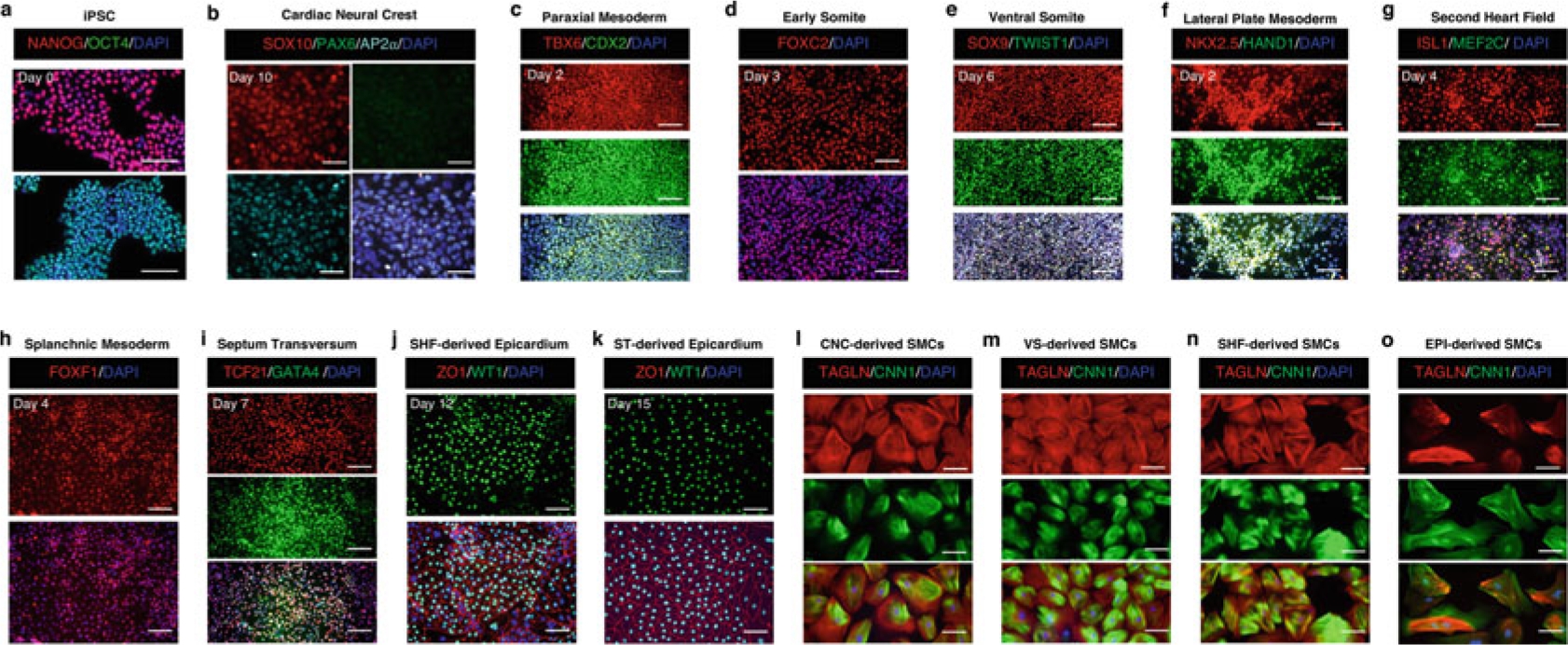
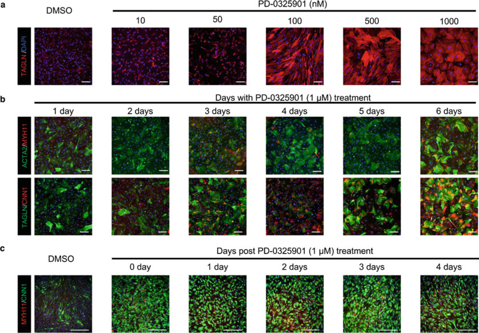
Vascular smooth muscle cells (VSMCs), a highly mosaic tissue, arise from multiple distinct embryonic origins and populate different regions of our vascular network with defined boundaries. Accumulating evidence has revealed that the heterogeneity of VSMC origins contributes to region-specific vascular diseases such as atherosclerosis and aortic aneurysm. These findings highlight the necessity of taking into account lineage-dependent responses of VSMCs to common vascular risk factors when studying vascular diseases. This chapter describes a reproducible, stepwise protocol for the generation of isogenic VSMC subtypes originated from proepicardium, second heart field, cardiac neural crest, and ventral somite using human induced pluripotent stem cells. By leveraging this robust induction protocol, patient-derived VSMC subtypes of desired embryonic origins can be generated for disease modeling as well as drug screening and development for vasculopathies with regional susceptibility.

Keywords: Embryonic origin; Induced pluripotent stem cells; Regional susceptibility; Smooth muscle cell; Vascular disease.


Figures

Fig. 1
Fig. 1
Schematic diagram of the conditions for deriving embryonic origin-specific VSMCs from human iPSCs. Lineage-specific intermediate cell types that give rise to each VSMC subtype are differentiated in a stepwise fashion. Each intermediate cell population is subjected to TGF-β1 and PDGF-BB treatment for 6 days before switching to a smooth muscle cell growth medium (Medium 231) for another 14–21 days. Phenotypic markers for specific cell types are represented in italics at the bottom of each bright-field image. NC neural crest, CNC cardiac neural crest, APS anterior primitive streak, PM paraxial mesoderm, ES early somite, VS ventral somite, MPS mid-primitive streak, LPM lateral plate mesoderm, SHF second heart field, SM splanchnic mesoderm, ST septum transversum, PE proepicardium, EPI epicardium. Scale bars represent 50 μm
Fig. 2
Fig. 2
Representative immunofluorescence images showing phenotypic markers for specific cell types during embryonic origin-specific VSMC differentiation. (a–k) The time point labeled on each set of cell marker-positive fluorescence images is consistent with that shown on the bright-field image of the same cell type in Fig. 1. (l–o) Embryonic origin-specific VSMCs are positive for TAGLN and CNN1 after being cultured in the SMC growth medium for 14 days. Scale bars represent 100 μm
Fig. 3
Fig. 3
PD0325901, a MEK inhibitor, can upregulate the expression of SMC contractile proteins. (a) Human iPSC-derived VSMCs show a dose-dependent response to PD0325901-mediated upregulation of SMC contractile proteins. (b) Human iPSC-derived VSMCs show a time-dependent upregulation of SMC contractile proteins with the treatment of PD0325901 at a concentration of 1 μM. (c) Human iPSC-derived VSMCs can preserve a contractile phenotype for at least 4 days after the withdrawal of PD0325901. Because PD0325901 application demonstrates the same effect on different VSMC subtypes, only representative images generated from CNC-specific VSMCs are shown in this figure. Scale bars represent 100 μm

References

    1. Owens GK, Kumar MS, Wamhoff BR (2004) Molecular regulation of vascular smooth muscle cell differentiation in development and disease. Physiol Rev 84(3):767–801. 10.1152/physrev.00041.2003 - DOI - PubMed
    1. Berk BC (2001) Vascular smooth muscle growth: autocrine growth mechanisms. Physiol Rev 81(3):999–1030. 10.1152/physrev.2001.81.3.999 - DOI - PubMed
    1. Basatemur GL, Jorgensen HF, Clarke MCH, Bennett MR, Mallat Z (2019) Vascular smooth muscle cells in atherosclerosis. Nat Rev Cardiol 16(12):727–744. 10.1038/s41569-019-0227-9 - DOI - PubMed
    1. Majesky MW (2007) Developmental basis of vascular smooth muscle diversity. Arterioscler Thromb Vasc Biol 27(6):1248–1258. 10.1161/ATVBAHA.107.141069 - DOI - PubMed
    1. Cai CL, Martin JC, Sun Y, Cui L, Wang L, Ouyang K, Yang L, Bu L, Liang X, Zhang X, Stallcup WB, Denton CP, McCulloch A, Chen J, Evans SM (2008) A myocardial lineage derives from Tbx18 epicardial cells. Nature 454(7200):104–108. 10.1038/nature06969 - DOI - PMC - PubMed

Publication types