Review
. 2022 Dec;16(4):1019-1033.
doi: 10.1007/s12105-022-01457-2. Epub 2022 May 4.

Combined Neuroendocrine and Squamous Cell Carcinoma of the Sinonasal Tract: A Morphologic and Immunohistochemical Analysis and Review of Literature

Affiliations
Review

Combined Neuroendocrine and Squamous Cell Carcinoma of the Sinonasal Tract: A Morphologic and Immunohistochemical Analysis and Review of Literature

Shweta Agarwal et al. Head Neck Pathol. 2022 Dec.

Abstract

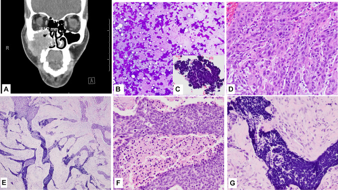
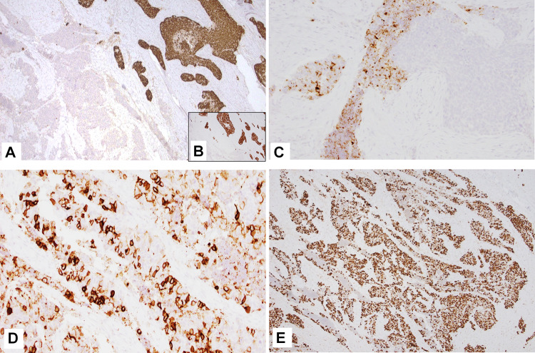
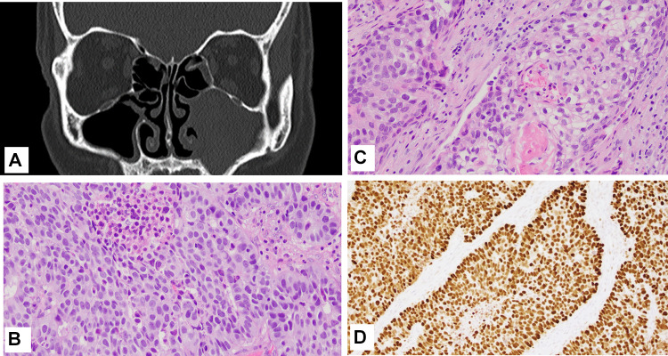
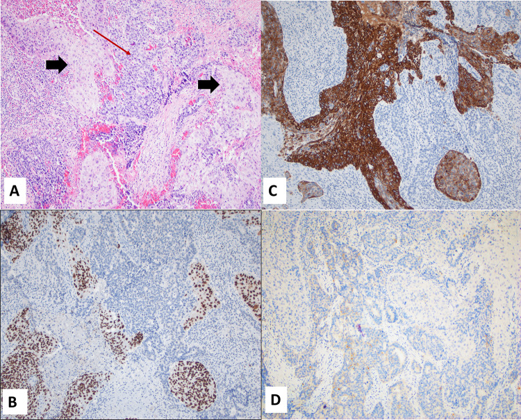
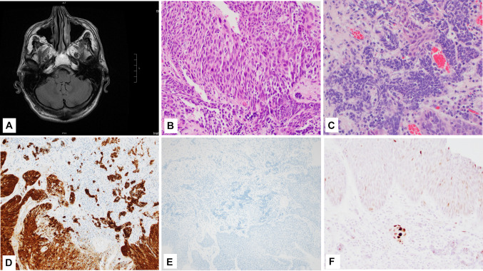
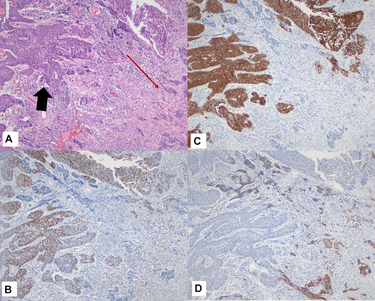
Sinonasal malignancies constitute 3% of head and neck cancers, with squamous cell carcinoma (SCC) the most common histology. Neuroendocrine carcinomas (NEC) are rare, with a subset showing neuroendocrine carcinoma and a non-neuroendocrine component. The pathogenesis of these combined tumors is largely unknown, and TP53 driver mutations may play a role. A database search for combined NEC was performed across two institutions (UNM and UCSF) spanning 15 years. Excluding NUT midline carcinoma, 3 cases met inclusion criteria. All were morphologically NEC + SCC and underwent a comprehensive immunohistochemical evaluation. Tumors demonstrated two components histologically: moderately to poorly differentiated SCC and high-grade NEC. Divergent differentiation was confirmed with lineage-specific markers. Only one patient received neoadjuvant chemotherapy prior to surgery, with a remarkable response (a marked decrease in the size of the primary lesion and resolution of liver metastases). Immunohistochemical staining for p53 was increased in 2 of 3 cases (both components), suggesting a role in the carcinogenesis of these tumors. Aberrant expression of beta-catenin was not identified. One case tested positive for p16, which can be seen in high grade NECs due to inactivation of Rb gene. Additionally, both cases with a small cell NEC component expressed PD-L1, suggesting that immunotherapy may be an effective treatment. Findings in this study support the role of p53 mutation in a subset of combined NEC + SCC of the sinonasal tract. Recognition of this rare entity is essential for optimal management of these aggressive neoplasms.

Keywords: Carcinoma; Combined; Neuroendocrine; Squamous cell; TP53; p53.


Conflict of interest statement

No disclosures.

Figures

Fig. 1
Fig. 1
Case 1 (Clinical and morphology) A CT with contrast shows a 4.1 cm mass in the right maxillary sinus; B, C Fine needle aspirate smear showed small blue cell morphology, Diff-Quick (C Inset shows the small blue cell clusters in the cell block, H&E); D Presurgical biopsy shows a moderately differentiated squamous cell carcinoma, H&E; E Histologic section from the resection specimen (post neo-adjuvant therapy) showing both squamous cell and neuroendocrine components, H&E; F High power view of the more basaloid appearing squamous cell component in the resection specimen, H&E; G High power view of the small cell NEC component in the resection specimen, H&E. Magnification in B is ×10; C is ×40; D, F, G is ×20; and E is ×4
Fig. 2
Fig. 2
Case 1 (IHC) A CK5/6-p40 (cocktail stain) and p63 (B, inset) stain the squamous cell carcinoma component; C and D Chromogranin and CD56 (respectively) stain the small cell NEC component; E Tumor showed high expression of p53. Magnification in A, B and E is ×4 while C and D is ×10
Fig. 3
Fig. 3
Case 2 A Maxillofacial CT showed opacification of the left maxillary sinus; B High power view of the moderately differentiated SCC component, H&E; C High power view of the large cell NEC component; D p53 expression. Magnification in B and C is ×20 and D is ×10
Fig. 4
Fig. 4
Case 2 A Combined SCC and NEC juxtaposed, H&E (Black arrows: SCC, Red arrow: NEC); B and C p63 and CK5/6 expression in SCC component (respectively); D Synaptophysin (weak) expression in NEC. Magnification in AD is ×10
Fig. 5
Fig. 5
Case 3 A A post biopsy CT showed residual tumor involving the left inferior turbinate and medial wall of the left maxillary sinus; B High power view of the SCC component, H&E; C High power view of the small cell NEC component, H&E; D p16 expression; E HPV hr ISH; F p53 expression. Magnification in B, C, and F is ×20 while D and E is ×10
Fig. 6
Fig. 6
Case 3 A Combined SCC and NEC juxtaposed, H&E (Black arrows: SCC, Red arrow: NEC); B and C p63 and CK5/6 expression in SCC component (respectively); D Synaptophysin (weak) expression in NEC. Magnification in AD is ×10
Fig. 7
Fig. 7
A Representative image of PD-L1 expression (Case 1); B Representative image of Beta-catenin (Case 1). Magnification in both A and B is ×10

References

    1. Youlden DR, Cramb SM, Peters S, et al. International comparisons of the incidence and mortality of sinonasal cancer. Cancer Epidemiol. 2013;37(6):770–779. doi: 10.1016/j.canep.2013.09.014. - DOI - PubMed
    1. Dutta R, Dubal PM, Svider PF, et al. Sinonasal malignancies: a population-based analysis of site-specific incidence and survival. Laryngoscope. 2015;125(11):2491–2497. doi: 10.1002/lary.25465. - DOI - PubMed
    1. Bishop JA, Guo TW, Smith DF, et al. Human papillomavirus-related carcinomas of the sinonasal tract. Am J Surg Pathol. 2013;37(2):185–192. doi: 10.1097/PAS.0b013e3182698673. - DOI - PMC - PubMed
    1. Bishop JA. Newly described tumor entities in Sinonasal Tract Pathology. Head Neck Pathol. 2016;10(1):23–31. doi: 10.1007/s12105-016-0688-7. - DOI - PMC - PubMed
    1. Turner JH, Reh DD. Incidence and survival in patients with sinonasal cancer: a historical analysis of population-based data. Head Neck. 2012;34(6):877–885. doi: 10.1002/hed.21830. - DOI - PubMed

Substances