. 2022 Aug;24(4):488-496.
doi: 10.1111/cid.13097. Epub 2022 May 4.

Tooth as graft material: Histologic study

Affiliations

Tooth as graft material: Histologic study

Elio Minetti et al. Clin Implant Dent Relat Res. 2022 Aug.

Abstract

Background: An effective regenerative protocol is key to reestablish and maintain the hard and soft tissue dimensions over time. The choice of the graft material and its properties also could have an impact on the results. To prevent alveolar ridge dimensional changes, since numerous graft materials have been suggested and in the past years, a growing interest in teeth material has been observed as a valuable alternative to synthetic biomaterials.

Aim: The aim of the study was to explore the histomorphometric outcomes of tooth derivative materials as used as bone substitute material in socket preservation procedure.

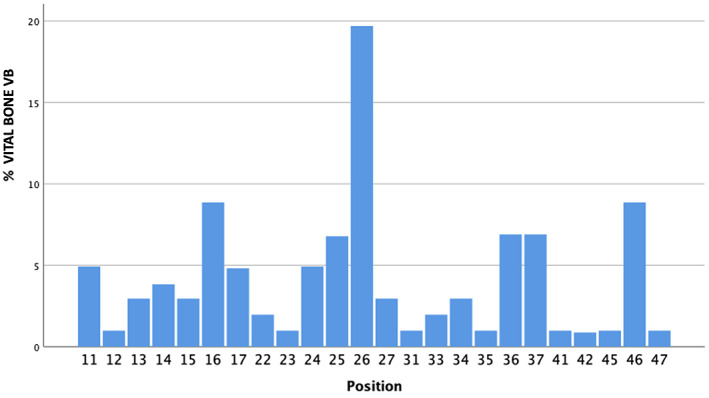
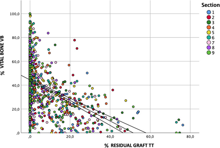
Methods: After alveolar socket preservation (ASP) procedures using autologous demineralized tooth as graft material prepared by means of an innovative device, was evaluated. A total of 101 histological samples, from 96 subjects, were analyzed by evaluating the total amount of bone (BV), residual tooth material (residual graft, TT), and vital bone (VB). The section from each sample was then split in nine subsections, resulting in 909 subsections, to allow statistical comparison between the different areas.

Results: It was not noticed a statistically significant difference between maxillary and mandibular sites, being the amount of VB in upper jaw sites 37.9 ± 21.9% and 38.0 ± 22.0% in lower jaw sites and the amount of TT was 7.7 ± 12.2% in maxilla and 7.0 ± 11.1% in mandibles. None of the other considered parameters, including defect type and section position, were statistically correlated to the results of the histomorphometric analysis.

Conclusions: ASP procedure using demineralized autologous tooth-derived biomaterial may be a predictable procedure to produce new vital bone potentially capable to support dental implant rehabilitation.

Keywords: alveolar ridge reconstruction; autogenous; biomaterials; bone; bone augmentation; bone grafting; bone substitutes; histological analysis; implantology; prospective.


Conflict of interest statement

The authors declare no conflict of interest.

Figures

FIGURE 1
FIGURE 1
A total of 101 biopsies belonging to different sites from 96 subjects. The figure shows the distribution of sites. There is a strong prevalence of the first molar sites
FIGURE 2
FIGURE 2
The figure represents the percentage of bone in relation to healing time. A total of 101 biopsies were compared at the collecting time.
FIGURE 3
FIGURE 3
Example of histologic analysis. The sample was subdivided in nine subsections following the marginal dimension of the image. The dentin granules (indicated with X) are homogeneously present in all sections of the image. Magnification 50×: stain hematoxylin and eosin
FIGURE 4
FIGURE 4
Example of a single subsection histologic analysis. X refers to the dentin granules. The bone is pink colored. The yellow lines surrounding the surfaces are useful to evaluate the relative percentage of individual histology components. Magnification 100×: stain hematoxylin and eosin
FIGURE 5
FIGURE 5
Newly formed tissue is closely linked to the tooth bone graft substitute (indicated with X). It is possible to see how osteoblasts are able to deposit new tissue around the entire perimeter of the granule, regardless the presence of irregularities. Magnification 200×: stain hematoxylin and eosin
FIGURE 6
FIGURE 6
The new bone matrix deeply penetrates into the non‐resorbed dentin granules. In the upper part of the image, there is newly formed bone. In the underlying part there is dentin, recognizable by the dentinal tubules firmly attached to the bone. As you can see, the junction between the two tissues does not present any kind of discontinuity. Magnification 1000×: stain hematoxylin and eosin (reproduced with permission from Minetti E. Bone regeneration in implantology: tooth as a graft. ©Edra, Milano 2021)
FIGURE 7
FIGURE 7
Characteristic signs of resorption on dentin granule profile (indicated with X) due to osteoclasts (indicated with O). The granule is completely surrounded by the new bone with total continuity between the two different types of tissue (graft and bone) called cement line 200× magnification: stain hematoxylin and eosin (reproduced with permission from Minetti E. Bone regeneration in implantology: tooth as a graft. ©Edra, Milano 2021).
FIGURE 8
FIGURE 8
Dispersion graph showing the distribution of residual %TT dentin graft values related to %Bone. The colors represent the different subsections of the samples. It is noticeable that the higher the proportion of residual TT the lower the proportion of new bone. As it is observable, the vast majority of the results are located in the area that represents a percentage of residual TT lower than 20%. We found no differences among different sections.
FIGURE 9
FIGURE 9
Dispersion graph showing the distribution of residual %TT values related to %Bone. The colors represent different healing times. It is noticeable that the higher the proportion of residual TT the lower the proportion of new bone. As it is observable, the vast majority of the results are located in the area that represents a percentage of residual TT lower than 20%.

References

    1. Van Der Weijden F, Dell'Acqua F, Slot DE. Alveolar bone dimensional changes of post‐extraction sockets in humans: a systematic review. J Clin Periodontol. 2009;36:1048‐1058. - PubMed
    1. Schropp L, Wenzel A, Kostopoulos L, Karring T. Bone healing and soft tissue contour changes following single‐tooth extraction: a clinical and radiographic 12‐month prospective study. Int J Periodontics Restorative Dent. 2003;23:313‐323. - PubMed
    1. Ten Heggeler JM, Slot DE, Van der Weijden GA. Effect of socket preservation therapies following tooth extraction in non‐molar regions in humans: a systematic review. Clin Oral Implants Res. 2011;22(8):779‐788. - PubMed
    1. Chappuis V, Engel O, Reyes M, Shahim K, Nolte L‐P, Buser D. Ridge alterations post‐extraction in esthetic zone:a 3D analysis with CBCT. J Dent Res. 2013;92(12 Supl):195S‐201S. - PMC - PubMed
    1. Del Fabbro M, Bucchi C, Lolato A, Corbella S, Testori T, Taschieri S. Healing of postextraction sockets preserved with autologous platelet concentrates. A systematic review and meta‐analysis. J Oral Maxillofac Surg. 2017;75(8):1601‐1615. doi:10.1016/j.joms.2017.02.009 - DOI - PubMed

Substances