. 2022 May 10;119(19):e2120098119.
doi: 10.1073/pnas.2120098119. Epub 2022 May 4.

Structural convergence for tubulin binding of CPAP and vinca domain microtubule inhibitors

Affiliations

Structural convergence for tubulin binding of CPAP and vinca domain microtubule inhibitors

Valérie Campanacci et al. Proc Natl Acad Sci U S A. .

Abstract

Microtubule dynamics is regulated by various cellular proteins and perturbed by small-molecule compounds. To what extent the mechanism of the former resembles that of the latter is an open question. We report here structures of tubulin bound to the PN2-3 domain of CPAP, a protein controlling the length of the centrioles. We show that an α-helix of the PN2-3 N-terminal region binds and caps the longitudinal surface of the tubulin β subunit. Moreover, a PN2-3 N-terminal stretch lies in a β-tubulin site also targeted by fungal and bacterial peptide-like inhibitors of the vinca domain, sharing a very similar binding mode with these compounds. Therefore, our results identify several characteristic features of cellular partners that bind to this site and highlight a structural convergence of CPAP with small-molecule inhibitors of microtubule assembly.

Keywords: centrioles; cytoskeleton; microtubule dynamics; peptide inhibitor; structural biology.


Conflict of interest statement

The authors declare no competing interest.

Figures

Fig. 1.
Fig. 1.
Overview of tubulin–CPAP structures. (A) Constructs used in this study and sequence conservation of the PN2-3 domain of CPAP. The PN2-3 sequence (residues 311 to 422 of CPAP, with an additional alanine after the N-terminal methionine) is aligned with that of constructs used in crystallization experiments. These constructs are based on the CPAP 321 to 397 region and include an additional valine after the N-terminal methionine. The residues 321 to 387 were traced in the different structures. This region has been divided into five structural motifs, as indicated under the sequence. Residues that were mutated for affinity measurements are highlighted in yellow in the sequence of the 321 to 397 construct. The PN2-3 sequence conservation scores range from 1 (not conserved; blue) to 9 (highly conserved; magenta) according to the ConSurf color code. (B) Crystal form 1: structure of the 321 to 397 CPAP fragment (cyan) in complex with tubulin (dark and light gray). Tubulin is further bound to two molecules (orange and yellow) of the iiH5 αRep used as a crystallization helper. (C) ITC of the interaction between tubulin and the 321 to 397 CPAP fragment or the Δloop construct (see also SI Appendix, Fig. S1 A and B and Table 1). (D) Crystal form 2: structure of tubulin bound to the CPAP Δloop fragment (magenta). The complex was crystallized with two molecules of the iE5 αRep. (B and D) The αReps in orange are those bound to tubulin in two structures of tubulin–αRep previously determined (20); the ones in yellow define new binding sites. N-ter, N terminal; Tub, tubulin.
Fig. 2.
Fig. 2.
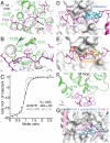
PN2-3 wraps around β-tubulin. (A) PN2-3 (cyan and magenta, from crystal forms 1 and 2, respectively) is modeled at the plus end of a microtubule (PDB ID 6DPU) by superposing β-tubulin of tubulin–CPAP complexes to β-tubulin at the plus end of a protofilament. Three protofilaments are shown and CPAP is modeled on the middle one. The five structural motifs of PN2-3 are indicated (see also panel B and Fig. 1). (B) The PN2-3 N-terminal α-helix binding site. The β-tubulin elements interacting with this α-helix are highlighted in green. The side chains of residues Leu342 and Ile346, replaced in the LI > EE mutant, are shown in crystal form 2, whereas the main chain of residues 347 to 349, targeted in the QLE > GGG mutant, is in blue in crystal form 1. (C) Structural basis for PN2-3 competition with a DARPin (PDB ID 5EIB) and with maytansine (PDB ID 4TV8) for tubulin binding. The side chain of Gln345, which would overlap with maytansine, is shown. N-ter, N terminal.
Fig. 3.
Fig. 3.
The interaction of the N-terminal stretch of PN2-3 with tubulin. (A) Overview of the interaction. The PN2-3 Δloop construct (from crystal form 2) is in magenta, and the tubulin-interacting elements are in green. The side chain of CPAP Lys328, which is weakly defined in the electron density maps, has been truncated after its Cβ atom. (B) Close-up image of the interaction with tubulin of the CPAP residue Ile327. (C) ITC analysis of the tubulin–I327R and tubulin–EE > RR interactions (see also SI Appendix, Fig. S1 E and G and Table 1). (D and E) The binding sites of vinblastine (PDB ID 1Z2B; panel D) and of phomopsin A (PDB ID 3DU7; panel E) overlap with that of the N-terminal stretch of PN2-3. (F) Close-up image of the interaction with tubulin of the CPAP residues Glu323 and Glu324. (G) In crystal form 1, the linker region of a symmetry-related CPAP molecule (cyan) occupies the tubulin binding site of the N-terminal stretch of PN2-3.

References

    1. Desai A., Mitchison T. J., Microtubule polymerization dynamics. Annu. Rev. Cell Dev. Biol. 13, 83–117 (1997). - PubMed
    1. Steinmetz M. O., Prota A. E., Microtubule-targeting agents: Strategies to hijack the cytoskeleton. Trends Cell Biol. 28, 776–792 (2018). - PubMed
    1. Mühlethaler T., et al. , Comprehensive analysis of binding sites in tubulin. Angew. Chem. Int. Ed. Engl. 60, 13331–13342 (2021). - PMC - PubMed
    1. Amos L. A., What tubulin drugs tell us about microtubule structure and dynamics. Semin. Cell Dev. Biol. 22, 916–926 (2011). - PubMed
    1. Risinger A. L., Giles F. J., Mooberry S. L., Microtubule dynamics as a target in oncology. Cancer Treat. Rev. 35, 255–261 (2009). - PMC - PubMed

Publication types