. 2022 May 2;37(2):e370204.
doi: 10.1590/acb370204. eCollection 2022.

Protective effect of polysaccharides isolated from the seeds of Cuscuta chinensis Lam. on 5-fluorouracil-induced intestinal mucositis in mice

Affiliations

Protective effect of polysaccharides isolated from the seeds of Cuscuta chinensis Lam. on 5-fluorouracil-induced intestinal mucositis in mice

Yanzhao Ji et al. Acta Cir Bras. .

Abstract

Purpose: To evaluate the protective effect of Cuscuta chinensis Lam. polysaccharides (PCCL) on 5-fluorouracil-(5-FU)-induced intestinal mucositis (IM) in mice.

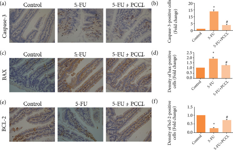
Methods: PCCL was orally administered at a dose of 20 mg·kg-1 for 7 days and its protective effect on 5-FU-induced IM (5-FU, 50 mg·kg-1 for 5 days) was evaluated by monitoring changes in body weight, degree of diarrhea, levels of tissue inflammatory factors (tumor necrosis factor α, interleukin 6, and interleukin 1β levels), apoptosis rates, and the expression levels of caspase-3, Bax and Bcl-2.

Results: The severity of mucosal injury (as reflected by body weight changes, degree of diarrhea, height of villi, and damage to crypts) was significantly attenuated by PCCL administration. PCCL also reduced the levels of tissue inflammatory factors, the apoptosis rate, and the expression of caspase-3 and Bax, and increased Bcl-2 expression.

Conclusions: PCCL administration may be significantly protective against 5-FU-induced IM by inhibiting apoptosis and regulating the abnormal inflammation associated with it.


Conflict of interest statement

Conflict of interest: Nothing to declare.

Figures

Figure 1
Figure 1. HPLC analysis of PCCL. Chemical structures of the monosaccharides comprising PCCL. Carbohydrates identified from reference to the peak numbers indicated on chromatograms. 1: PMP (PubChem CID: 4021); MW: 174.203 g·mol–1. MF: C10H10N2O; 2: Mannose (PubChem CID: 18950); MW: 180.156 g·mol–1. MF: C6H12O6; 3 Glucose (PubChem CID: 107526); MW: 180.156 g·mol–1. MF: C6H12O6; 4 Galactose (PubChem CID: 6036); MW: 180.156 g·mol–1 MF: C6H12O6.
Figure 2
Figure 2. Effects of PCCL on changes in body weight and on diarrhea during 5-FU treatment. 5-FU (50 mg·kg–1) was injected intraperitoneally once daily, and PCCL (20 mg·kg–1) were administered orally twice daily for 6 days (days 0–5). (a) Body weight is shown as a percentage of initial body weight; (b) and the severity of diarrhea is scored using the four-grade scale (0 to 3). Data are the mean ± SEM of 8–10 mice.
Figure 3
Figure 3. Effects of PCCL on the shortening of villus height and on crypt destruction induced by 5-FU in mouse small intestine. 5-FU (50 mg·kg–1) was injected intraperitoneally once daily, and PCCL (20 mg·kg–1) were administered orally twice daily for 6 days (days 0–5). (a) The jejunum was excised on day 6, sectioned, and stained with H&E (400×); (b) The height of villi; (c) the length of crypts; (d) and the villi height per crypt were measured. Data are the mean ± SEM of 8–10 mice.
Figure 4
Figure 4. Effects of PCCL on TNF-α, IL-6, and IL-1β production in the jejunum. Cytokine (TNF-α, IL-6 and IL-1β)concentrations were determined using the appropriate enzyme-linked immunosorbent assay (ELISA) kit: (a) TNF-α levels;(b) IL-6 levels; (c) IL-1β levels. (ANOVA followed by Bonferroni’s test.) TNF-α: tumor necrosis factor α; IL-6: interleukin6; IL-1β: interleukin 1β; PCCL: C. chinensis polysaccharides; ANOVA, analysis of variance.
Figure 5
Figure 5. Effect of PCCL on apoptosis in intestinal crypts induced by 5-FU. 5-FU (50 mg·kg–1) was injected intraperitoneally, and PCCL (20 mg·kg–1) was administered orally twice, 30 min before and 8 h after 5-FU injection. (a) At the end of the experiment, the jejunum was excised 24 h after the final 5-FU injection, sectioned, and a TUNEL assay (400×) was performed; (b) The number of apoptotic cells was counted. Data are presented as the mean ± SEM of 8 mice.
Figure 6
Figure 6. Immunohistochemistry of caspase-3, BAX, and BCL-2 in the jejunum Expression levels of caspase-3, BAX, and BCL-2 in the jejunum were assessed by immunofluorescent staining (400 ×). Positive cell numbers and staining intensities were assessed by the semi-quantitative method for evaluation as described in the Materials and Methods. (a) Images showing caspase-3 expression in each experimental group. (b) Data for caspase-3 expression (means ± SD). (c) Images showing BAX expression in each experimental group. (d) Data for BAX expression (means ± SD). (e) Images showing BCL-2expression in each experimental group. (f) Data for BCL-2 expression (means ± SD).

Similar articles

Cited by

References

    1. Elting LS, Cooksley C, Chambers M, Cantor SB, Manzullo E, Rubenstein EB. The burdens of cancer therapy. Cancer. 2003;98(7):1531–1539. doi: 10.1002/cncr.11671. - DOI - PubMed
    1. Longley DB, Harkin DP, Johnston PG. 5-fluorouracil: Mechanisms of action and clinical strategies. Nat Rev Cancer. 2003;3(5):330–338. doi: 10.1038/nrc1074. - DOI - PubMed
    1. Bowen JM, Gibson RJ, Cummins AG, Keefe DM. Intestinal mucositis: The role of the Bcl-2 family, p53 and caspases in chemotherapy-induced damage. Support Care Cancer. 2006;14(7):713–731. doi: 10.1007/s00520-005-0004-7. - DOI - PubMed
    1. De Rosa M, Pace U, Rega D, Costabile V, Duraturo F, Izzo P, Delrio P. Genetics, diagnosis and management of colorectal cancer (Review) Oncol Rep. 2015;34(3):1087–1096. doi: 10.3892/or.2015.4108. - DOI - PMC - PubMed
    1. Symonds RP. Treatment-induced mucositis: An old problem with new remedies. Br J Cancer. 1998;77(10):1689–1695. doi: 10.1038/bjc.1998.279. - DOI - PMC - PubMed