Review
. 2022 Jul 8:45:581-601.
doi: 10.1146/annurev-neuro-110920-040422. Epub 2022 May 4.

Synaptic Mechanisms Regulating Mood State Transitions in Depression

Affiliations
Review

Synaptic Mechanisms Regulating Mood State Transitions in Depression

Puja K Parekh et al. Annu Rev Neurosci. .

Abstract

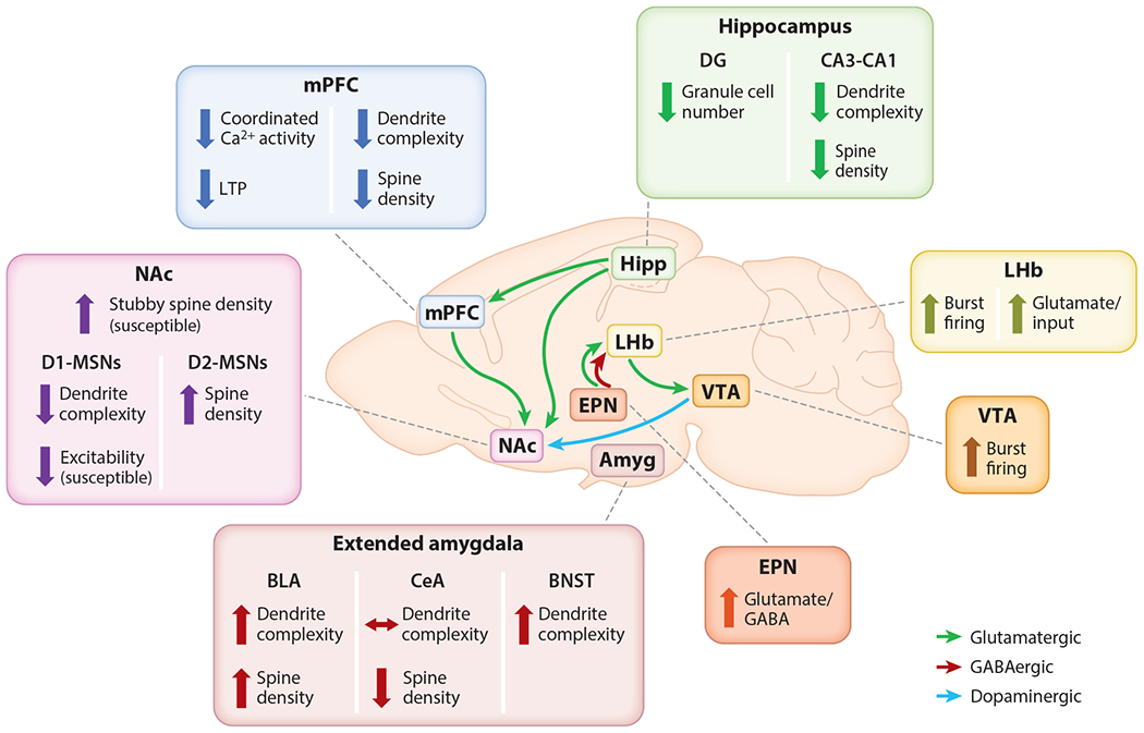
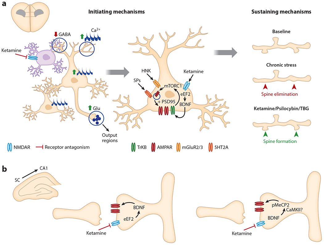
Depression is an episodic form of mental illness characterized by mood state transitions with poorly understood neurobiological mechanisms. Antidepressants reverse the effects of stress and depression on synapse function, enhancing neurotransmission, increasing plasticity, and generating new synapses in stress-sensitive brain regions. These properties are shared to varying degrees by all known antidepressants, suggesting that synaptic remodeling could play a key role in depression pathophysiology and antidepressant function. Still, it is unclear whether and precisely how synaptogenesis contributes to mood state transitions. Here, we review evidence supporting an emerging model in which depression is defined by a distinct brain state distributed across multiple stress-sensitive circuits, with neurons assuming altered functional properties, synapse configurations, and, importantly, a reduced capacity for plasticity and adaptation. Antidepressants act initially by facilitating plasticity and enabling a functional reconfiguration of this brain state. Subsequently, synaptogenesis plays a specific role in sustaining these changes over time.

Keywords: dendritic spines; depression; ketamine; rapid-acting antidepressants; stress; synaptic plasticity.


Figures

Figure 1
Figure 1
Regional synaptic effects of stress and depression models. Hipp and mPFC undergo dendritic atrophy and spine loss. NAc experiences an overall proliferation of stubby spines, but D1-MSNs may experience dendritic atrophy relative to D2-MSNs. Other regions, including amygdala and Amyg, EPN, VTA, and LHb, experience spine formation (spinogenesis) and increased excitability following stress. Abbreviations: Amyg, extended amygdala; BLA, basolateral amygdala; BNST, bed nucleus of the stria terminalis; Ca2+, calcium; CeA, central amygdala; D1-MSN, dopamine receptor 1 medium spiny neuron; D2-MSN, dopamine receptor 2 medium spiny neuron; DG, dentate gyrus; EPN, entopeduncular nucleus; GABA, γ-aminobutyric acid; Hipp, hippocampus; LHb, lateral habenula; LTP, long-term potentiation; mPFC, medial prefrontal cortex; NAc, nucleus accumbens; VTA, ventral tegmental area.
Figure 2
Figure 2
The initiating and sustaining mechanisms of rapid-acting antidepressants. (a) In the mPFC, ketamine acts to elevate glutamatergic tone of excitatory pyramidal neurons (tan), potentially through disinhibitory mechanisms involving tonically firing interneurons (purple). Ketamine leads to enhanced somatic and spine Ca2+ transients in layer 2/3 pyramidal cells within 1 h and increases multicellular ensemble activity within 3 h of administration (left). Ketamine, its metabolite HNK, and SPs each increase neurotrophic signaling and AMPAR insertion in mPFC excitatory neurons. The sustained antidepressant effects of ketamine and psychedelics involve targeted dendritic spinogenesis on excitatory projection neurons (right). Ketamine-induced spine formation is detected 12 h after administration and follows circuit reorganization. (b) In hippocampal SC to CA1 synapses, ketamine rapidly enhances BDNF release and increases postsynaptic glutamatergic transmission (left) while a sustained effect involves the delayed increase in pMeCP2 through a BDNF-dependent mechanism (right). Abbreviations: 5HT2A, 5-hydroxy-tryptamine receptor 2A; AMPAR, α-amino-3-hydroxy-5-methyl-4-isoxazole propionic acid receptor; BDNF, brain-derived neurotrophic factor; Ca2+, calcium; CaMKII, calcium-calmodulin kinase II; eEF2, eukaryotic elongation factor 2; GABA, γ-aminobutyric acid; Glu, glutamate; HNK, hydroxynorketamine; mGluR2/3, metabotropic glutamate receptor; mPFC, medial prefrontal cortex; mTORC1, mammalian target of rapamycin complex 1; NMDAR, N-methyl-d-aspartate receptor; pMeCP2, phosphorylated methyl-CpG-binding protein 2; PSD95, postsynaptic density protein 95; SC, Schaffer collateral; SP, serotonergic psychedelic; TBG, tabernanthalog; TrkB, tropomyosin receptor kinase B.

References

    1. Abdallah CG, Averill LA, Gueorguieva R, Goktas S, Purohit P, et al. 2020. Modulation of the antidepressant effects of ketamine by the mTORC1 inhibitor rapamycin. Neuropsychopharmacology 45(6):990–97 - PMC - PubMed
    1. Abdallah CG, De Feyter HM, Averill LA, Jiang L, Averill CL, et al. 2018. The effects of ketamine on prefrontal glutamate neurotransmission in healthy and depressed subjects. Neuropsychopharmacology 43(10):2154–60 - PMC - PubMed
    1. Abdallah CG, Jackowski A, Salas R, Gupta S, Sato JR, et al. 2017. The nucleus accumbens and ketamine treatment in major depressive disorder. Neuropsychopharmacology 42(8):1739–46 - PMC - PubMed
    1. Ali F, Gerhard DM, Sweasy K, Pothula S, Pittenger C, et al. 2020. Ketamine disinhibits dendrites and enhances calcium signals in prefrontal dendritic spines. Nat. Commun 11(1):72. - PMC - PubMed
    1. Ampuero E, Rubio FJ, Falcon R, Sandoval M, Diaz-Veliz G, et al. 2010. Chronic fluoxetine treatment induces structural plasticity and selective changes in glutamate receptor subunits in the rat cerebral cortex. Neuroscience 169(1):98–108 - PubMed

Publication types

MeSH terms

Substances

LinkOut - more resources