Review
. 2022 Aug 22:42:311-335.
doi: 10.1146/annurev-nutr-062320-112625. Epub 2022 May 4.

Molecular Mechanisms of Iron and Heme Metabolism

Affiliations
Review

Molecular Mechanisms of Iron and Heme Metabolism

Sohini Dutt et al. Annu Rev Nutr. .

Abstract

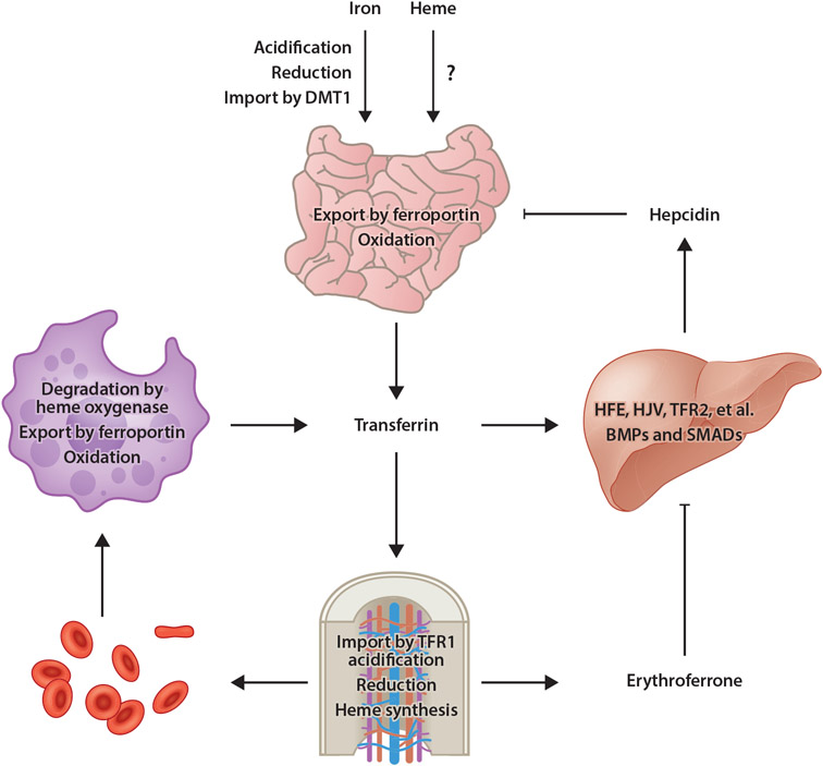
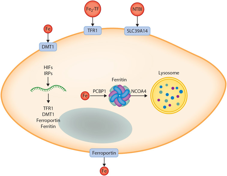
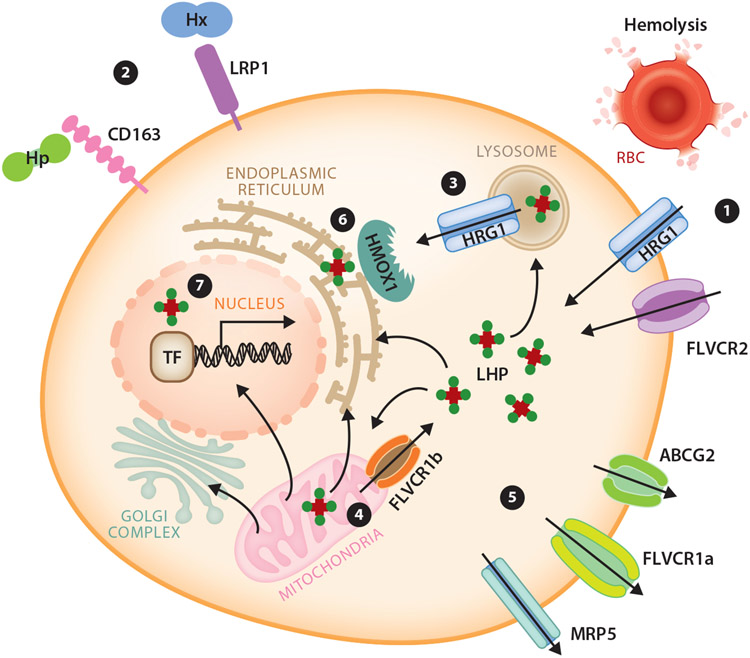
An abundant metal in the human body, iron is essential for key biological pathways including oxygen transport, DNA metabolism, and mitochondrial function. Most iron is bound to heme but it can also be incorporated into iron-sulfur clusters or bind directly to proteins. Iron's capacity to cycle between Fe2+ and Fe3+ contributes to its biological utility but also renders it toxic in excess. Heme is an iron-containing tetrapyrrole essential for diverse biological functions including gas transport and sensing, oxidative metabolism, and xenobiotic detoxification. Like iron, heme is essential yet toxic in excess. As such, both iron and heme homeostasis are tightly regulated. Here we discuss molecular and physiologic aspects of iron and heme metabolism. We focus on dietary absorption; cellular import; utilization; and export, recycling, and elimination, emphasizing studies published in recent years. We end with a discussion on current challenges and needs in the field of iron and heme biology.

Keywords: heme; iron; metabolism; porphyrin; tetrapyrrole; trafficking.


Figures

Figure 1
Figure 1
Model of mammalian iron homeostasis. Both nonheme and heme iron are absorbed in the small intestine. Transporters involved with heme iron absorption have yet to be identified. Nonheme iron is acidified in the stomach, reduced by gastric acid or DCYTB in the small intestine, then imported into enterocytes by DMT1. Iron is then utilized by or stored in enterocytes or exported into circulation by ferroportin. After oxidation by hephaestin, iron binds to transferrin for distribution throughout the body. Most transferrin-bound iron is imported by TFR1-mediated endocytosis into erythroid precursors, a process that requires acidification to liberate iron from transferrin then export into the cytosol followed by reduction by STEAP3. Iron is then utilized for heme synthesis. Senescent or damaged RBCs are degraded by RES macrophages, in which heme oxygenase degrades heme, thereby liberating iron for export by ferroportin into circulation. Under conditions of iron excess, transferrin (and other factors) stimulates BMP expression in sinusoidal endothelial cells. BMPs then stimulate hepcidin expression by hepatocytes in a pathway dependent upon HFE, HJV, TFR2, and other membrane factors, as well as SMAD1/5/8 transcription factors. Hepcidin then posttranslationally inhibits ferroportin activity and expression. Under conditions of anemia, erythroid progenitors produce the hormone erythroferrone, which inhibits BMP-dependent hepcidin expression, thereby ensuring continued dietary iron absorption for erythropoiesis. Abbreviations: BMP, bone morphogenetic protein; DCYTB, duodenal cytochrome b reductase 1; DMT1, divalent metal transporter 1; HFE, homeostatic iron regulator; HJV, hemojuvelin; RBC, red blood cell; RES, reticuloendothelial system; TFR1, transferrin receptor 1; TFR2, transferrin receptor 2. Figure adapted from images created with BioRender.com.
Figure 2
Figure 2
Model of main pathways of cellular iron import and export and intracellular distribution. Iron can be imported into cells via one of several pathways including import of ferrous iron by DMT1; import of Fe2-TF by TFR1-mediated endocytosis, followed by reduction by STEAP3 and export into the cytoplasm; and import of NTBI by SLC39A14. Iron is exported by ferroportin, the only known mammalian iron export protein, then oxidized by hephaestin or ceruloplasmin and bound to transferrin for distribution to other cellular targets. Within the cell, expression of iron transporters, the iron storage protein ferritin, and other factors is regulated by HIFs and IRPs to ensure sufficient but nontoxic levels of iron for cellular metabolism and adequate storage in settings of iron excess. The iron metallochaperone PCBP1 delivers iron to multiple protein targets within the cell including ferritin. Under conditions of iron limitation, ferritin is mobilized to the lysosome by NCOA4 for degradation and liberation of stored iron. For simplicity, not all pathways of iron import are shown, nor are all known intracellular destinations or targets for iron indicated. Abbreviations: DMT1, divalent metal transporter 1; Fe2-TF, diferric transferrin; HIF, hypoxia-inducible factor; IRP, iron regulatory protein; NCOA4, nuclear receptor coactivator 4; NTBI, nontransferrin-bound iron; PCBP1, poly(rC)-binding protein; TFR1, transferrin receptor 1. Figure adapted from images created with BioRender.com.
Figure 3
Figure 3
Model of known metazoan heme transport mediators and pathways for intracellular heme trafficking. A cell can autonomously meet its heme requirements by de novo synthesis in mitochondria. (①) It can also uptake heme through the plasma membrane heme importer HRG1, FLVCR2, or via endocytosis of senescent RBCs. Additionally, (②) Hp and Hx are scavenged by membrane-bound CD163 and LRP1 receptor-mediated endocytosis, respectively. Intracellular heme can be imported into the cytosol via HRG1 on endolysosomal membranes (③) and FLVCR1b on mitochondrial membranes (④). (⑤) Export of heme can be mediated by plasma membrane exporters FLVCR1a, ABCG2, and MRP5. To prevent free heme cytotoxicity, heme is degraded by HMOX1 (⑥. Cellular LHP may facilitate intracellular heme trafficking. (⑦) In the nucleus, TFs bind heme to regulate transcription of downstream target genes. Abbreviations: ABCG2, ATP-binding cassette subfamily G member 2; FLVCR, feline leukemia virus subgroup C receptor; HMOX1, heme oxygenase 1; Hp, haptoglobin; HRG, heme responsive gene; Hx, hemopexin; LHP, labile heme pool; LRP1, LDL receptor related protein 1; MRP, multidrug resistance protein; RBC, red blood cell; TF, transcription factor. Figure adapted from images created using smart.servier.com and motifolio.com.

References

    1. Adams PA, Berman MC. 1980. Kinetics and mechanism of the interaction between human serum albumin and monomeric haemin. Biochem. J 191(1):95–102 - PMC - PubMed
    1. Altamura S, Vegi NM, Hoppe PS, Schroeder T, Aichler M, et al. 2020. Glutathione peroxidase 4 and vitamin E control reticulocyte maturation, stress erythropoiesis and iron homeostasis. Haematologica 105(4):937–50 - PMC - PubMed
    1. Andolfo I, Rosato BE, Manna F, Rosa GD, Marra R, et al. 2020. Gain-of-function mutations in PIEZO1 directly impair hepatic iron metabolism via the inhibition of the BMP/SMADs pathway. Am. J. Hematol 95(2):188–97 - PubMed
    1. Ascenzi P, Bocedi A, Visca P, Altruda F, Tolosano E, et al. 2005. Hemoglobin and heme scavenging. IUBMB Life 57(11):749–59 - PubMed
    1. Ashouri R, Fangman M, Burris A, Ezenwa MO, Wilkie DJ, Doré S. 2021. Critical role of hemopexin mediated cytoprotection in the pathophysiology of sickle cell disease. Int. J. Mol. Sci 22(12):6408. - PMC - PubMed

Publication types