Review
. 2022 Apr:86:101165.
doi: 10.1016/j.plipres.2022.101165. Epub 2022 May 1.

Polyunsaturated fatty acids and fatty acid-derived lipid mediators: Recent advances in the understanding of their biosynthesis, structures, and functions

Affiliations
Review

Polyunsaturated fatty acids and fatty acid-derived lipid mediators: Recent advances in the understanding of their biosynthesis, structures, and functions

Simon C Dyall et al. Prog Lipid Res. 2022 Apr.

Abstract

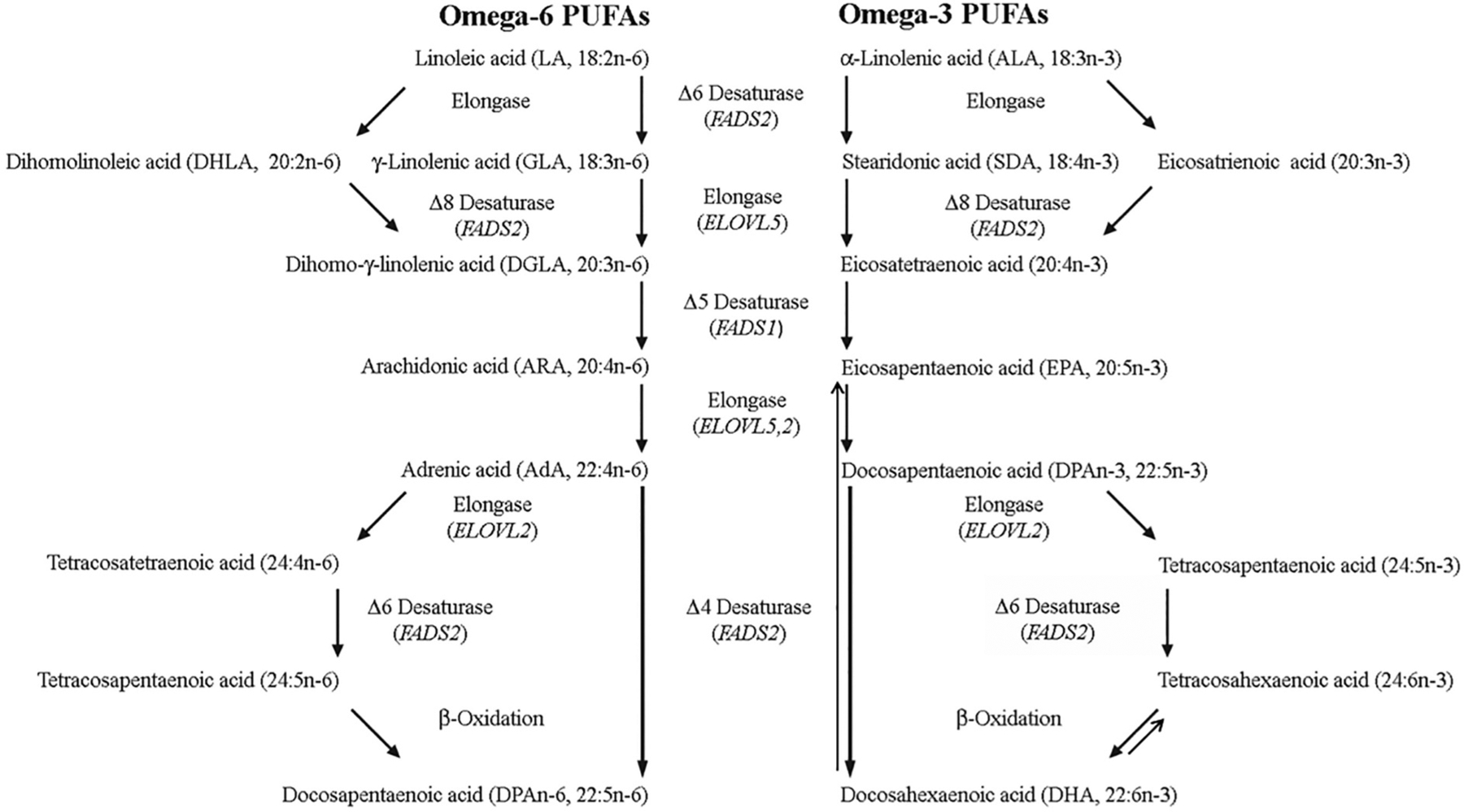
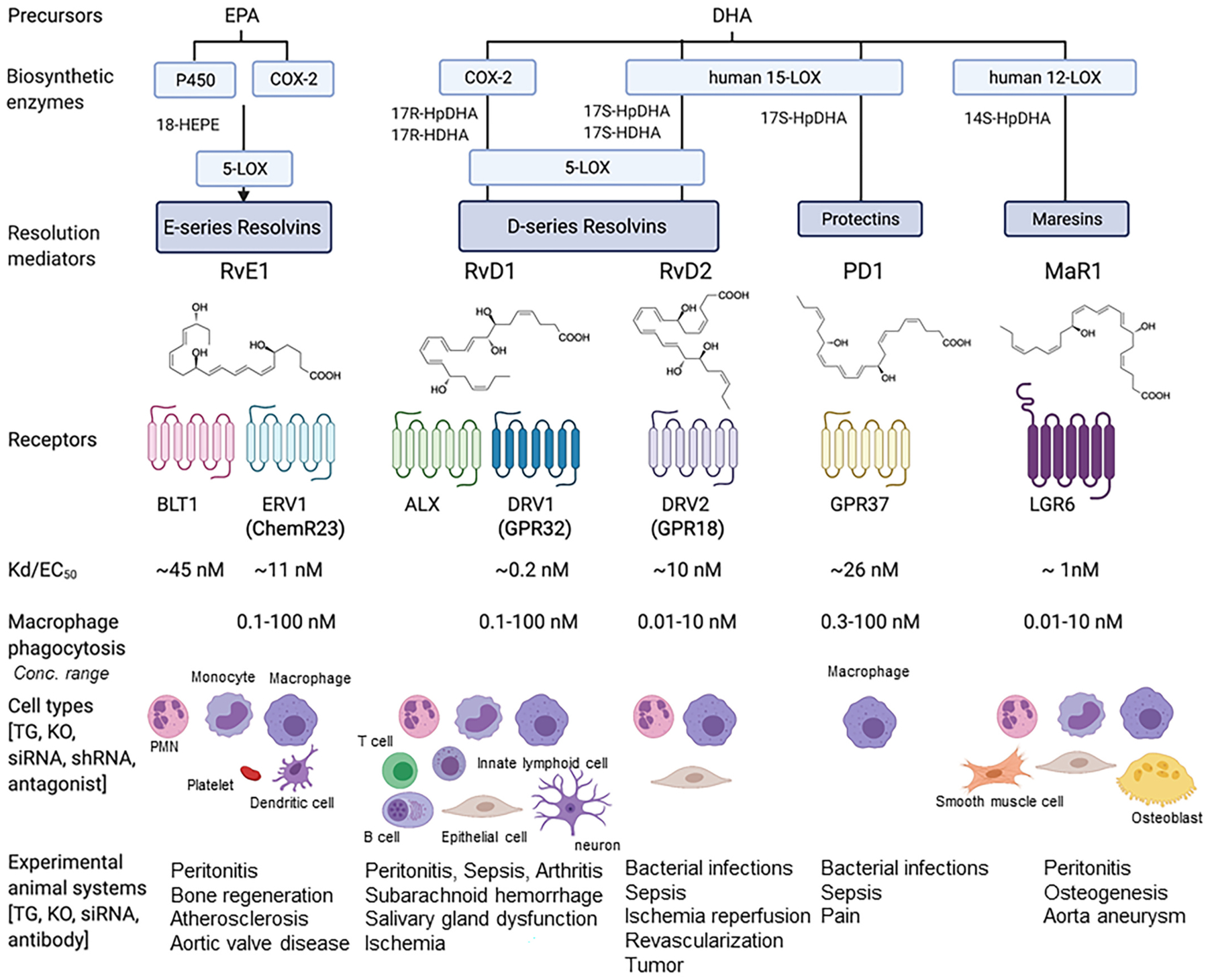
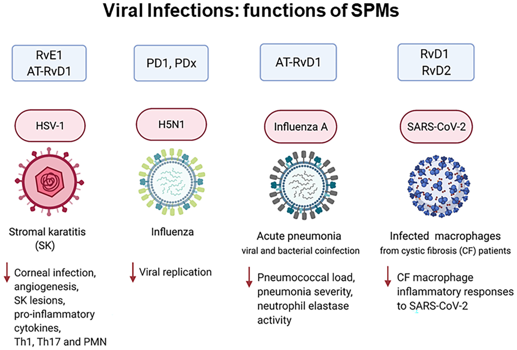
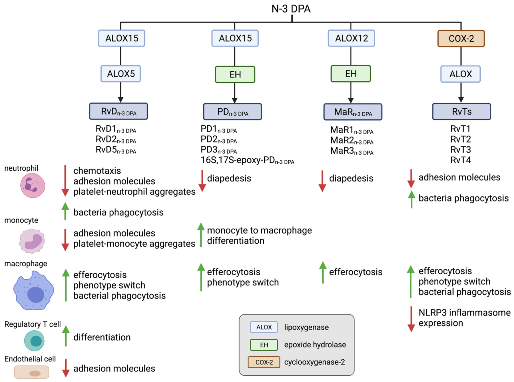
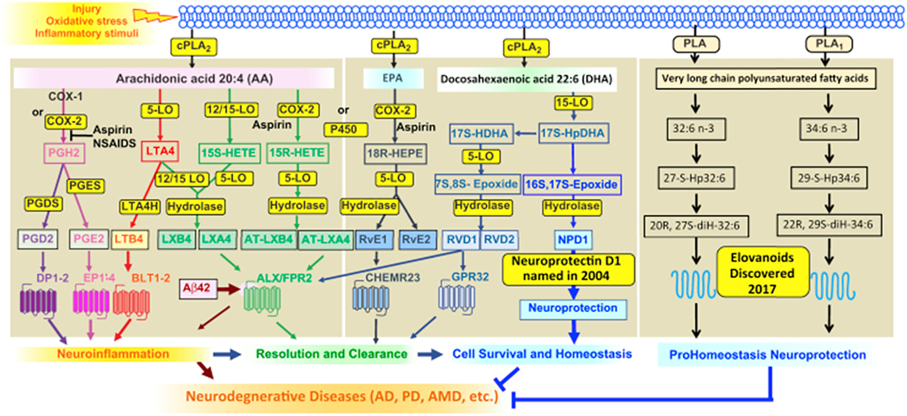
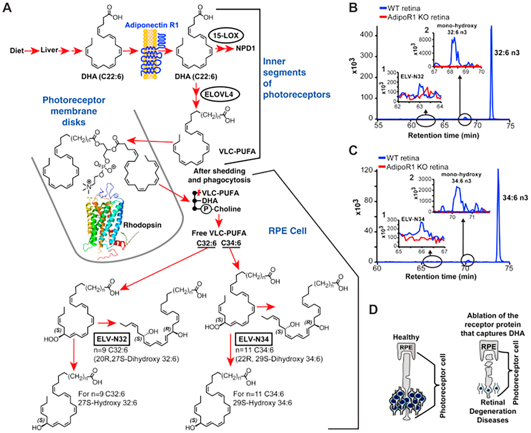
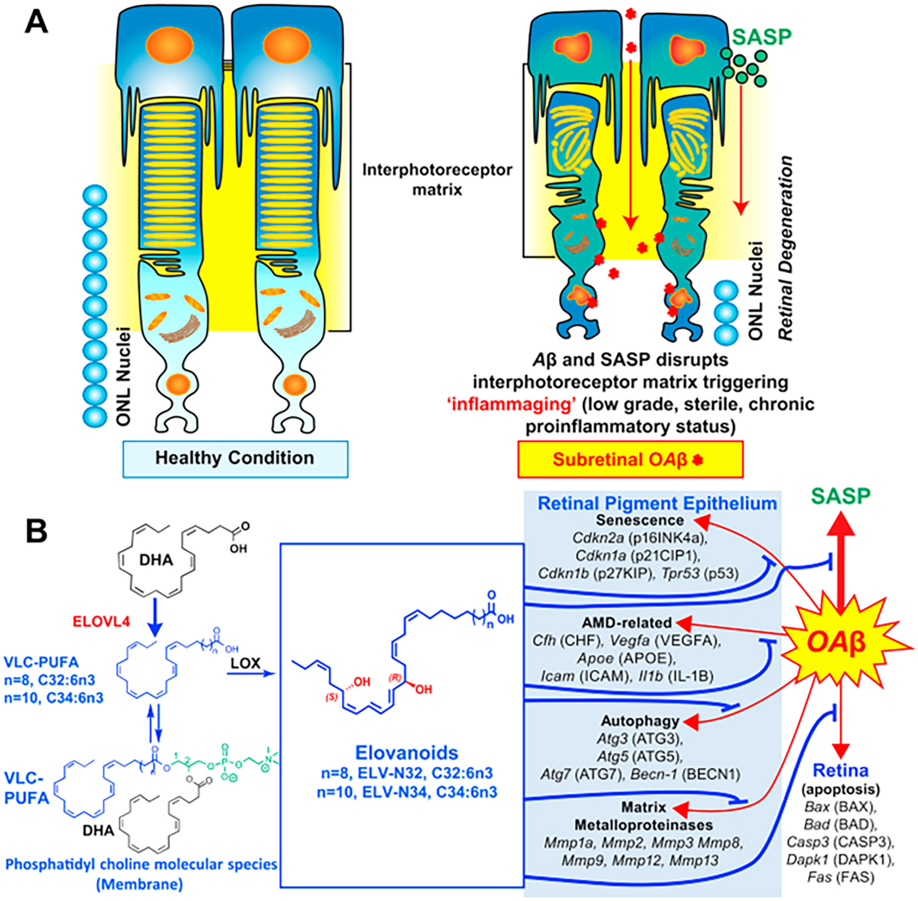
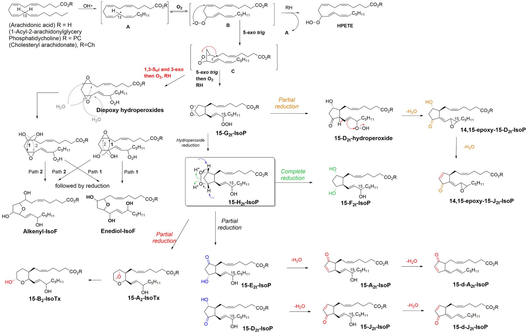
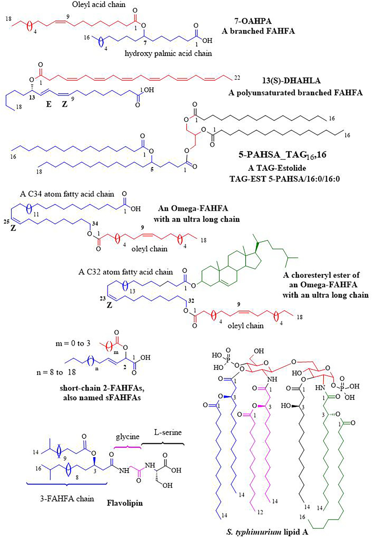
Polyunsaturated fatty acids (PUFAs) are structural components of membrane phospholipids, and influence cellular function via effects on membrane properties, and also by acting as a precursor pool for lipid mediators. These lipid mediators are formed via activation of pathways involving at least one step of dioxygen-dependent oxidation, and are consequently called oxylipins. Their biosynthesis can be either enzymatically-dependent, utilising the promiscuous cyclooxygenase, lipoxygenase, or cytochrome P450 mixed function oxidase pathways, or nonenzymatic via free radical-catalyzed pathways. The oxylipins include the classical eicosanoids, comprising prostaglandins, thromboxanes, and leukotrienes, and also more recently identified lipid mediators. With the advent of new technologies there is growing interest in identifying these different lipid mediators and characterising their roles in health and disease. This review brings together contributions from some of those at the forefront of research into lipid mediators, who provide brief introductions and summaries of current understanding of the structure and functions of the main classes of nonclassical oxylipins. The topics covered include omega-3 and omega-6 PUFA biosynthesis pathways, focusing on the roles of the different fatty acid desaturase enzymes, oxidized linoleic acid metabolites, omega-3 PUFA-derived specialized pro-resolving mediators, elovanoids, nonenzymatically oxidized PUFAs, and fatty acid esters of hydroxy fatty acids.

Keywords: Elovanoids; FAHFA; Fatty acid desaturase; Lipid mediators; Maresins; Omega-3 PUFA; Oxylipins; Protectins; Resolvins; SPM.


Conflict of interest statement

Declaration of Competing Interest

N.G. Bazan is the founder of two start-up companies that have exclusively licensed technologies from LSUHSC involving elovanoids and related lipids for clinical applications: NeuResto Therapeutics, LLC and CurVirBiotech, LLC. All other authors declare no conflicts of financial interest.

Figures

Fig. 1.
Fig. 1.
Biosynthesis of omega-3 and omega-6 PUFAs. The biosynthesis of longer-chain omega-3 and omega-6 PUFAs proceeds via a series of alternating position-specific desaturation and elongation steps from ALA and LA, respectively. FADS1 and FADS2 appear responsible for all omega-3 and omega-6 PUFA desaturation in mammals, with FADS1 exhibiting Δ5-desaturase activity, and although FADS2 was originally identified as the Δ6-desaturase, it has subsequently been shown to also possess Δ4- and Δ8-desaturase activities. Octadecanoids are lipid mediators derived from C18 PUFAs, such as ALA or LA, eicosanoids are derived from C20 PUFAs such as DGLA, ARA or EPA, and docosanoids are derived from C22 PUFAs such as DPAn-3, and DHA. See text for further details.
Fig. 2.
Fig. 2.
Illustration of resolution metabolome: SPM biosynthesis, receptors and functions. Precursors EPA and DHA are converted via biosynthetic enzymes to SPMs, which in turn activate their specific receptors to stimulate pro-resolving innate immune functions. Each SPM demonstrates stereoselective activation of its cognate GPCR on select cell types, leading to intracellular signals, pathways and pro-resolving functions The affinities of SPMs for their respective recombinant GPCRs (i.e., Kd or EC 50 values) are consistent with their bioactive concentration ranges, e.g. macrophage phagocytosis (picomolar to low nanomolar) in vitro and dose ranges (picograms to low nanograms) in vivo.The in vivo functions of these SPM receptors were demonstrated using transgenic and/or knock-out mice, as well as specific blockage of the receptor, e.g., siRNA, antibodies or receptor antagonists (see text and recent reviews [98,116] for details).
Fig. 3.
Fig. 3.
SPM actions in viral infections. See text for details.
Fig. 4.
Fig. 4.
Illustration of the omega-3 DPA derived SPM families and the biological activities exerted on immune and stromal cells. For details of the stereochemistry of the structures of these SPMs please see [162].
Fig. 5.
Fig. 5.
Eicosanoids, docosanoids, and elovanoids. PLAs that release ARA, EPA, DHA, or VLC-PUFAs are depicted at the top. Synthesis of mediators and receptors involved are illustrated. The outcome is modulation of inflammatory responses and homeostasis. AD, Alzheimer’s disease; AMD, age-related macular degeneration; VLC-PUFA, very long-chain PUFA. Reproduced, with permission from the Journal of Lipid Research [178].
Fig. 6.
Fig. 6.
Genetic ablation of adiponectin receptor 1 leads to depletion of VLC-PUFAs and its derivatives in retina. A: Dietary DHA, or DHA derived from dietary 18:3n3, is supplied by the liver and taken by the Adiponectin Receptor 1 (AdipoR1), followed by elongation in the inner segment of photoreceptor cell (PRC) by Elongation of Very Long chain fatty acids-4 (ELOVL4) to VLC-PUFA and incorporation into PC molecular species, which contains DHA at sn-2. During daily PRC outer segment renewal, these PC molecular species interact with rhodopsin and, after shedding of the PRC tips and phagocytosis, become part of retinal pigment epithelium (RPE) cells. Uncompensated oxidative stress (UOS) or other disruptors of homeostasis trigger the release of VLC-PUFAs. 32:6n-3 and 34:6n-3 are depicted generating hydroperoxyl forms, and then elovanoid (ELV)-N32 or ELV-N34, respectively. B: The pool size of free 32:6n-3 in retinas of AdipoR1 KO mice (red) is decreased as compared with that in wild type (WT) (blue). Insert (1) shows ELV-N32 in KO (red) and WT (blue); insert (2) shows monohydroxy 32:6n3, the stable derivative of the hydroperoxyl precursor of ELV-N32, in WT (blue) and lack of detectable signal in the KO (red). C: Similarly, the pool size of free 34:6n-3 in retinas of AdipoR1 KO mice (red) is decreased as compared with that in WT (blue). Insert (1) shows ELV-N32 in KO (red) and WT (blue); insert (2) shows mono-hydroxy 34:6n-3, the stable derivative of the hydroperoxyl precursor of ELV-N34, in WT (blue) and lack of detectable signal in the KO (red). D: RPE cells sustain PRC functional integrity (left); right, the ablation of AdipoR1 switches off DHA availability, and PRC degeneration ensues. Reproduced, with permission, from Scientific Reports [190]. (For interpretation of the references to colour in this figure legend, the reader is referred to the web version of this article.)
Fig. 7.
Fig. 7.
ELVs effect on oligomeric amyloid-β (OAβ)-induced RPE and PRC damage. A: OAβ induces a senescence gene program and disrupts RPE tight junctions. OAβ penetrates the retina, causing PRC cell death in our in vivo WT mice study, as reflected in less outer nuclear layer (ONL) nuclei (Fig. 5 from [198]). OAβ activates the senescence-associated secretome (SASP) that contributes to perturbing the interphotoreceptor matrix (IPM), triggering inflammaging in PRC and also likely in Mueller glia, which limits the IPM. Therefore, senescence paracrine expression takes place. ELVs restore RPE morphology and PRC integrity. B: OAβ induces expression of senescence, autophagy, matrix metalloproteinases, and age-related macular degeneration (AMD)-related genes in the RPE and apoptosis genes in retina in addition to p16INK4a protein abundance. ELVs downregulated the OAβ-gene inductions and p16INK4a protein abundance. Pathways for the ELV synthesis are outlined. ELV, elovanoid; PRC, photoreceptor cell; RPE, retinal pigment epithelium. Reproduced, with permission, from the Proceedings of the National Academy of Sciences of the United States [198].
Fig. 8.
Fig. 8.
Mechanism of the free radical chain process leading to cyclic NEO-PUFAs. (Only one H-atom abstraction is shown for clarity, as well as stereoisomers, and ARA was chosen as the PUFA). See text for details.
Fig. 9.
Fig. 9.
Examples of Fatty acid esters of hydroxy fatty acids (FAHFAs) and derivatives. See text for details.

Similar articles

Cited by

References

    1. Gomez-Larrauri A, Presa N, Dominguez-Herrera A, Ouro A, Trueba M, Gomez-Munoz A. Role of bioactive sphingolipids in physiology and pathology. Essays Biochem 2020;64(3):579–89. - PubMed
    1. Hannun YA, Obeid LM. Principles of bioactive lipid signalling: lessons from sphingolipids. Nat Rev Mol Cell Biol 2008;9(2):139–50. - PubMed
    1. Shimizu T Lipid mediators in health and disease: enzymes and receptors as therapeutic targets for the regulation of immunity and inflammation. Annu Rev Pharmacol Toxicol 2009;49:123–50. - PubMed
    1. Dyall SC. Interplay between n-3 and n-6 long-chain polyunsaturated fatty acids and the endocannabinoid system in brain protection and repair. Lipids 2017;52 (11):885–900. - PMC - PubMed
    1. von Euler US. History and development of prostaglandins. Gen Pharmacol 1983; 14(1):3–6. - PubMed

Publication types