. 2022 Aug;163(2):495-506.e8.
doi: 10.1053/j.gastro.2022.04.037. Epub 2022 May 1.

Postacute COVID-19 is Characterized by Gut Viral Antigen Persistence in Inflammatory Bowel Diseases

Affiliations

Postacute COVID-19 is Characterized by Gut Viral Antigen Persistence in Inflammatory Bowel Diseases

Andreas Zollner et al. Gastroenterology. 2022 Aug.

Abstract

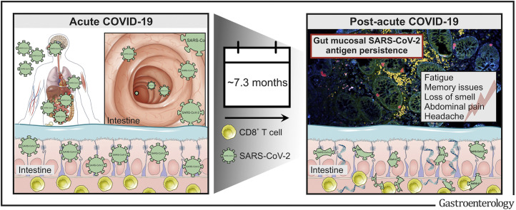
Background & aims: The coronavirus disease 2019 (COVID-19) pandemic has affected populations, societies, and lives for more than 2 years. Long-term sequelae of COVID-19, collectively termed the postacute COVID-19 syndrome, are rapidly emerging across the globe. Here, we investigated whether severe acute respiratory syndrome coronavirus 2 (SARS-CoV-2) antigen persistence underlies the postacute COVID-19 syndrome.

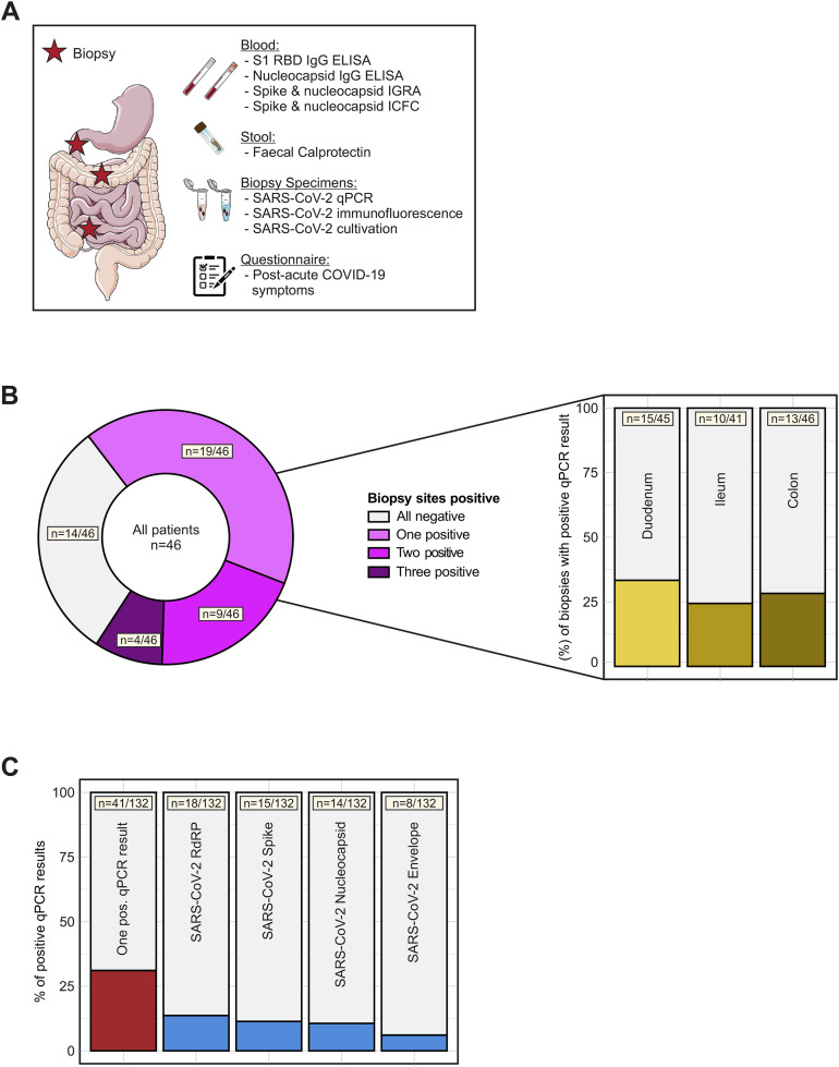
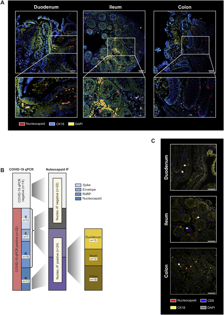
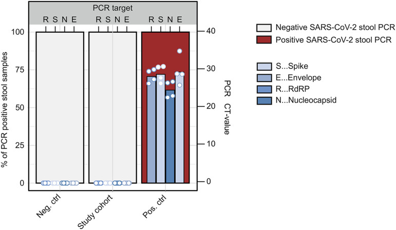
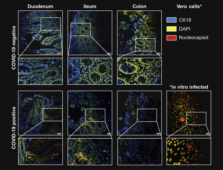
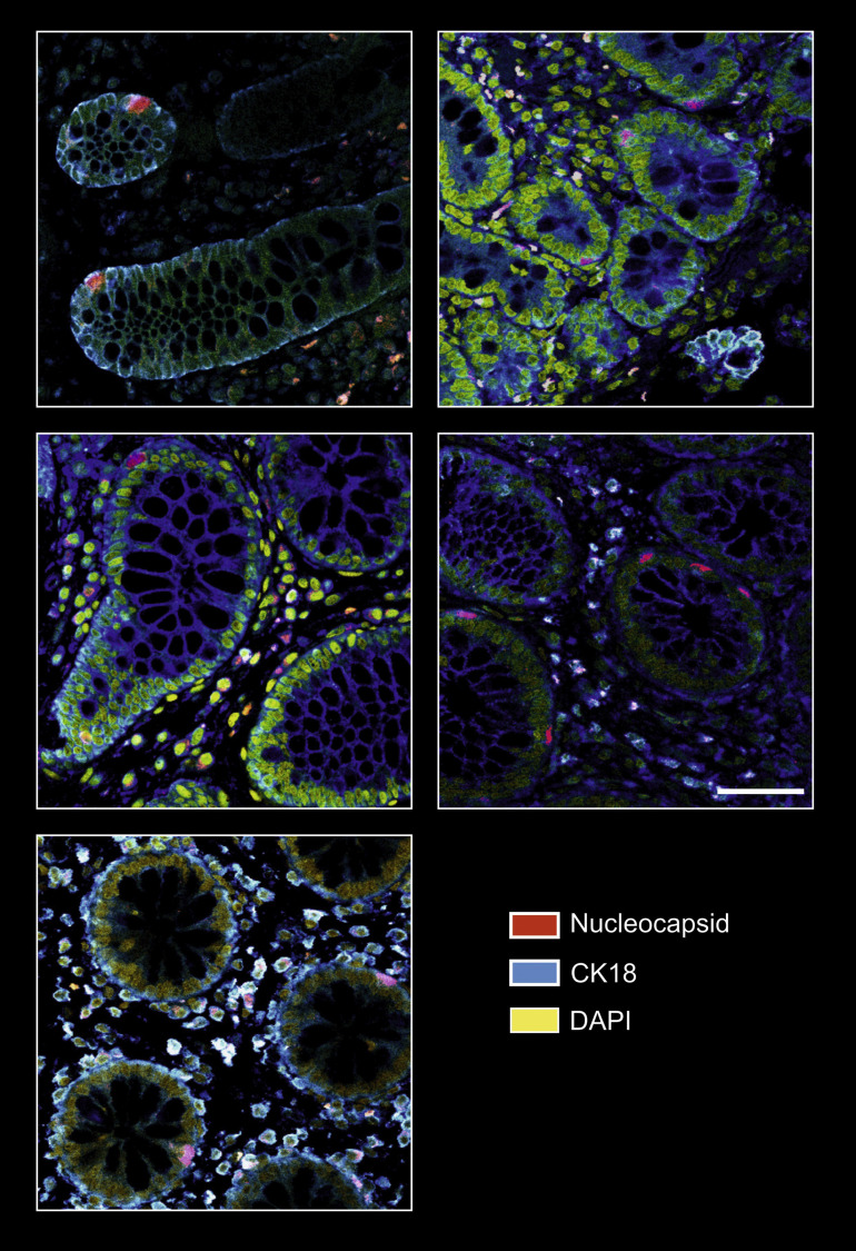
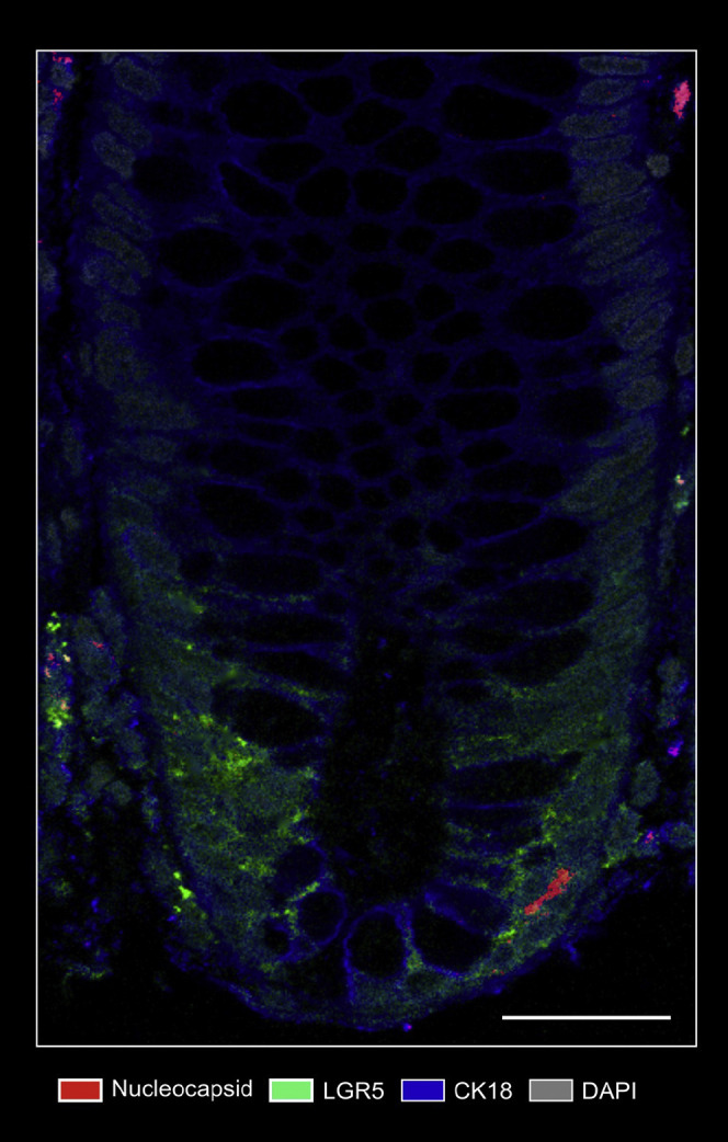
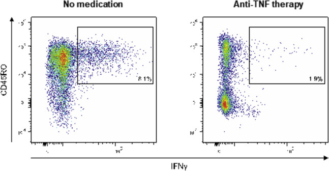
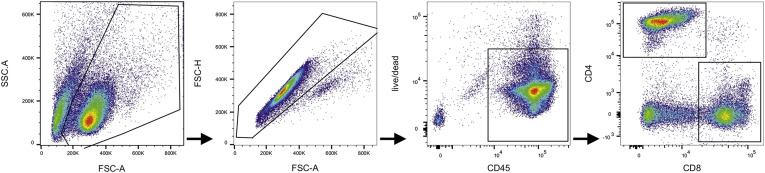
Methods: We performed an endoscopy study with 46 patients with inflammatory bowel disease (IBD) 219 days (range, 94-257) after a confirmed COVID-19 infection. SARS-CoV-2 antigen persistence was assessed in the small and large intestine using quantitative polymerase chain reaction of 4 viral transcripts, immunofluorescence of viral nucleocapsid, and virus cultivation from biopsy tissue. Postacute COVID-19 was assessed using a standardized questionnaire, and a systemic SARS-CoV-2 immune response was evaluated using flow cytometry and enzyme-linked immunosorbent assay at endoscopy. IBD activity was evaluated using clinical, biochemical, and endoscopic means.

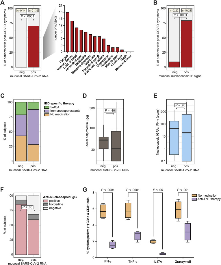
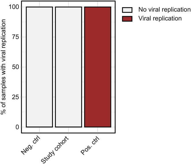
Results: We report expression of SARS-CoV-2 RNA in the gut mucosa ∼7 months after mild acute COVID-19 in 32 of 46 patients with IBD. Viral nucleocapsid protein persisted in 24 of 46 patients in gut epithelium and CD8+ T cells. Expression of SARS-CoV-2 antigens was not detectable in stool and viral antigen persistence was unrelated to severity of acute COVID-19, immunosuppressive therapy, and gut inflammation. We were unable to culture SARS-CoV-2 from gut tissue of patients with viral antigen persistence. Postacute sequelae of COVID-19 were reported from the majority of patients with viral antigen persistence, but not from patients without viral antigen persistence.

Conclusion: Our results indicate that SARS-CoV-2 antigen persistence in infected tissues serves as a basis for postacute COVID-19. The concept that viral antigen persistence instigates immune perturbation and postacute COVID-19 requires validation in controlled clinical trials.

Keywords: COVID-19; Postacute COVID-19; SARS-CoV-2; Viral Antigen Persistence.


Figures

None
Graphical abstract
Figure 1
Figure 1
SARS-CoV-2 antigen persistence frequently occurs in the gut mucosa. (A) Study design. In a cohort of 46 patients with IBD with history of COVID-19, 219 days (range, 94–257) after the first positive PCR test endoscopy was performed and biopsy-derived tissue was phenotyped for SARS-CoV-2 mucosal antigen persistence and IBD activity. Patients were also evaluated for symptoms compatible with postacute COVID-19 sequelae using a questionnaire. The questionnaire was adapted according to the AWMF Post-Covid/long-Covid S1 guidelines, and systemic SARS-CoV-2–directed immune responses were analyzed. (B) SARS-CoV-2 RNA was detected in 32/46 patients using qPCR. Viral RNA was detected in 1 biopsy in 19 patients, in 2 biopsies in 9 patients, and in all 3 biopsies collected in 4 patients. Detection was unrelated to the intestinal location and viral RNA was detected in 15/46 duodenal, 10/46 ileal, and 13/46 colonic samples. (C) Biopsies were analysed for 4 viral transcripts using qPCR (ie, RNA polymerase [RdRP], nucleocapsid phosphoprotein [Nucleocapsid], surface glycoprotein [Spike], and envelope protein). The red bar (left) indicates the proportion of biopsies with at least 1 positive qPCR signal, the blue bars illustrate the proportions for the respective transcript.
Figure 2
Figure 2
SARS-CoV-2 nucleocapsid immunofluorescence in the intestine. (A) Representative confocal microscopy images of viral nucleocapsid (red) in the duodenum, ileum, and colon from patients with IBD ∼219 days after acute COVID-19. Cytokeratin 18 (blue) visualizes the epithelial cytoskeleton and 4′,6-diamidino-2-phenylindole (DAPI) (yellow) depicts the nucleus. Scale bar indicates 50 μm. (B) Association between viral qPCR positivity and antinucleocapsid immunoreactivity based on immunofluorescence. SARS-CoV-2 RNA was detected in 32/46 patients with RdRP in 16 patients, the surface glycoprotein (Spike) in 12 patients, the nucleocapsid phosphoprotein (Nucleocapsid) in 13 patients, and the envelope protein in 7 patients. All patients with a positive nucleocapsid qPCR displayed nucleocapsid immunoreactivity based on immunofluorescence in at least 1 of 3 gut segments. (C) Representative confocal microscopy images of viral nucleocapsid (red) colabelled with anti-CD8 (blue) in the duodenum, ileum, and colon. Cytokeratin 18 (yellow) visualizes the cytoskeleton and DAPI (grey) depicts the nucleus. Scale bar indicates 50 μm.
Figure 3
Figure 3
SARS-CoV-2 antigen persistence links to postacute sequelae of COVID-19. (A) Patients were stratified based on viral qPCR positivity in any gut segment. Patients without evident mucosal viral RNA by PCR (negative [neg.]) were compared with patients with detectable viral RNA (positive [pos.]). Proportion of patients who reported symptoms compatible with postacute COVID-19 syndrome is shown in red. Note that 0/14 patients without detectable mucosal viral RNA (based on qPCR in any gut segment) reported symptoms, whereas postacute COVID-19 symptoms were reported from 21/32 patients with detectable RNA (with specific symptoms listed). Statistical significance was calculated using the Fisher exact test. (B) Patients were stratified according to nucleocapsid immunoreactivity as assessed using immunofluorescence and data is depicted equivalent to (A). (C) Proportion of patients without IBD medication, with 5-aminosalycilates, and with immunosuppressive therapy (ie, azathioprine, anti-TNF therapy, or vedolizumab) in both groups (defined using qPCR positivity). (D) Quantification of fecal calprotectin concentrations in both groups (defined using qPCR positivity) using ELISA. (E) Quantification of SARS-CoV-2 nucleocapsid specific T-cell responses defined using IGRA (according to qPCR positivity). (F) Proportion of patients with positive, borderline, and negative nucleocapsid IgG in both groups (defined using qPCR positivity). Borderline and negative results were pooled, and statistical significance was calculated using the Fisher exact test. (G) Relative quantification of indicated cytokines defined using ICFC of T cells from patients with or without anti-TNF therapy after exposure to peptides of the SARS-CoV-2 nucleocapsid (n = 5). Boxplots represent median percentages of cytokine-positive CD4+ and CD8+ cells. Differences between groups were calculated using the Fisher exact test (A and B), 2-tailed Student t tests (D, E, and F), and 2-way analysis of variance (ANOVA) with Sidak multiple comparison post-hoc test (G). Boxplots represent values as median (bold horizontal line), interquartile range (IQR) (box), and 1.5 x IQR (whiskers). Bars represent the mean and whiskers the standard deviation.
Supplementary Figure 1
Stool SARS-CoV-2 PCR. RNA was isolated from stool of patients before the pandemic (neg. ctrl; n = 4), from the study cohort (n = 46), and stool from COVID-19 subjects (pos. ctrl; n = 4). The red bars indicate percentages of samples with positive SARS-CoV-2 PCR. The blue bars show CT values for the RNA-dependent RNA polymerase (R), the spike (S), the nucleocapsid (N), and the envelope (E).
Supplementary Figure 2
SARS-CoV-2 nucleocapsid immunofluorescence of intestinal tissue. Representative confocal microscopy images of viral nucleocapsid (red) in the duodenum, ileum and colon mucosa from patients with IBD ∼219 days after acute COVID-19 (lower panel). Immunostaining of sections from patients with no history of SARS-CoV-2 infection (retrieved before the pandemic) is shown in the upper panel and serves as a negative control. Cytokeratin 18 (blue) visualizes the cytoskeleton and DAPI (yellow) depict the nucleus. Scale bar indicates 50 μm.
Supplementary Figure 3
Additional SARS-CoV-2 nucleocapsid immunofluorescence from intestinal tissue. As in Supplementary Figure 1, representative confocal microscopy images of viral nucleocapsid (red) in the duodenum, ileum, and colon mucosa from 5 patients with IBD ∼219 days after acute COVID-19. Cytokeratin 18 (blue) visualizes the cytoskeleton and DAPI (yellow) depict the nucleus. Scale bar (bottom left) indicates 50 μm.
Supplementary Figure 4
Nucleocapsid/Lgr5 immunofluorescence. Representative confocal microscopy image of costainings of viral nucleocapsid (red) and Lgr5 (green) in the mucosa from a patient with IBD ∼219 days after acute COVID-19. Cytokeratin 18 (blue) visualizes the cytoskeleton and DAPI (grey) depicts the nucleus. Scale bar indicates 100 μm.
Supplementary Figure 5
SARS-CoV-2 cultivation. Lysates of negative controls (n = 4), the study cohort (n = 46), and positive controls (n = 4) were used to infect ACE-2 and TMPRSS2 overexpressing Vero cells (Vero-TMPRSS2/ACE2) and cytopathic effect was analyzed. The percentages of samples showing a cytopathic effect are indicated in red.
Supplementary Figure 6
Representative flow cytometry plots of cellular nucleocapsid reactivity. PBMCs were cocultivated with peptide pools covering the N protein for 60 h, restimulated, and analyzed using intracellular cytokine staining. Dot plots show CD4+ and CD8+ T cells that produced IFN-γ in response to stimulation nucleocapsid peptides in a patient with IBD without medication (left) and a patient with anti-TNF therapy (right).
Supplementary Figure 7
Representative gating strategy. First cells were gated based on FSC and SSC; singlets were excluded by FSC-A vs FSC-H gate; live CD45+ high leukocytes were gated; live CD45+ high cells were separated in CD4+ and CD8+ cells. These cell populations were analyzed for intracellular IFN-γ, TNFα, IL-17, and GranzymeB. FSC, forward scatter; SSC, side scatter.

Comment in

References

    1. Gupta A., Madhavan M.V., Sehgal K., et al. Extrapulmonary manifestations of COVID-19. Nat Med. 2020;26:1017–1032. - PMC - PubMed
    1. Jackson C.B., Farzan M., Chen B., et al. Mechanisms of SARS-CoV-2 entry into cells. Nat Rev Molec Cell Biol. 2022;23:3–20. - PMC - PubMed
    1. Penninger J.M., Grant M.B., Sung J.J.Y. The role of angiotensin converting enzyme 2 in modulating gut microbiota, intestinal inflammation, and coronavirus infection. Gastroenterology. 2021;160:39–46. - PMC - PubMed
    1. Effenberger M., Grabherr F., Mayr L., et al. Faecal calprotectin indicates intestinal inflammation in COVID-19. Gut. 2020;69:1543. - PMC - PubMed
    1. Lamers Mart M., Beumer J., van der Vaart J., et al. SARS-CoV-2 productively infects human gut enterocytes. Science. 2020;369:50–54. - PMC - PubMed

Publication types