. 2022 Jun 8;42(23):4693-4710.
doi: 10.1523/JNEUROSCI.2257-21.2022. Epub 2022 May 4.

The Dorsal Visual Pathway Represents Object-Centered Spatial Relations for Object Recognition

Affiliations

The Dorsal Visual Pathway Represents Object-Centered Spatial Relations for Object Recognition

Vladislav Ayzenberg et al. J Neurosci. .

Abstract

Although there is mounting evidence that input from the dorsal visual pathway is crucial for object processes in the ventral pathway, the specific functional contributions of dorsal cortex to these processes remain poorly understood. Here, we hypothesized that dorsal cortex computes the spatial relations among an object's parts, a process crucial for forming global shape percepts, and transmits this information to the ventral pathway to support object categorization. Using fMRI with human participants (females and males), we discovered regions in the intraparietal sulcus (IPS) that were selectively involved in computing object-centered part relations. These regions exhibited task-dependent functional and effective connectivity with ventral cortex, and were distinct from other dorsal regions, such as those representing allocentric relations, 3D shape, and tools. In a subsequent experiment, we found that the multivariate response of posterior (p)IPS, defined on the basis of part-relations, could be used to decode object category at levels comparable to ventral object regions. Moreover, mediation and multivariate effective connectivity analyses further suggested that IPS may account for representations of part relations in the ventral pathway. Together, our results highlight specific contributions of the dorsal visual pathway to object recognition. We suggest that dorsal cortex is a crucial source of input to the ventral pathway and may support the ability to categorize objects on the basis of global shape.SIGNIFICANCE STATEMENT Humans categorize novel objects rapidly and effortlessly. Such categorization is achieved by representing an object's global shape structure, that is, the relations among object parts. Yet, despite their importance, it is unclear how part relations are represented neurally. Here, we hypothesized that object-centered part relations may be computed by the dorsal visual pathway, which is typically implicated in visuospatial processing. Using fMRI, we identified regions selective for the part relations in dorsal cortex. We found that these regions can support object categorization, and even mediate representations of part relations in the ventral pathway, the region typically thought to support object categorization. Together, these findings shed light on the broader network of brain regions that support object categorization.

Keywords: dorsal stream; object recognition; shape perception; two visual streams; ventral stream; visual cortex.


Figures

Figure 1.
Figure 1.
Example stimuli from the (A) object-centered part relations, (B) allocentric relations (C) depth, (D) and tool localizers used in experiment 1.
Figure 2.
Figure 2.
Object stimuli presented in experiment 2. Participants viewed five exemplars from five categories in an event-related design.
Figure 3.
Figure 3.
Significant activation to part relations (vs features) condition from the object-centered part relations localizer displayed (A) for each individual participant and in (B) a group average map inflated (above) and flattened (below). Values reflect the standardized parameter estimate.
Figure 4.
Figure 4.
Activation to the part relations (left-out runs), allocentric distance, 3D shape, and tools conditions in (A) left pIPS and (B) right pIPS, (C) left aIPS, and (D) right aIPS. Activation values reflect the standardized parameter estimate. Error bars reflect SEM.
Figure 5.
Figure 5.
Conjunction maps illustrating areas of distinct and overlapping coding for object-centered part relations and (A) allocentric relations, (B) depth, and (C) tools. A value closer 1 indicates a greater response to part relations; a value closer to 0 indicates a greater response to the control localizer. Maps are zoomed in on the visual cortex for easier inspection.
Figure 6.
Figure 6.
Task-based functional connectivity results. A, B, Functional connectivity map (zoomed in on the visual cortex) for (A) right pIPS and (B) right aIPS. Seed regions are displayed as white circles. There was no functional connectivity above the cluster corrected threshold in left pIPS, left aIPS, or the left allocentric ROI. C, Plots comparing the connectivity between pIPS, aIPS, and the other ROIs in left LOC and right LOC ROIs. Error bars reflect SEM.
Figure 7.
Figure 7.
Plots comparing the task-based effective connectivity between left and right pIPS and aIPS with left LOC and right LOC ROIs. Error bars reflect SEM.
Figure 8.
Figure 8.
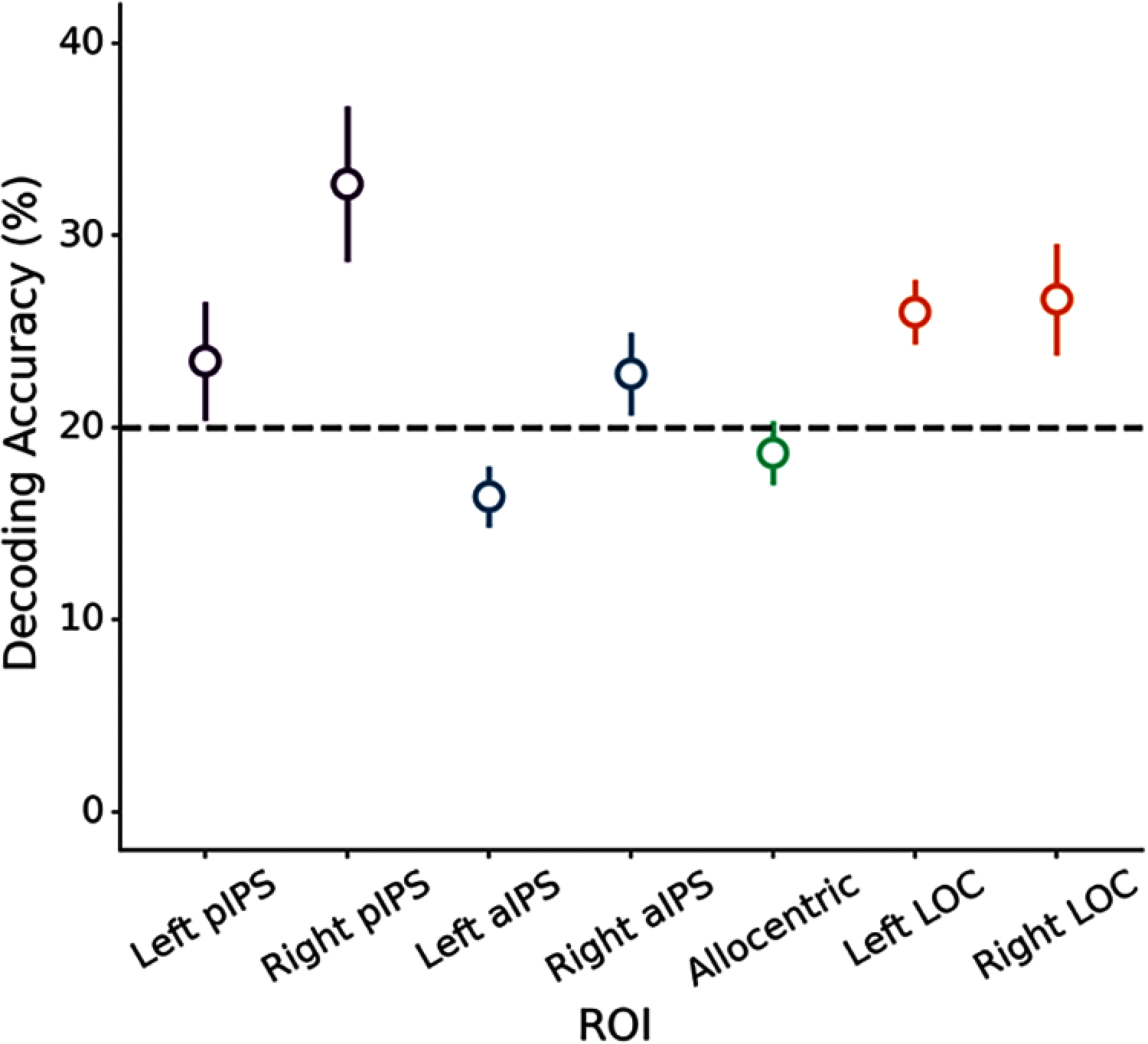
Object categorization accuracy for pIPS, aIPS, the left allocentric ROI, and LOC. Error bars reflect SEM.
Figure 9.
Figure 9.
RDMs and a schematic illustration of the (left) the skeletal model, (middle) GBJ model, and (right) CorNet-S.
Figure 10.
Figure 10.
Results of the representational similarity analyses. A–C, Standardized coefficients (βs) from the linear regression analyses examining the fit of the skeletal, GBJ, and CorNet-S models for left and right (A) pIPS, (B) aIPS, and (C) LOC.
Figure 11.
Figure 11.
Multivariate functional connectivity results. A–D, Functional connectivity map for (A) left pIPS, (B) right pIPS, (C) left aIPS, and (D) right aIPS. Seed regions are displayed as a white circle. E, Plots comparing the connectivity between ROIs in left LOC and right LOC ROIs. Error bars reflect SEM.
Figure 12.
Figure 12.
Plots illustrating the multivariate effective connectivity between pIPS and aIPS with left LOC and right LOC ROIs. Error bars reflect SEM.

References

    1. Abraham A, Pedregosa F, Eickenberg M, Gervais P, Mueller A, Kossaifi J, Gramfort A, Thirion B, Varoquaux G (2014) Machine learning for neuroimaging with scikit-learn. Front Neuroinform 8:14. 10.3389/fninf.2014.00014 - DOI - PMC - PubMed
    1. Anzellotti S, Caramazza A, Saxe R (2017) Multivariate pattern dependence. PLoS Comput Biol 13:e1005799. - PMC - PubMed
    1. Ayzenberg V, Lourenco SF (2019) Skeletal descriptions of shape provide unique perceptual information for object recognition. Sci Rep 9:9359. 10.1038/s41598-019-45268-y - DOI - PMC - PubMed
    1. Ayzenberg V, Lourenco SF (2021) The shape skeleton supports one-shot categorization in human infants: behavioral and computational evidence. PsyArxiv
    1. Ayzenberg V, Chen Y, Yousif SR, Lourenco SF (2019) Skeletal representations of shape in human vision: evidence for a pruned medial axis model. J Vis 19:6. 10.1167/19.6.6 - DOI - PMC - PubMed

Publication types

LinkOut - more resources