. 2022 May 4;79(5):281.
doi: 10.1007/s00018-022-04296-0.

The mechanism of activation of MEK1 by B-Raf and KSR1

Affiliations

The mechanism of activation of MEK1 by B-Raf and KSR1

Ryan C Maloney et al. Cell Mol Life Sci. .

Abstract

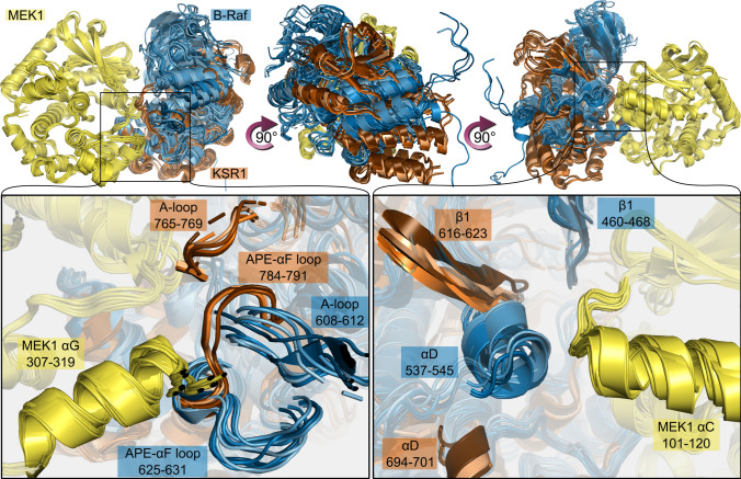
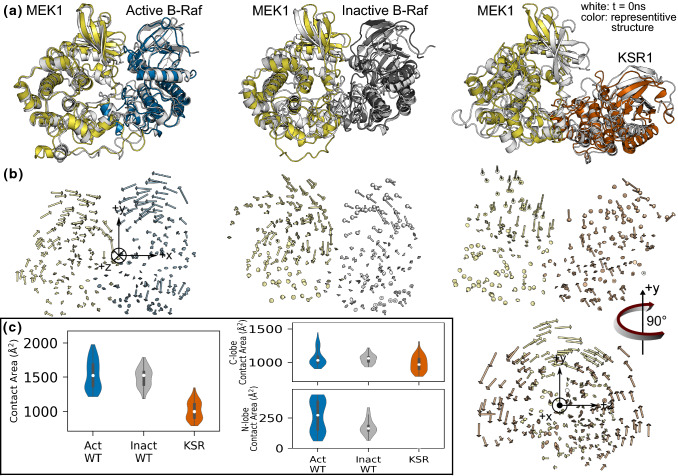
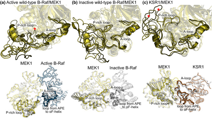
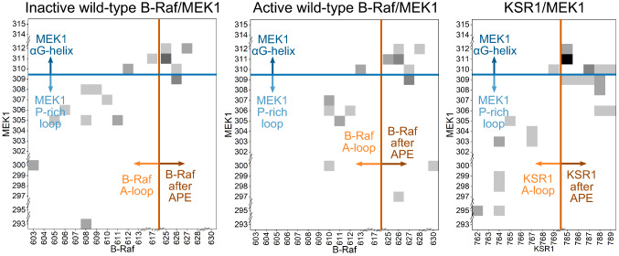
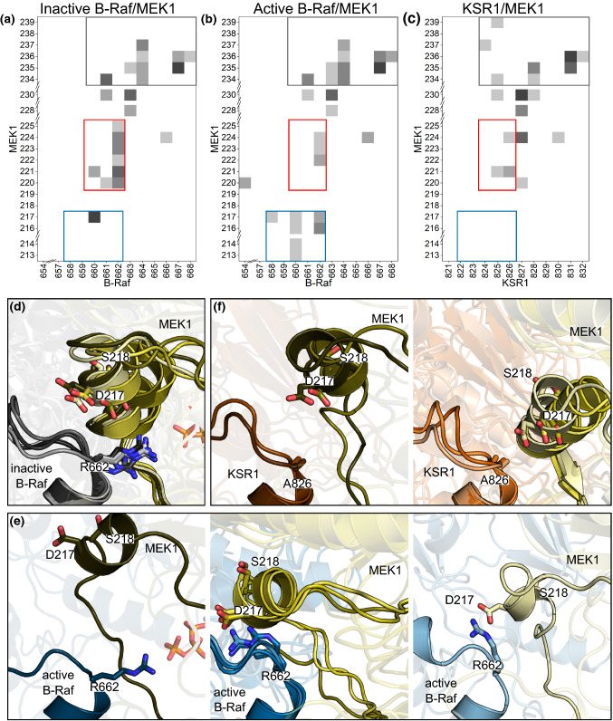
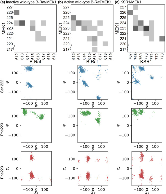
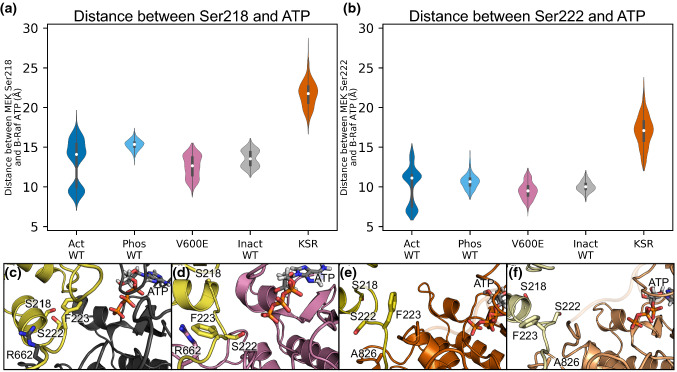
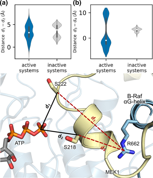
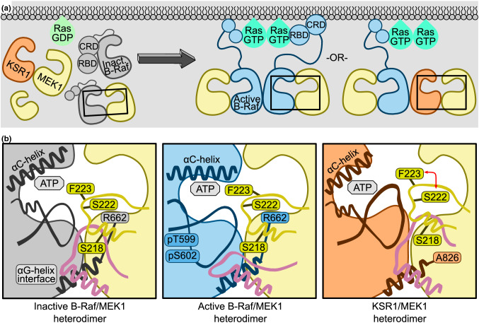
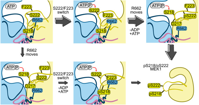
MEK1 interactions with B-Raf and KSR1 are key steps in Ras/Raf/MEK/ERK signaling. Despite this, vital mechanistic details of how these execute signal transduction are still enigmatic. Among these is why, despite B-Raf and KSR1 kinase domains similarity, the B-Raf/MEK1 and KSR1/MEK1 complexes have distinct contributions to MEK1 activation, and broadly, what is KSR1's role. Our molecular dynamics simulations clarify these still unresolved ambiguities. Our results reveal that the proline-rich (P-rich) loop of MEK1 plays a decisive role in MEK1 activation loop (A-loop) phosphorylation. In the inactive B-Raf/MEK1 heterodimer, the collapsed A-loop of B-Raf interacts with the P-rich loop and A-loop of MEK1, minimizing MEK1 A-loop fluctuation and preventing it from phosphorylation. In the active B-Raf/MEK1 heterodimer, the P-rich loop moves in concert with the A-loop of B-Raf as it extends. This reduces the number of residues interacting with MEK1 A-loop, allowing increased A-loop fluctuation, and bringing Ser222 closer to ATP for phosphorylation. B-Raf αG-helix Arg662 promotes MEK1 activation by orienting Ser218 towards ATP. In KSR1/MEK1, the KSR1 αG-helix has Ala826 in place of B-Raf Arg662. This difference results in much fewer interactions between KSR1 αG-helix and MEK1 A-loop, thus a more flexible A-loop. We postulate that if KSR1 were to adopt an active configuration with an extended A-loop as seen in other protein kinases, then the MEK1 P-rich loop would extend in a similar manner, as seen in the active B-Raf/MEK1 heterodimer. This would result in highly flexible MEK1 A-loop, and KSR1 functioning as an active, B-Raf-like, kinase.

Keywords: Assemblies; Autoinhibition; Cancer; ERK; KSR; MAPK; MD simulations.


Conflict of interest statement

The authors have no relevant financial or non-financial interests to disclose.

Figures

Fig. 1
Fig. 1
Crystal structures show structural differences in the intermolecular interfaces between B-Raf/MEK1 and KSR1/MEK1. MEK1 is shown in yellow, B-Raf in blue, and KSR1 in orange. PDB IDs for B-Raf/MEK1 structures: 4MNE, 6NYB, 6PP9, 6Q0J, 6Q0T, 6V2W, 7M0T, 7M0U, 7M0V, 7M0W, 7M0Z. PDB IDs for KSR1/MEK1: 7JUW, 7JUX, 7JUY, 7JUZ, 7JV0, 7JV1
Fig. 2
Fig. 2
a Snapshots comparing the initial structure with the most representative structure for the B-Raf/MEK1 and KSR1/MEK1 heterodimers. The N-lobes in B-Raf and MEK1 move towards each other while the N-lobes of KSR1 and MEK1 move apart. Cartoon drawings of the initial configuration (white) and a representative snapshot of the active wild-type B-Raf/MEK1 (blue/yellow), inactive wild-type B-Raf/MEK1 (gray/yellow) and KSR1/MEK1 (orange/yellow) systems. b First normal mode of every other residue for the same systems as above. c Total contact area, C-lobe to C-lobe contact area, and N-lobe to N-lobe contact area for active wild-type B-Raf/MEK1 (Act WT), inactive wild-type B-Raf/MEK1 (Inact WT) and KSR1/MEK1 (KSR) systems
Fig. 3
Fig. 3
MEK1 P-rich loop adopts different configurations based on if it interacts with active B-Raf, inactive B-Raf, or KSR1. Representative structures for the five most populated configuration subfamilies of the ensemble trajectories for a the active wild-type B-Raf/MEK1, b inactive wild-type B-Raf/MEK1, and c KSR1/MEK1 systems (top row). The first normal mode motion of the MEK1 P-rich loop and B-Raf (or KSR1) A-loop (up to the αF-helix) is shown in the bottom row
Fig. 4
Fig. 4
MEK1 P-rich loop makes more contact with inactive B-Raf or KSR1 A-loop residues than with active B-Raf A-loop residues. Contact maps for MEK1 P-rich loop and αG-helix resides versus B-Raf (or KSR1) A-loop residues and the residues between the APE motif and αF-helix for the inactive wild-type B-Raf/MEK1 (left panel), active wild-type B-Raf/MEK1 (middle panel), and (c) KSR1/MEK1 (right panel) systems
Fig. 5
Fig. 5
MEK1 A-loop displays different interactions with the αG-helix of inactive B-Raf, active B-Raf, and KSR1. Contact maps for MEK1 A-loop through αF-helix residues versus B-Raf (or KSR1) αG-helix residues including the residues from the N-terminal end of αG-helix for a inactive wild-type B-Raf/MEK1, b active wild-type B-Raf/MEK1, c and KSR1/MEK1 systems. Representative snapshots of d inactive B-Raf/MEK1, e active B-Raf/MEK1, and f KSR1/MEK1 systems, aligned with respect to the B-Raf (or KSR) αG-helix position
Fig. 6
Fig. 6
Increased flexibility in the MEK1 A-loop leads to increased rotation of Ser222 and Phe223 and more contact between MEK and B-Raf (or KSR1) A-loop. Contact map (top), main chain dihedral angles of Ser222 (blue), main chain dihedral angle of Phe223 (green), and sidechain dihedral angles of Phe223 (red) of MEK1 for a inactive B-Raf/MEK1, b active B-Raf/MEK1, and c KSR1/MEK1 systems
Fig. 7
Fig. 7
Active B-Raf/MEK1 dimer interface allows the -OH group of MEK Ser222 to be positioned for phosphorylation. Distance between ATP in B-Raf (or KSR1) and MEK1 a Ser218 and b Ser222 for all systems. c Representative snapshots of inactive B-Raf/MEK1 with Ser222 oriented towards MEK1 and Phe223 oriented towards B-Raf. d Representative snapshot of active B-Raf V600E with Ser222 oriented towards ATP and Phe223 oriented towards MEK1. e, f Ser222 and Phe223 can switch orientations in the KSR1/MEK1 system despite KSR1 not adopting an active kinase configuration
Fig. 8
Fig. 8
Ser218 can move closer to ATP when B-Raf Arg662 moves to the outside of the N-terminal helix of the A-loop in MEK1. a Distance between Ser222 and ATP (d1) minus the distance between Ser218 and ATP (d2). Positive values indicate that Ser222 is closer to ATP than Ser218. b Distance between Arg662 and Ser222 (d3) minus the distance between Arg662 and Ser218 (d4). Positive values indicate Arg662 is inside the A-loop of MEK1. Bottom: diagram indicating the distances measured in (a, b)
Fig. 9
Fig. 9
Features of B-Raf/MEK1 and KSR1/MEK1 dimer interfaces that play key role in MEK activation scenarios by B-Raf (shown in Fig. 10) and their comparison with KSR1. (a, Left) Ras is inactive, as is B-Raf (in gray color), MEK1 (yellow) and KSR1 (orange). The monomers are in equilibrium with B-Raf/MEK1 and KSR1/MEK1 dimers. (Right) Ras is active; B-Raf and KSR1 are recruited to the membrane, B-Raf is activated through side-to-side dimerization with either B-Raf or KSR1. Active B-Raf (blue), KSR1 and MEK1 can then form quaternary complexes. b Close up views of the dimer interfaces outlined in black boxes in (a). (Left) Inactive B-Raf is unable to phosphorylate MEK1 due to the collapsed MEK1 A-loop (yellow loop) stabilized by interactions with B-Raf A-loop (black loop), MEK1 P-rich loop (pink loop) residues, and with B-Raf Arg662. (Middle) The B-Raf A-loop extends and the MEK1 P-rich loop relocates, allowing increased MEK1 A-loop flexibility. (Left) In the KSR1/MEK1 dimer, the MEK1 P-rich loop interacts with both KSR1 and MEK1 A-loops, as in inactive B-Raf/MEK1. Flexibility of the MEK1 A-loop is due to the small Ala826 in KSR1 replacing the large Arg826 in B-Raf, allowing MEK1 Phe223 and Ser222 to switch positions
Fig. 10
Fig. 10
Proposed mechanism for the activation of MEK1 by B-Raf. B-Raf can activate MEK1 through two routes. The primary route involves phosphorylation of Ser222 first, followed by phosphorylation of Ser218. The secondary route reverses the order of phosphorylation. Route preference, as to whether S222/F223 flip their orientation first, or R662 moves, reflects the relative populations of the conformational states, which depends on the relative stabilities. We suggest that route shown on top is more highly populated

Similar articles

Cited by

References

    1. Nussinov R, Tsai CJ, Jang H. Signaling in the crowded cell. Curr Opin Struct Biol. 2021;71:43–50. doi: 10.1016/j.sbi.2021.05.009. - DOI - PMC - PubMed
    1. Pylayeva-Gupta Y, Grabocka E, Bar-Sagi D. RAS oncogenes: weaving a tumorigenic web. Nat Rev Cancer. 2011;11:761–774. doi: 10.1038/nrc3106. - DOI - PMC - PubMed
    1. Simanshu DK, Nissley DV, McCormick F. RAS proteins and their regulators in human disease. Cell. 2017;170:17–33. doi: 10.1016/j.cell.2017.06.009. - DOI - PMC - PubMed
    1. Gillies TE, et al. Oncogenic mutant RAS signaling activity is rescaled by the ERK/MAPK pathway. Mol Syst Biol. 2020;16:e9518. doi: 10.15252/msb.20209518. - DOI - PMC - PubMed
    1. Hymowitz SG, Malek S. Targeting the MAPK pathway in RAS mutant cancers. Cold Spring Harb Perspect Med. 2018 doi: 10.1101/cshperspect.a031492. - DOI - PMC - PubMed

MeSH terms