. 2022 May 4;12(1):7282.
doi: 10.1038/s41598-022-11204-w.

A comprehensive atlas of Aggrecan, Versican, Neurocan and Phosphacan expression across time in wildtype retina and in retinal degeneration

Affiliations

A comprehensive atlas of Aggrecan, Versican, Neurocan and Phosphacan expression across time in wildtype retina and in retinal degeneration

A Matsuyama et al. Sci Rep. .

Abstract

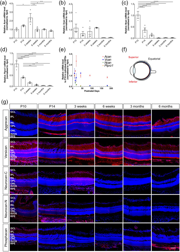
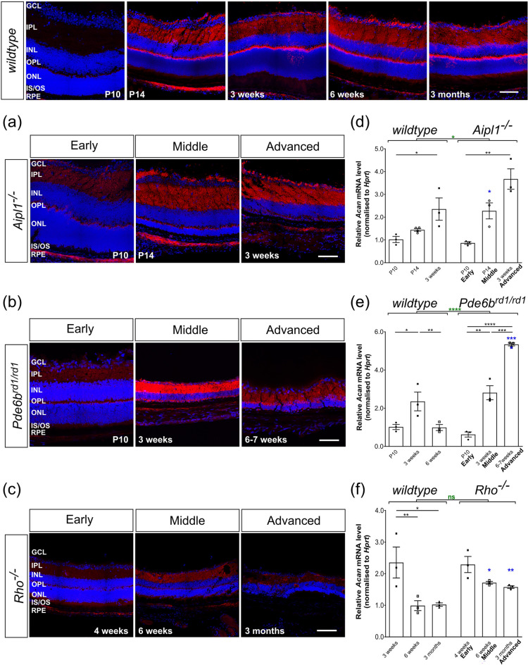
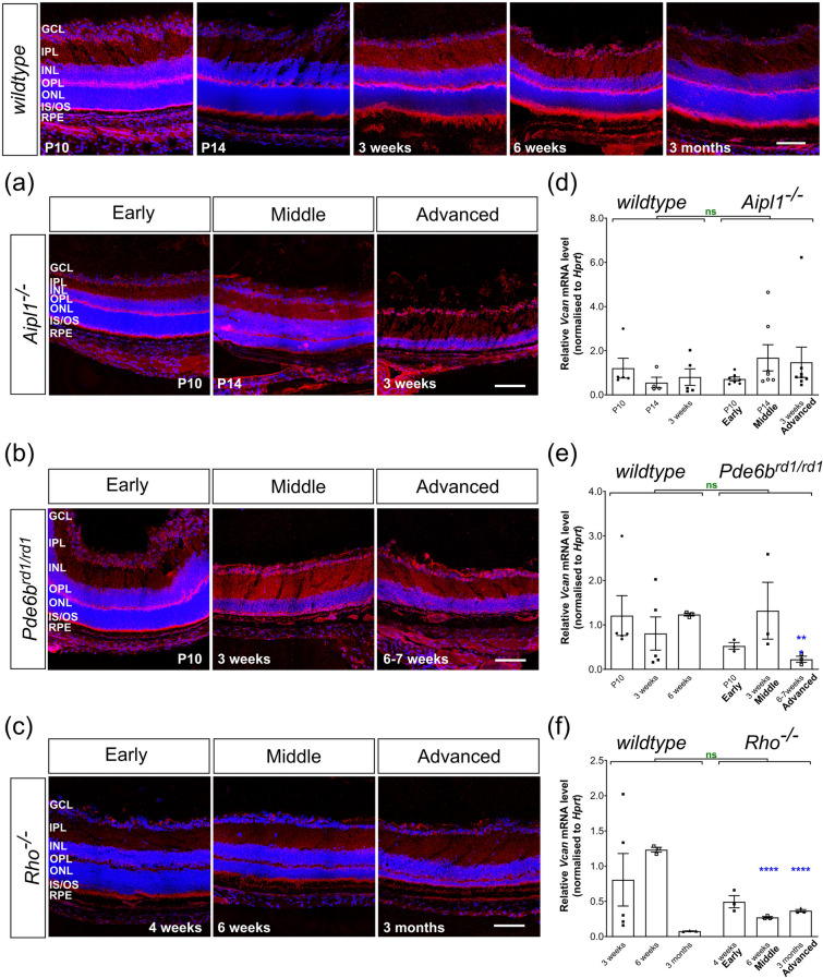
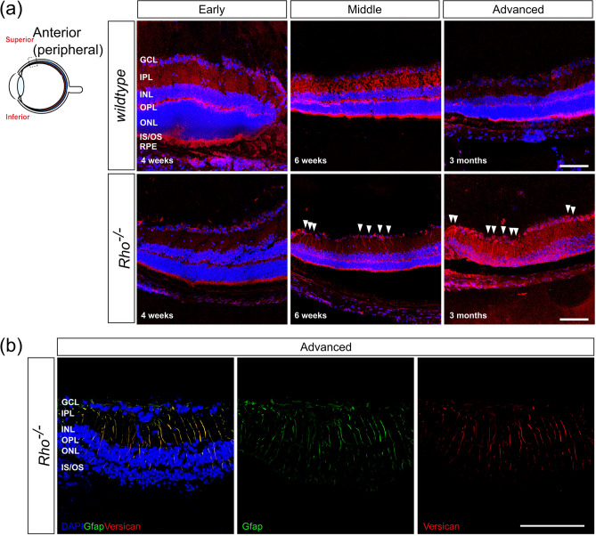
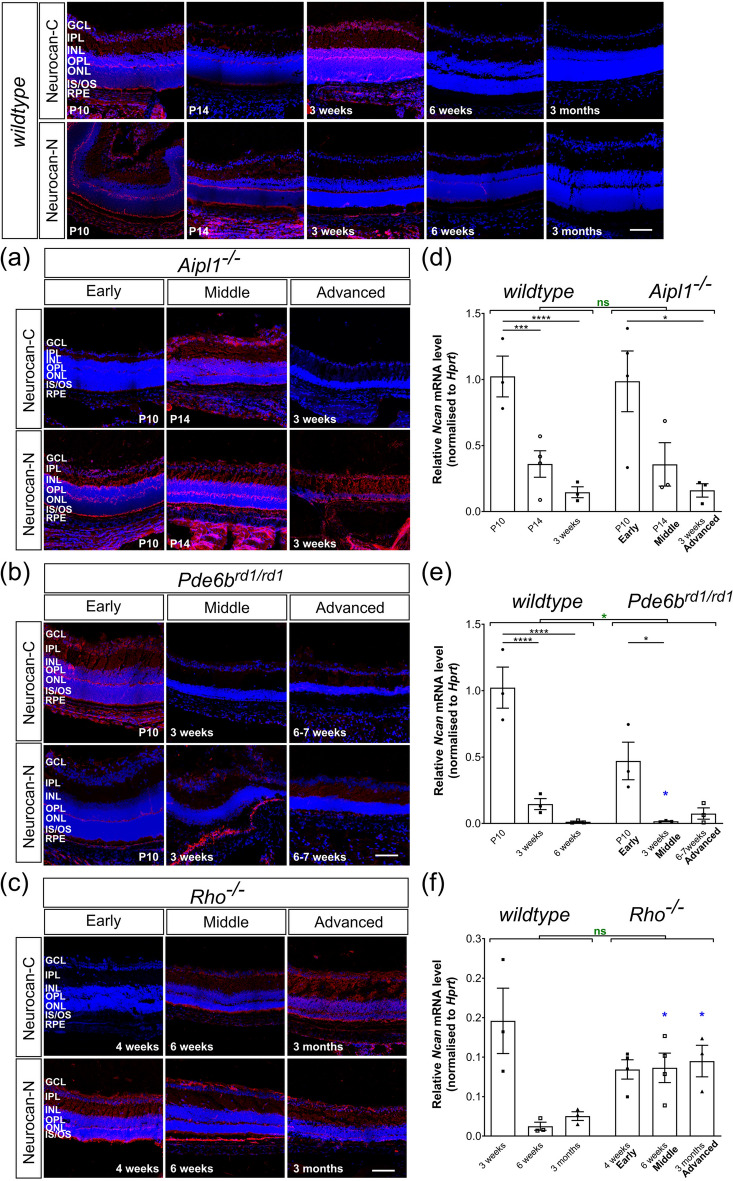
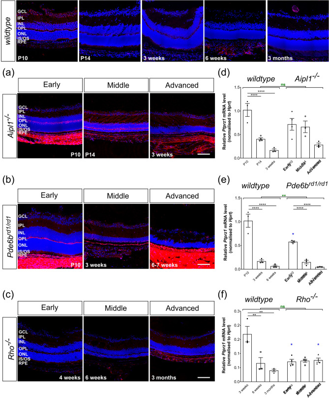
As photoreceptor cells die during retinal degeneration, the surrounding microenvironment undergoes significant changes that are increasingly recognized to play a prominent role in determining the efficacy of therapeutic interventions. Chondroitin Sulphate Proteoglycans (CSPGs) are a major component of the extracellular matrix that have been shown to inhibit neuronal regrowth and regeneration in the brain and spinal cord, but comparatively little is known about their expression in retinal degeneration. Here we provide a comprehensive atlas of the expression patterns of four individual CSPGs in three models of inherited retinal degeneration and wildtype mice. In wildtype mice, Aggrecan presented a biphasic expression, while Neurocan and Phosphacan expression declined dramatically with time and Versican expression remained broadly constant. In degeneration, Aggrecan expression increased markedly in Aipl1-/- and Pde6brd1/rd1, while Versican showed regional increases in the periphery of Rho-/- mice. Conversely, Neurocan and Phosphacan broadly decrease with time in all models. Our data reveal significant heterogeneity in the expression of individual CSPGs. Moreover, there are striking differences in the expression patterns of specific CSPGs in the diseased retina, compared with those reported following injury elsewhere in the CNS. Better understanding of the distinct distributions of individual CSPGs will contribute to creating more permissive microenvironments for neuro-regeneration and repair.


Conflict of interest statement

The authors declare no competing interests.

Figures

Figure 1
Figure 1
Expression patterns of Aggrecan, Versican, Neurocan and Phosphacan in wildtype mice over time. (a)–(d) histograms showing mean (+ /− SEM) mRNA levels of Acan (a), Vcan (b), Ncan (c) and Ptprz1 (d) in whole retinae. (e) best-fit curves summarizing the changes in expression of all four CSPGs over time. (Lognormal for Acan and Vcan, one phase decay for Ncan and Ptprz1.) (f) a schematic of the regions where immunostaining images were taken. (g) Immunostaining for Aggrecan, Versican, Neurocan and Phosphacan (red) over time. (a) Relative expression of Acan mRNA increased between P10 and 3 weeks, decreasing thereafter and remained at a similar level throughout adulthood. (b) Vcan expression was fairly constant across the time points examined, reducing at 3 and 6 months, although inter-sample variation was high. (c) (d) Ncan and Ptprz1 mRNA expression decreased significantly with time. (e) Neurocan-C and -N fractions and Versican were sparsely distributed throughout all the layers of retina when they are expressed. Phosphacan and Aggrecan were restricted mostly to the GCL, IPL and OPL. Images show confocal maximum projection images (MIPs) of the superior retina in the equatorial region. Scale bar, 100 µm. *p < 0.05, **p < 0.01, ***p < 0.001, ****p < 0.0001 (one-way ANOVA test with Bonferroni’s correction). ONL—outer nuclear layer; OPL—outer plexiform layer; INL—inner nuclear layer; IPL—inner plexiform layer; GCL—ganglion cell layer. Nuclei are counter stained with Dapi -blue; CSPGs -red.
Figure 2
Figure 2
Aggrecan expression is increased in the Aipl1-/- and Pde6brd1/rd1 but not Rho-/- models of retinal degeneration. (a)–(c), Immunostaining for Aggrecan (red) was mainly seen in the OPL, IPL, and GCL with relatively little staining in ONL in all models. Increasing expression was most notable in the IPL, compared to early time-points, in wildtype, Aipl1-/- and Pde6brd1/rd1 retinae. (d)–(f), Acan mRNA levels were dramatically elevated with disease progression, showing increased expression in (d) P14 (mid-stage) Aipl1-/- and (e) 6 week (advanced-stage) Pde6brd1/rd1 retinae compared to age-matched wildtype mice. (f) Acan mRNA levels decreased in adulthood in Rho-/- retinae but remained higher than age-matched wildtype retinae. N.B. mRNA levels shown for wildtype are the same as in Fig. 1b. Scale bar, 100 µm. *p < 0.05, **p < 0.01, ***p < 0.001, ****p < 0.0001 (one-way ANOVA test with Bonferroni’s correction, black; unpaired t-test for age-matched comparisons between wildtype and disease model, blue; two-way ANOVA test applied for assessments of change over time, red). ONL—outer nuclear layer; OPL—outer plexiform layer; INL—inner nuclear layer; IPL—inner plexiform layer; GCL—ganglion cell layer. Nuclei are counter stained with Dapi (blue).
Figure 3
Figure 3
Versican expression remains similar to wildtype during degeneration. (a)–(c), Immunolabelling for Versican (red) was detected in all the layers of the retina in wildtype, Aipl1-/- and Pde6brd1/rd1 with relatively strong signals in the INL and IPL, particularly in mid and advanced stage Pde6brd1/rd1 and advanced-stage Rho-/- retinae. (d)–(f) Vcan mRNA levels were largely unchanged in Aipl1-/- and Pde6brd1/rd1 retinae (with the exception of a reduction at 6 weeks age of Pde6brd1/rd1 mice). (f) In Rho-/-, Vcan mRNA expression remained similar between P10 and 3 months of age, decreasing slightly, but remained significantly higher than age-matched wildtype mice. N.B. mRNA levels shown for wildtype are the same as in Fig. 1c. Scale bar, 100 µm. *p < 0.05, **p < 0.01, ***p < 0.001, ****p < 0.0001 (one-way ANOVA test with Bonferroni’s correction, black; unpaired t-test for age-matched comparisons between wildtype and disease model, blue; two-way ANOVA test applied for assessments of change over time, red). ONL—outer nuclear layer; OPL—outer plexiform layer; INL—inner nuclear layer; IPL—inner plexiform layer; GCL—ganglion cell layer. Nuclei are counter stained with Dapi (blue).
Figure 4
Figure 4
Versican colocalizes with Müller glia in the peripheral retina in advanced stage degeneration of Rho-/- mice. (a) Versican + radial processes (red) were observed in mid- to advanced-stage degeneration of Rho-/- mice in the peripheral retina (white arrows) but not in age-matched wildtype mice, or in any other model examined. (b) Versican + processes (red) co-labelled for the reactive glial marker, Glial Fibrillary Acidic Protein (GFAP; green). Image was acquired at × 63 magnification and a single optical section is shown. Scale bar, 100 µm. Nuclei are counter stained with Dapi (blue).
Figure 5
Figure 5
Neurocan expression decreases with time and is unaffected by degeneration. Neurocan immunolabelling (red) was distributed throughout all the layers of the retina and low levels of mRNA transcripts were observed in adult retinae in all models. mRNA levels shown for wildtype are the same as in Fig. 1d. (a) (b) Immunolabelling of the Neurocan C-terminal fraction typically decreased in intensity in Aipl1-/- and Pde6brd1/rd1 mice with time, while that of the Neurocan N-terminal fraction remained constant in these models. (d) (e) mRNA levels of Ncan decreased with time in Aipl1-/-, Pde6brd1/rd1 and wildtype mice. (f) Ncan mRNA and (c) immunolabelling for both Neurocan-C and -N were largely unchanged across degeneration in Rho-/- mice. Scale bar, 100 µm. *p < 0.05, **p < 0.01, ***p < 0.001, ****p < 0.0001 (one-way ANOVA test with Bonferroni’s correction, black; unpaired t-test for age-matched comparisons between wildtype and disease model, blue; two-way ANOVA test applied for assessments of change over time, red). ONL—outer nuclear layer; OPL—outer plexiform layer; INL—inner nuclear layer; IPL—inner plexiform layer; GCL—ganglion cell layer. Nuclei are counter stained with Dapi (blue).
Figure 6
Figure 6
Phosphacan expression decreases with time and is unaffected by degeneration. (a)–(c) Immunolabelling for Phosphacan (red) was mostly restricted to GCL, IPL and OPL. In P10 wildtype, labelling was particularly evident at the outer margin of the OPL and the edge of the ONL itself. In (a) Aipl1-/- and (b) Pde6brd1/rd1 mice, labelling was most prominent in the OPL and RPE, compared to age-matched wildtype. (c) In Rho-/- retinae, labelling was weak at all time-points. (d)–(f) Ptprz1 mRNA expression was significantly decreased with time in all models including wildtype retinae. N.B. mRNA levels shown for wildtype are the same as in Fig. 1e. Scale bar, 100 µm. *p < 0.05, **p < 0.01, ***p < 0.001, ****p < 0.0001 (one-way ANOVA test with Bonferroni’s correction, black; parametric t-test for age-matched comparisons between wildtype and disease model, blue; two-way ANOVA test applied for assessments of change over time, red). ONL—outer nuclear layer; OPL—outer plexiform layer; INL—inner nuclear layer; IPL—inner plexiform layer; GCL—ganglion cell layer. Nuclei are counter stained with Dapi (blue).

References

    1. Barber AC, et al. Repair of the degenerate retina by photoreceptor transplantation. Proc. Natl. Acad. Sci. 2013;110:354–359. doi: 10.1073/pnas.1212677110. - DOI - PMC - PubMed
    1. Nori S, et al. Human oligodendrogenic neural progenitor cells delivered with chondroitinase ABC facilitate functional repair of chronic spinal cord injury. Stem Cell Rep. 2018;11:1433–1448. doi: 10.1016/j.stemcr.2018.10.017. - DOI - PMC - PubMed
    1. Fawcett JW, Oohashi T, Pizzorusso T. The roles of perineuronal nets and the perinodal extracellular matrix in neuronal function. Nat. Rev. Neurosci. 2019;20:451–465. doi: 10.1038/s41583-019-0196-3. - DOI - PubMed
    1. Burns ME, Stevens B. Report on the National Eye Institute’s audacious goals initiative: Creating a cellular environment for neuroregeneration. eneuro. 2018 doi: 10.1523/ENEURO.0035-18.2018. - DOI - PMC - PubMed
    1. Muir E, De Winter F, Verhaagen J, Fawcett J. Recent advances in the therapeutic uses of chondroitinase ABC. Exp. Neurol. 2019;321:113032. doi: 10.1016/j.expneurol.2019.113032. - DOI - PubMed

Publication types

MeSH terms