. 2022 May;605(7911):767-773.
doi: 10.1038/s41586-022-04695-0. Epub 2022 May 4.

Molecular basis for the initiation of DNA primer synthesis

Affiliations

Molecular basis for the initiation of DNA primer synthesis

Arthur W H Li et al. Nature. 2022 May.

Abstract

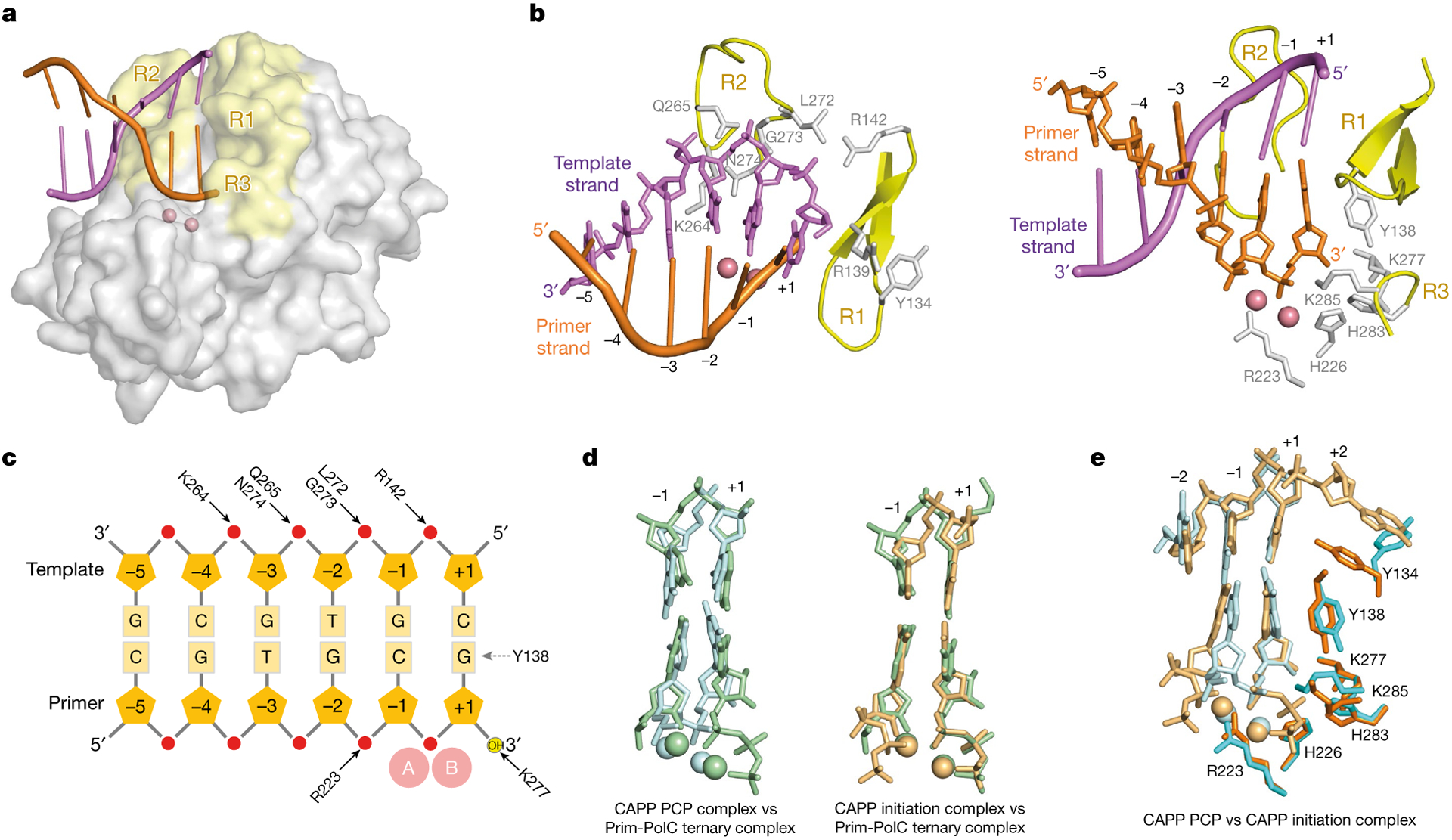
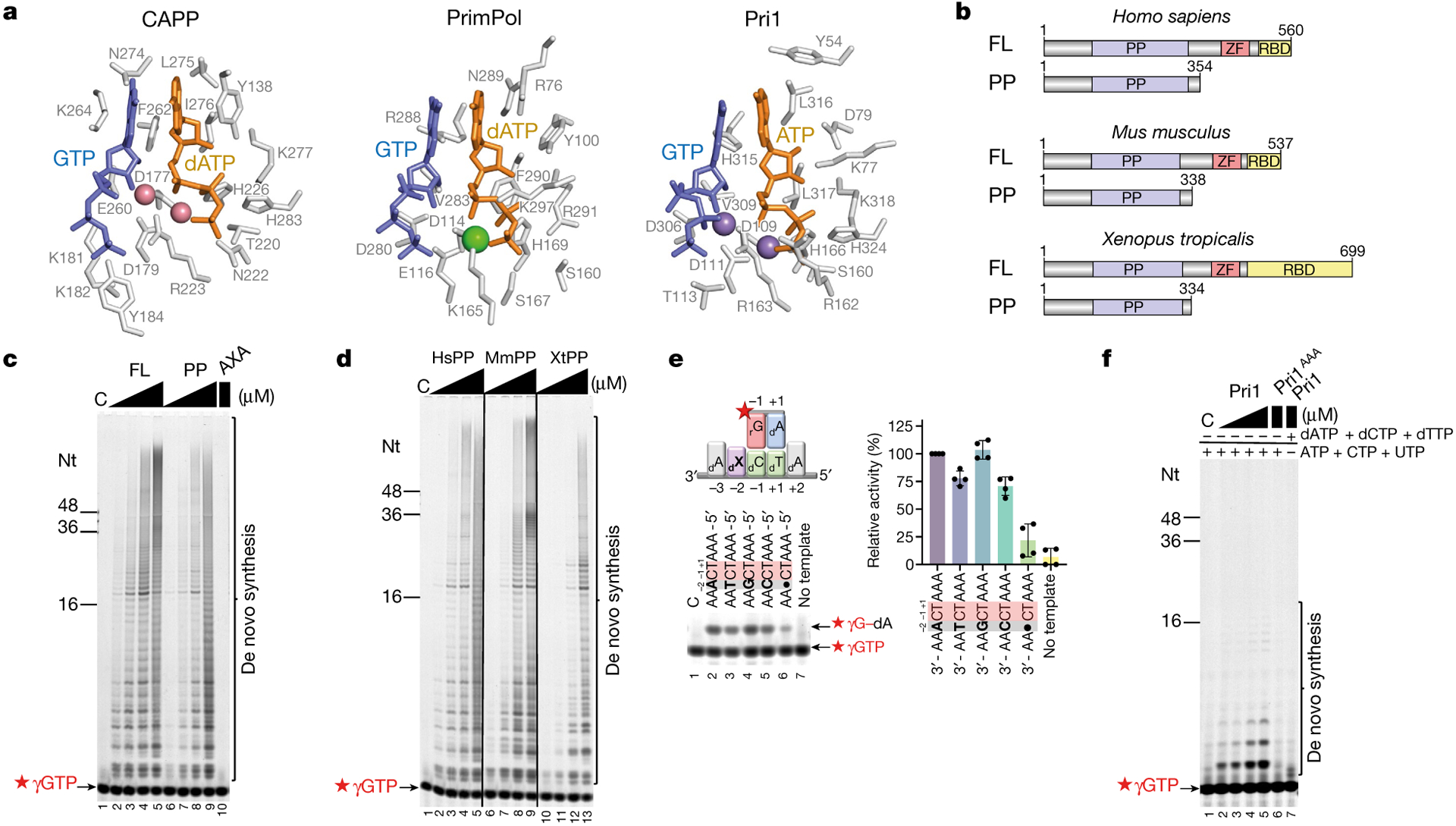
During the initiation of DNA replication, oligonucleotide primers are synthesized de novo by primases and are subsequently extended by replicative polymerases to complete genome duplication. The primase-polymerase (Prim-Pol) superfamily is a diverse grouping of primases, which includes replicative primases and CRISPR-associated primase-polymerases (CAPPs) involved in adaptive immunity1-3. Although much is known about the activities of these enzymes, the precise mechanism used by primases to initiate primer synthesis has not been elucidated. Here we identify the molecular bases for the initiation of primer synthesis by CAPP and show that this mechanism is also conserved in replicative primases. The crystal structure of a primer initiation complex reveals how the incoming nucleotides are positioned within the active site, adjacent to metal cofactors and paired to the templating single-stranded DNA strand, before synthesis of the first phosphodiester bond. Furthermore, the structure of a Prim-Pol complex with double-stranded DNA shows how the enzyme subsequently extends primers in a processive polymerase mode. The structural and mechanistic studies presented here establish how Prim-Pol proteins instigate primer synthesis, revealing the requisite molecular determinants for primer synthesis within the catalytic domain. This work also establishes that the catalytic domain of Prim-Pol enzymes, including replicative primases, is sufficient to catalyse primer formation.


Conflict of interest statement

Competing interests The authors declare no competing interests.

Figures

Extended Data Fig. 1 |
Extended Data Fig. 1 |. CAPP’s Prim-Pol domain alone is sufficient for polymerase and primase synthesis.
a. Alignment of the C-terminal domains (CTD) of different primases or CAPPs. Sc: Saccharomyces cerevisiae PriL (Pri2p), Ls: Lokiarchaeum sp. GC14_75 PriL, Mp: Marinitoga piezophila CAPP, Tp: Thermotoga profunda CAPP, Fg: Fervidobacterium gondwanense CAPP. Conserved motifs: cysteines – red; basic residues – blue; hydrophobic residues – green; prolines – yellow. b. Visualisation of MpCAPP full length wild-type (WT) and C462S, C464S (CC) proteins after two-step purification. c. UV-visible absorption spectrum of MpCAPP WT and CC. CAPP WT, but not CC, exhibited a major absorbance peak at 412 nm. d. Comparison of three different MpCAPP constructs using an Iron assay kit (MAK025, Sigma). MpCAPP FL WT sample had a significantly higher concentration of iron than the MpCAPP CC mutant or CAPP ΔCTD fragment (aa 1–360). Data are the mean of four measurements, except reduced FL WT and FL CC – three measurements. Error bars indicate the mean ± standard deviation. Individual values are presented as black dots. Reduced – sample after treatment with an iron reducing agent; Non-reduced – sample without treatment. e. MpCAPP CC mutant has similar polymerase activity as WT. 1, 5, 25 and 125 nM MpCAPP FL WT (lanes 2–5) or FL CC (lanes 6–9) was added into 30 nM DNA substrate (DNA template − oPK404 + FAM-labelled DNA primer − oPK405) and 100 μM dNTPs. Reactions were incubated at 37 °C for 30 min. f. MpCAPP CC mutant has comparable priming activity as WT. 0.5, 1, 2 and 4 μM MpCAPP FL WT (lanes 2–5) or FL CC (lanes 6–9) was added into reactions containing 1 μM ssDNA template (oKZ388), 2.5 μM non-labelled dATP, dTTP, dGTP, 2.5 μM FAM-dCTP (⋆dCTP) and 100 μM GTP. The reactions were incubated at 50 °C for 30 min. The products were resolved on 20% urea-PAGE gel. g. MpCAPP PP domain exhibits efficient polymerase activity. 1, 5, 25 and 125 nM MpCAPP full length wild-type (FL) (lanes 2–5) and its fragments (lanes 6–17) or 125 nM D177A, D179A full-length mutant (FL AxA) (lane 18) were tested as described in panel e. h. MpCAPP PP domain exhibits strong primase activity. 0.25, 0.5, 1 and 2 μM of MpCAPP FL (lanes 2–5) and its fragments (lanes 6–17) or 2 μM FL AxA (lane 18) were tested as described in panel f. ‘C’ indicates a control reaction without protein. Oligonucleotide (Nts) length markers are shown on the left of the gel. Results shown in panels e-h are representative of three independent repeats, except PP and FL AxA in polymerase assay – four independent repeats.
Extended Data Fig. 2 |
Extended Data Fig. 2 |. MsCAPP PP structure and its primase activity.
a. Structure of MsCAPP PP domain in cartoon representation showing the apo protein (left) and the dGTP complex with Mn(II) ions (right). Protein – grey, dGTP – orange, Mn(II) – purple spheres. b. Protein sequence alignment of MpCAPP111–328 (MpPP) and Marinitoga sp. 1137 CAPP111–328 (MsPP). Conserved amino acids mutated in this study are shown in yellow. Non-conserved amino acids are shown in red. Conserved catalytic motifs I, II and III are in black rectangles as indicated. c. MsPP possesses priming activity. 0.5, 1, 2 and 4 μM MpCAPP full-length (MpFL) (lanes 2–5) or MsCAPP111–328 (MsPP) (lanes 6–9) was added into the reaction containing 1 μM ssDNA mixed sequence template (oKZ388), 100 μM non-labelled dNTP mix and 10 μM FAM-γGTP (⋆γGTP). The reactions were incubated at 50 °C for 30 min. ‘C’ indicates a control reaction without protein. Oligonucleotide (Nts) length markers are shown on the left of the gel. Results shown are representative of three independent repeats.
Extended Data Fig. 3 |
Extended Data Fig. 3 |. MsCAPP PP domain structure analyses and comparison.
a. Overall structure of dGTP-complexed MsCAPP PP domain, with N-terminal α/β domain coloured in green and RRM-like domain coloured in yellow (left), and a close-up view showing dGTP (orange), Mn(II) ions (spheres) and residues lining the active site pocket. b. Architecture of MsCAPP PP domain showing the secondary structural elements within the α/β domain (aa 111–164; aa 274–278) in green and the RRM-like domain (aa 169–262; aa 291–328) in yellow. c. Side by side comparison of PP domains from various Prim-Pols with a single nucleotide (ball-and-stick model) bound to the elongation site. N-terminal RRM-like domain (yellow), α/β domain (green) and helical domain (red). d. Simulated annealing Fo-Fc omit map of Co(II) ions in the active site of MsCAPP (contoured at 5 σ-level at 1.90 Å resolution), along with GTP (blue), dATP (orange), and acidic residues D177, D179 and E260 in stick representation. e. Close-up view of Mn(II) bound in the A site of the MsCAPP dGTP complex, showing octahedral coordination to dGTP, ethylene glycol and surrounding acidic residues with distances labelled in Å (left). Close-up view of Mn(II) bound in the B site of MsCAPP dGTP complex, showing octahedral coordination to dGTP, DxD motif and a water molecule, with distances labelled in Å (right). Mn(II) – purple spheres. f. Overlay of Region 1 (aa 130–142) (left), Region 2 (aa 263–274) (middle) and residues around Region 3 (aa 280–289) (right) from the structures of MsCAPP apo (green) and primer initiation complex (grey). Residues 283–286 in Region 3 are flexible and not resolved in the apo structure. g. MsCAPP active site with surface coloured according to hydrophobicity, with regions of high hydrophobicity coloured in red. Three core residues that form part of the active site hydrophobic pocket (L275, I276 and F262) are shown in stick representation (red). I-site GTP (blue) and E-site dATP (orange) are shown in stick representation. Templating DNA (pink) is shown in cartoon representation. h. Comparison of CAPP primer initiation complex active site. Figure on the left – CAPP primer initiation complex shown in the main figures (PDB: 7P6J). Figure on the right shows the structure of an alternative CAPP primer initiation complex (PDB: 7QAZ), where the phosphate tail (red) of I-site GTP adopts a different conformation and coordinate to an extra Co(II) ion. The rest of the GTP molecule is shown in blue and dATP in orange. Templating DNA in pink, surface of α/β domain is coloured in green and RRM-like domain is shown in yellow.
Extended Data Fig. 4 |
Extended Data Fig. 4 |. Intermolecular interaction analyses of the active site of the primer initiation complex.
a. Non-Covalent Interaction (NCI) analysis of the active site (isosurface = 0.35, cut-off = 8 Å). b. Representation of the active site used for Symmetry-Adapted Perturbation Theory (SAPT0) calculations. For dA, dC, dT, Y138 and R223, only the side chains have been considered. c. Results from SAPT0 calculations for the indicated pairs using the def2-SV(P) basis set in kcal/mol. The final row indicates the SAPT0/def2-SV(P) calculation for GTP interacting with all other fragments in the system (dATP, Y138, dT and both Mg(II) ions).
Extended Data Fig. 5 |
Extended Data Fig. 5 |. Comparison of the different crystal structures of the MsCAPP PP domain.
From left to right, top to bottom; apo, dGTP-bound, primer initiation complex, primer initiation complex (alternative conformation) and post-ternary complex. Protein – grey cartoon, deoxyribonucleotide (dGTP / dATP) – orange sticks, ribonucleotide (GTP) blue sticks, templating DNA strand – pink cartoon, primer DNA strand – orange cartoon, Mn(II) – purple spheres, Co(II) – pink spheres.
Extended Data Fig. 6 |
Extended Data Fig. 6 |. Structure-function and binding studies on the PP domain of MsCAPP.
a. Effect of mutations of MpCAPP100–360 (MpPP) on its polymerase activity. 50 nM MpPP wild-type (WT) (lane 2) or its mutated variants (lanes 3–18) were added to 50 nM DNA substrate (DNA template − oNB1 + FAM-labelled DNA primer − oNB2) and 100 μM dNTPs. The reactions were incubated at 37 °C for 15 min. b. Primase activity of MpPP WT and its mutants. 2 μM MpPP WT protein (lane 2) or its mutants (lanes 3–18) were added to reactions containing 1 μM DNA substrate (oKZ388), 100 μM dNTP mix and 10 μM FAM-labelled GTP (fused via γ-phosphate) (⋆γGTP). The reactions were incubated at 50 °C for 30 min. The products were resolved on 20% urea-PAGE gel. ‘C’ indicates a control reaction without protein. AxA – D177A, D179A, RR – R142A, R143A, KK – K181A, K182A, KQN – K264A, Q265A, N274A. Results are representative of five (panel a) and four (panel b) independent repeats. Oligonucleotide length marker (Nts) is shown on the left of the gel. ‘C’ indicates control without protein. c. Fluorescence polarization assays (FP) reveal that the presence of dinucleotide (rG-dA) does not stimulate binding of MsCAPP111–328 (MsPP) to template. FP: 0–80 μM 5’−3prG-dA-3’ (ATDBio) dinucleotide was added to 5 μM MsPP and 50 nM FAM-DNA (✶ DNA, oKZ409) in presence or absence of 0.1 mM dTTP. d. Stimulation of PP DNA binding affinity in presence of nucleotides is dependent on the template sequence. FP: 0–20 μM MsPP was added to 50 nM ✶ DNA templates (oKZ409, oKZ413, oKZ414 and oKZ416) ± 1 mM GTP and 0.1 mM dATP. e. The efficiency of PP dinucleotide formation is dependent on the −2 base on the template. 1 μM MsPP was added into the reaction containing 1 μM template (lane 2 - oKZ435, lane 3 - oKZ447, lane 4 - oKZ449, lane 5 - oKZ450, lane 6 – oKZ448), 100 μM dATP and 10 μM ✶ γGTP. The reactions were incubated at 50 °C for 30 min. The gel is representative of three independent repeats (left). Signal of synthetized dinucleotides were normalized to signal of dinucleotide in presence of 3’-AAACTAAA-5’ ssDNA template (100%). Data represent the mean ± standard deviation from three independent experiments. Black dots – individual values. ‘C’ indicates control reaction without protein. f. Affinity of PP to template increases with the template length. FP – 0–20 μM MsPP was added to 50 nM ✶ DNA templates (oKZ408–oKZ412). Data representing the mean ± standard deviation from four independent experiments (Panels c, d and f). The mean values were used to calculate the dissociation constants (Kd) shown on the right (Panel d, f); SD – standard deviation of calculated Kd; ND – not determined (Panels d, f).
Extended Data Fig. 7 |
Extended Data Fig. 7 |. Comparison of MsCAPP and PP domains of human Prim-Pols.
a. Overlay of HsPrimPol (pink) and MsCAPP (grey) PP domains (left), and of HsPri1 (orange) and MsCAPP (grey) PP domains (right). Protein and DNA strands are displayed in cartoon representation, nucleic acids are displayed in stick representation, and metal ions are displayed as spheres. b. Close-up view of the E-site nucleotide, amino acid residues close to the 2’ position of the ribose ring, and Region 1 of MsCAPP (left), HsPrimPol (middle) and HsPri1 (right). Interaction between D79 of HsPri1 and 2’-OH is displayed with a dashed line.
Extended Data Fig. 8 |
Extended Data Fig. 8 |. Analyses of PrimPol mutations and how incoming nucleotides influence the primase activities of different Prim-Pols.
a. The primase activity of HsPrimPol1–354 (PP) is reduced compared to the full-length enzyme (WT) at lower concentrations. HsPrimPol1–354 D114A, E116A (AxA) (1 μM) exhibits no primase activity. Quantification of primase assays represented in Fig. 4c. Data represent the mean ± standard deviation from four independent experiments. b. Efficiency of priming by HsPrimPol1–354 is dependent on FAM-γGTP (⋆γGTP) concentration. Primase reactions contained varying concentrations of ⋆γGTP (0.02, 0.1, 0.5 and 2.5 μM), 100 μM dNTP mix and 1 μM ssDNA template (oKZ388). c. GTP is outcompeted by high concentrations of dATP from MsCAPP’s active site. FP – 10 μM MsCAPP111–328 (MsPP) was added to 100 nM ⋆γGTP ± 1 μM DNA template (oKZ435) with increasing concentrations of dATP (0.1 μM – 10 mM). d. The effect of different concentration of GTP and dATP on MsPP affinity to DNA. FP – 5 μM MsCAPP111–328 (MsPP) was added to 50 μM DNA template (oKZ409) in presence of 1 mM GTP/dATP and increasing concentrations (x mM) of dATP/GTP (15.625 μM – 1 mM). Data were obtained from four independent repeats (Panels c and d). Error bars in the graphs show the mean ± standard deviation. IC50 and EC50 values were calculated as described in materials and methods. e. Structurally equivalent residues of MpCAPP and HsPrimPol. f. Polymerase activity of HsPrimPol1–354 is severely disrupted by point mutations in key catalytic residues. AxA – D114A, E116A, RNR – R286A, N287R, R288A. 200 nM protein was incubated with 50 nM DNA substrate (oNB1 + oNB2) for 30 min at 37 °C. g. Primase activity of HsPrimPol1–354 is significantly disrupted by point mutations in key catalytic residues. The reactions contained 4 μM protein, 10 μM ⋆γGTP, 100 μM dNTP mix and 1 μM DNA template (oKZ388). h. HsPrimPol preferentially utilizes GTP to initiate primer synthesis. Primase assay reaction contained 1 μM HsPrimPol1–354 (HsPP), 1 μM DNA template (oKZ388), 2.5 μM FAM-dATP (⋆dATP), 2.5 μM dCTP, dGTP, and dTTP and 100 μM individual NTPs. i. HsPrimPol preferentially initiates primer synthesis with GTP over ATP. Primase assay reaction contained 1 μM HsPP, 1 μM DNA template (oKZ388), 100 μM dNTPs and 2.5 μM FAM-γATP (⋆γATP) or ⋆γGTP. j. Human Pri1 prefers GTP over ATP as the primer initiation base. Reactions containing 1, 2, 4 and 8 μM protein were incubated with 1 μM DNA template (oKZ388), 10 μM ⋆γGTP or ⋆γATP and 100 μM CTP, ATP and UTP or 100 μM CTP, GTP and UTP, respectively, at 25 °C for 30 min. Results shown in panel b and f-j are representative of three independent repeats. ‘C’ indicates control reaction without protein. Oligonucleotide (Nts) length marker is shown on the left of the gels.
Extended Data Fig. 9 |
Extended Data Fig. 9 |. Qualitative gel-based analysis of purified proteins.
a. MpCAPP fragments and FL mutants. 1 μg of each purified MpCAPP variant was resolved on 12% SDS-PAGE and Coomassie stained. Note: FL WT, FL CC and ΔTPR fragment are fused to MBP. FL WT – full-length wild type WT, FL AxA – full-length D177A, D179A, ΔCTD – aa1-360, ΔTPR – aa100–546, PP – aa100–360, FL CC – full-length C462S, C464S. b. Mutants of MpCAPP PP domain. 1 μg of each purified MpCAPP PP mutant was resolved using 12% SDS-PAGE and Coomassie stained. AxA – D177A, D179A, RR – R142A, R143A, KK – K181A, K182A, KQN – K264A, Q265A, N274A. c. MsCAPP fragments. 2 μg of purified fragments of MsCAPP100–359 and MsCAPPaa111–328 were resolved using 12% SDS-PAGE and Coomassie stained. d. HsPrimPol full-length (HsFL) and HsPrimPol1–354 fragment (HsPP) and mutants. 2 μg of each purified variant was resolved using 12% SDS-PAGE and Coomassie stained. e. HsPP WT and mutants. 2 μg of each purified mutant was resolved on 12% SDS-PAGE and Coomassie stained. Note: AxA – D114A, E116A, RNR – R286A, N287A, R288A. f. Eukaryotic PrimPols. 2 μg of each purified PP was resolved using 15% SDS-PAGE and Coomassie stained. g. HsPri1. 1 μg of HsPri1 wild-type (HsPri1) or HsPri1 D109A, D111A, D306A (HsPri1AAA) was resolved using 12% SDS-PAGE and Coomassie stained.
Fig. 1 |
Fig. 1 |. MpCAPP’s PP domain is polymerase and primase proficient.
a, Top, schematic representations of MpCAPP fragments with the highlighted domains. Bottom left, quantification of the polymerase activity of full-length wild-type MpCAPP (FL), its fragments or the full-length D177A/D179A mutant (FL AXA) (125 nM protein). Bottom right, quantification of the priming activity of MpCAPP FL, its fragments or the FL AXA mutant (2 μM protein). Data represent the mean ± s.d. from three independent experiments, except for FL and FL AXA in the primase assay, where four independent experiments were performed. Representative gels are shown in Extended Data Fig. 1g, h. Black dots represent individual values. WT, wild type. b, Left, overall structure of a primer initiation complex of MsCAPP (grey) bound to ssDNA (pink), GTP (blue), dATP (orange) and Co(II) (spheres) highlighting DNA-interacting regions R1–R3 (yellow). Right, MsCAPP active site showing a simulated annealing FoFc omit map of the ssDNA template, I-site GTP and E-site dATP (contoured at 2σ at a resolution of 1.90 Å), along with the side chains of Y134, Y138 and Co(II). c, Schematic showing the protein–DNA and protein–nucleotide interactions in the primer initiation complex. Pentagon, sugar; square, base; red sphere, phosphate; yellow sphere, OH group; peach sphere, metal ion. Black arrows indicates an interaction between an amino acid and DNA, nucleotides or metal ions; blue dashed arrows indicate π–π stacking. d, Residues interacting with nucleotides bound in the I-site (left) and E-site (right). e, Left, active site with the oxygen of the 3′-OH of GTP about to initiate a nucleophilic attack on the α-phosphate of dATP. Middle, A-site Co(II), showing octahedral coordination to nucleotides and surrounding acidic residues with distances labelled in angstroms. E260 interacts with oxygen from the 2′-OH of GTP. Right, B-site Co(II), showing octahedral coordination to dATP, the DXD motif and a water molecule, with distances labelled in angstroms.
Fig. 2 |
Fig. 2 |. Structure of a post-ternary complex of PP bound to dsDNA.
a, A surface representation of the MsCAPP PP domain (grey) bound to dsDNA (pink and orange) forming a post-ternary complex, highlighting important DNA-interacting regions R1–R3 (yellow). b, Molecular models showing protein–DNA interactions with the template strand (left) and the primer strand (right). c, Schematic showing the important protein–DNA and protein–nucleotide interactions formed in the PP–dsDNA complex. Pentagon, sugar (deoxyribose); square, base; red sphere, phosphate; yellow sphere, OH group; peach sphere, metal ion. Black arrows indicate interactions between indicated amino acids and DNA. d, Superposition of active site bases and metal ions of the CAPP–dsDNA (cyan) and Prim-PolC–DNA (PDB 6SA0) (green) complexes (left) and the CAPP primer initiation (orange) and Prim-PolC–DNA (green) complexes (right). e, Superposition of the active sites of the CAPP–dsDNA (cyan) and CAPP primer initiation (orange) complexes.
Fig. 3 |
Fig. 3 |. Structure–function analyses of MsCAPP PP primer synthesis and extension activities.
a, Schematic representation of the interactions of MsCAPP with template and nucleotides in the primer initiation complex. Red sphere, phosphate; yellow sphere, OH group; peach sphere, metal ion. Black arrows indicate interactions. b, Effects of MpCAPP PP domain mutations on polymerase (left) and primase (right) activities. AXA, D177A/D179A; RR, R142A/R143A; KK, K181A/K182A; KQN, K264A/Q265A/N274A. Data represent the mean ± s.d. from five (polymerase) and four (primase) independent experiments. Representative gels are shown in Extended Data Fig. 6a, b. Black circles indicate individual values. c, Affinity of MsCAPP PP for template, dATP and GTP. FP assays contained 0–20 μM protein, 25 nM FAM–dATP (⋆dATP) or FAM–γGTP (⋆γGTP) or 50 nM FAM–DNA (⋆DNA, oKZ409) ± 1 mM GTP and 0.1 mM dATP. d, Presence of GTP and dATP stimulates affinity of MsCAPP PP for template. FP assays contained 0–20 μM protein, 50 nM FAM–DNA (oKZ409) ± 0.1 mM dATP and/or 1 mM GTP/dGTP. e, Affinity of MsCAPP PP for template decreased over time in the presence of GTP and dATP. FP assays contained 10 μM protein, 50 nM FAM–DNA (oKZ409) ± 1 mM GTP and 0.1 mM dATP. FP was measured every 5 min for 2 h; 0.1 mM dATP was added at 60 min (dashed line indicated by a red arrowhead). f, The MsCAPP PP domain prefers to bind template containing a purine at the −2 position. FP assays contained 0–20 μM protein, 50 nM FAM–DNA (oKZ409, oKZ417 or oKZ418) ± 1 mM GTP and 0.1 mM dATP. A star indicates FAM labelling. Data in cf represent the mean ± s.d. from four independent experiments. ND, not determined. g, A model of Prim-Pol proteins’ primer initiation and extension mechanism. Green crescent, Prim-Pol; blue rectangle, ssDNA template; peach rectangle, newly primed strand; red R, purine ribonucleotide. Primed strand deoxyribonucleotides are shown in purple.
Fig. 4 |
Fig. 4 |. The PP domains of eukaryotic replicative primases are primase proficient.
a, I- and E-sites of CAPP (left), human PrimPol (middle) and human Pri1 (right). GTP (CAPP primer initiation complex) was superposed onto human Prim-Pol structures to identify their I-sites. b, Schematic representations of human, mouse and X. tropicalis full-length Prim-Pol proteins and their PP domains. ZF, zinc finger; RBD, RPA-binding domain. c, The human PrimPol PP is priming proficient. Primase assays contained 0.125, 0.25, 0.5 or 1 μM full-length HsPrimPol (lanes 2–5), 0.125, 0.25, 0.5 or 1 μM HsPrimPol1–354 (PP; lanes 6–9) or 1 μM HsPrimPol1–354 D114A/E116A (AXA; lane 10), 1 μM ssDNA (oKZ388), 2.5 μM FAM–γGTP (⋆γGTP) and 100 μM dNTP. d, Mouse and X. tropicalis PPs are priming proficient. Assays contained 0.125, 0.25, 0.5 or 1 μM HsPrimPol1–354 (HsPP; lanes 2–5), MmPrimPol1–338 (MmPP; lanes 6–9) or XtPrimPol1–334 (XtPP; lanes 10–13) under the conditions described in c. e, The HsPrimPol PP prefers a purine base at the −2 position (template) for dinucleotide synthesis. Reactions contained 0.25 μM HsPP, 1 μM ssDNA template (lane 2, oKZ435; lane 3, oKZ447; lane 4, oKZ449; lane 5, oKZ450; lane 6, oKZ448), 2.5 μM FAM–γGTP and 100 μM dNTP. Left, representative gel. Right, quantification. Dinucleotide signal was normalized to that in the presence of 3′-AAACTAAA-5′ (100%). Data represent the mean ± s.d. Black dots indicate individual values. f, Human Pri1 exhibits priming activity. Reactions contained 1, 2, 4 or 8 μM wild-type Pri1 (Pri1; lanes 2–5, 7) or 8 μM Pri1 D109A/D111A/D306A (Pri1AAA; lane 6), 1 μM ssDNA (oKZ388), 10 μM FAM–γGTP, and 100 μM ATP, CTP and UTP (lanes 1–6) or 100 μM dATP, dCTP and dTTP (lane 7). Results are representative of three (d, f) or four (c, e) independent experiments. Nt, oligonucleotide length marker; ‘C’ indicates control without protein.

References

    1. Guilliam TA, Keen BA, Brissett NC & Doherty AJ Primase-polymerases are a functionally diverse superfamily of replication and repair enzymes. Nucleic Acids Res. 43, 6651–6664 (2015). - PMC - PubMed
    1. Iyer LM, Koonin EV, Leipe DD & Aravind L Origin and evolution of the archaeo-eukaryotic primase superfamily and related palm-domain proteins: structural insights and new members. Nucleic Acids Res. 33, 3875–3896 (2005). - PMC - PubMed
    1. Zabrady K, Zabrady M, Kolesar P, Li AWH & Doherty AJ CRISPR-associated primase-polymerases are implicated in prokaryotic CRISPR–Cas adaptation. Nat. Commun 12, 3690 (2021). - PMC - PubMed
    1. Bouché JP, Zechel K & Kornberg A dnaG gene product, a rifampicin-resistant RNA polymerase, initiates the conversion of a single-stranded coliphage DNA to its duplex replicative form. J. Biol. Chem 250, 5995–6001 (1975). - PubMed
    1. Aravind L, Leipe DD & Koonin EV Toprim—a conserved catalytic domain in type IA and II topoisomerases, DnaG-type primases, OLD family nucleases and RecR proteins. Nucleic Acids Res. 26, 4205–4213 (1998). - PMC - PubMed

Publication types