. 2022 May;605(7909):298-303.
doi: 10.1038/s41586-022-04668-3. Epub 2022 May 4.

Tbx2 is a master regulator of inner versus outer hair cell differentiation

Affiliations

Tbx2 is a master regulator of inner versus outer hair cell differentiation

Jaime García-Añoveros et al. Nature. 2022 May.

Abstract

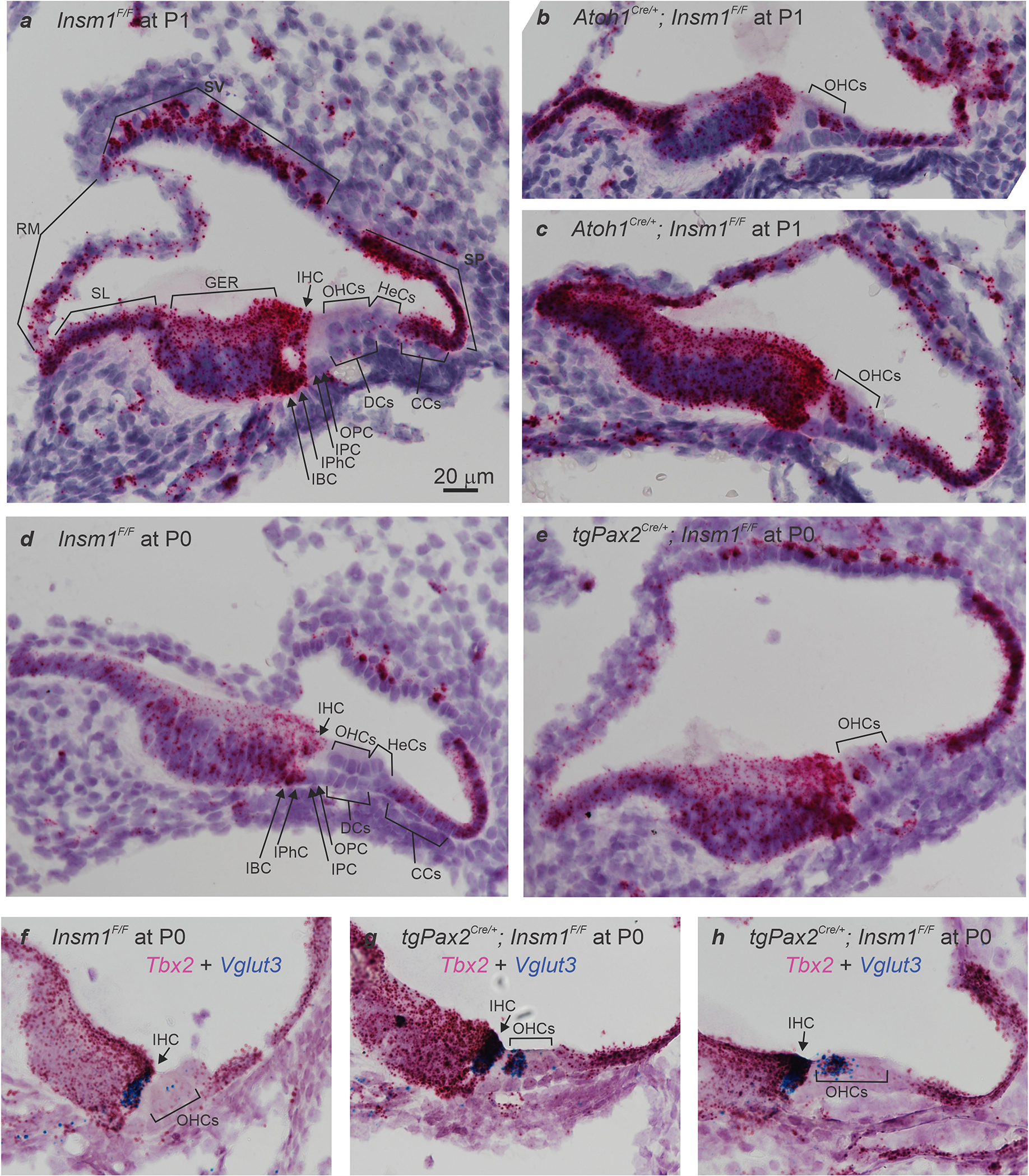
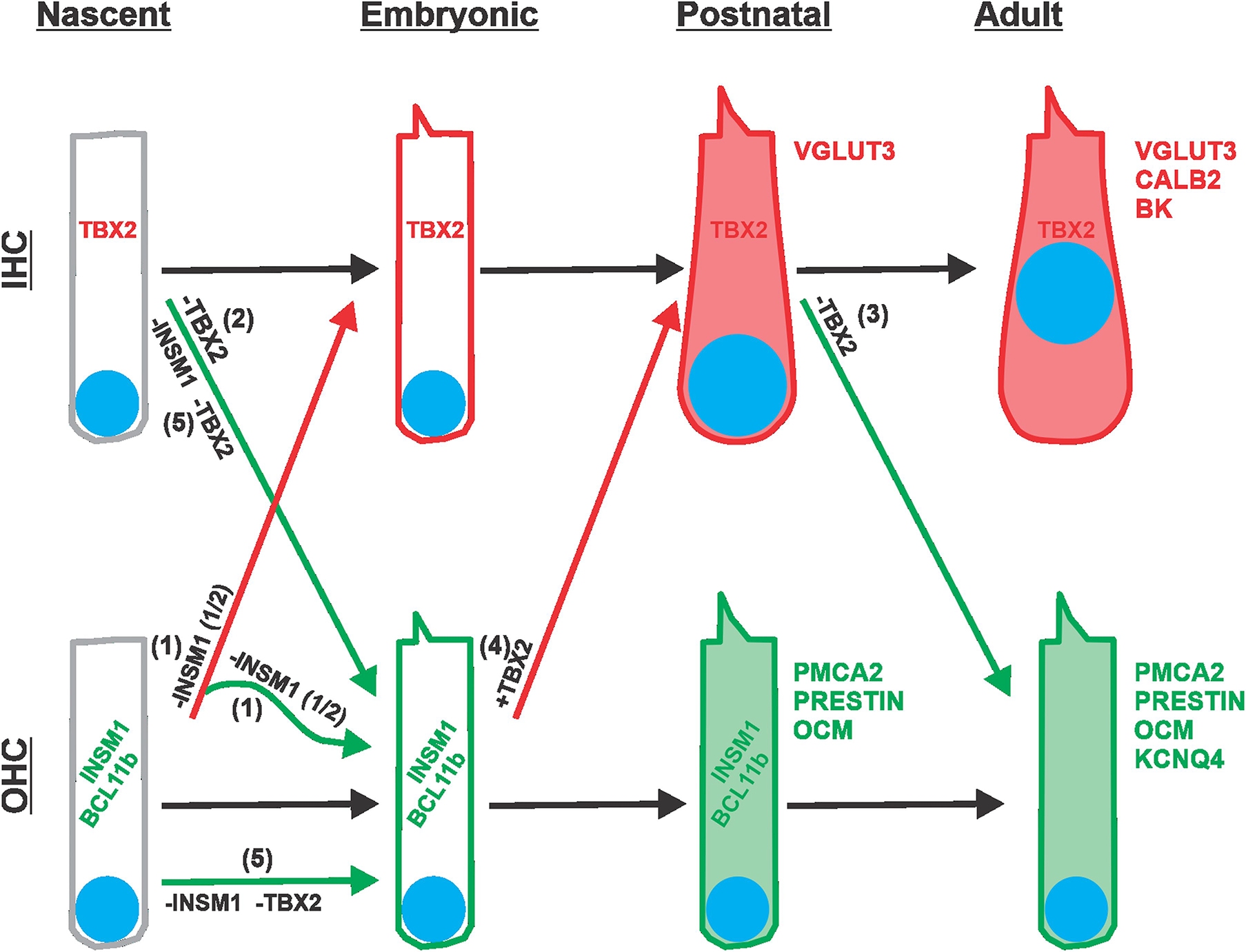
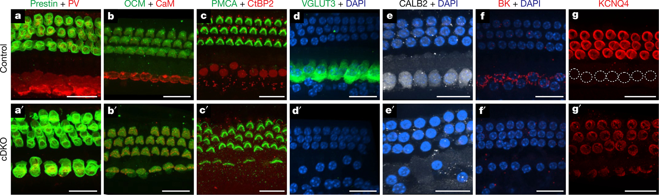
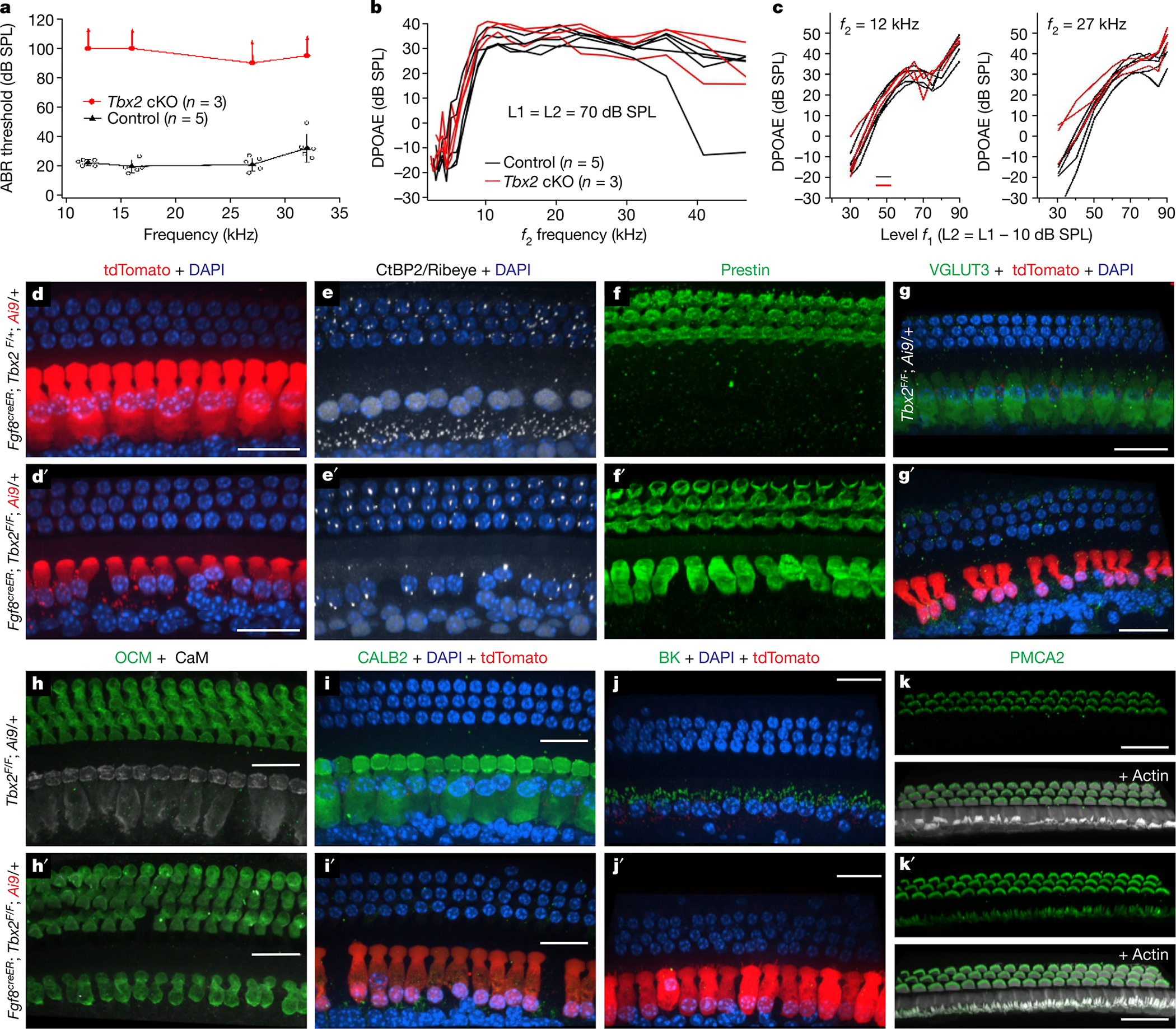
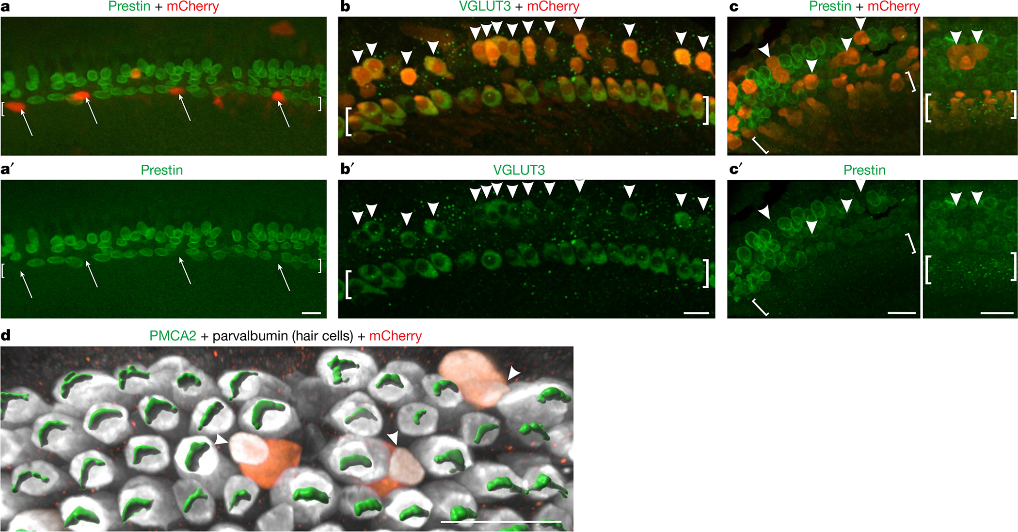
The cochlea uses two types of mechanosensory cell to detect sounds. A single row of inner hair cells (IHCs) synapse onto neurons to transmit sensory information to the brain, and three rows of outer hair cells (OHCs) selectively amplify auditory inputs1. So far, two transcription factors have been implicated in the specific differentiation of OHCs, whereas, to our knowledge, none has been identified in the differentiation of IHCs2-4. One such transcription factor for OHCs, INSM1, acts during a crucial embryonic period to consolidate the OHC fate, preventing OHCs from transdifferentiating into IHCs2. In the absence of INSM1, embryonic OHCs misexpress a core set of IHC-specific genes, which we predict are involved in IHC differentiation. Here we find that one of these genes, Tbx2, is a master regulator of IHC versus OHC differentiation in mice. Ablation of Tbx2 in embryonic IHCs results in their development as OHCs, expressing early OHC markers such as Insm1 and eventually becoming completely mature OHCs in the position of IHCs. Furthermore, Tbx2 is epistatic to Insm1: in the absence of both genes, cochleae generate only OHCs, which suggests that TBX2 is necessary for the abnormal transdifferentiation of INSM1-deficient OHCs into IHCs, as well as for normal IHC differentiation. Ablation of Tbx2 in postnatal, largely differentiated IHCs makes them transdifferentiate directly into OHCs, replacing IHC features with those of mature and not embryonic OHCs. Finally, ectopic expression of Tbx2 in OHCs results in their transdifferentiation into IHCs. Hence, Tbx2 is both necessary and sufficient to make IHCs distinct from OHCs and maintain this difference throughout development.


Conflict of interest statement

Competing interests The authors declare no competing interests.

Figures

Extended Data Fig. 1 |
Extended Data Fig. 1 |. Expression of Tbx2 mRNA in cochlea.
(a,d) In situ hybridizations in sections of neonatal (P0 and P1) cochleae from control mice (Insm1F/F) reveal Tbx2 expression in most cells lining the scala media including IHCs, Inner Phalangeal Cells (IPhC), Inner Border Cells (IBC), Greater Epithelial Ridge (GER), interdental cells of the Spiral Limbus (SL), Reissner’s Membrane (RM), some cells of the Stria Vascularis (SV), Spiral Prominence (SP), and Claudius’ Cells (CCs); but little or no expression (above background levels) of Tbx2 in OHCs or other cells of the outer compartment, namely Hensen’s Cells (HeCs), Deiters’ Cells (DCs), and Outer and Inner Pillar cells (OPC and IPC). (b,c,e) In situ hybridizations in sections of developing Insm1 mutant cochleae (Atoh1Cre/+; Insm1F/F in b and c; TgPax2Cre/+; Insm1F/F in e) reveal that Tbx2 is expressed in about half of the OHCs, presumably those transdifferentiating into IHCs. In controls, none of the OHCs (0/117 from N=4 Insm1F/F mice at E16.5, P0 and P1) expressed Tbx2, whereas in Insm1 mutants, in which nearly half of the OHCs transdifferentiate into IHCs (46.0±5.6%), an equivalent fraction of hair cells in the position of the OHCs expressed Tbx2 (46.6%; 62/133 cells from N=4 TgPax2Cre/+; Insm1F/F or Atoh1Cre/+; Insm1F/F mice at E16.5, P0 and P1). (f-h) Double in situ hybridizations in sections of neonatal (P0) cochleae for detection of Tbx2 (magenta) and Vglut3 (blue), the earliest marker of functional, mature IHCs. (f) In Insm1F/F controls, only IHCs express high levels of Vglut3 (a few dots label OHCs, perhaps representing a much lower level of expression). (g,h) In TgPax2Cre/+; Insm1F/F cochleae, only OHCs misexpressing Tbx2 (50/114 at P0, 14/30 at P2 and 9/21 at P4) also misexpressed high levels (those characteristic of IHCs) of Vglut3, whereas none of the OHCs misexpressed either gene alone (at IHC-like levels). Hence, early expression of Tbx2 in OHCs lacking INSM1 appears to correlate perfectly with their transdifferentiating into IHCs. Images shown are examples of results obtained in three separate tissue samples (n=3 biologically independent samples). Scale bar of 20 μm in (a) applies to all other panels.
Extended Data Fig. 2 |
Extended Data Fig. 2 |. Nuclear volumes of hair cells in the inner or outer compartment of controls and in various Tbx2 conditional KOs.
Blue dots represent nuclear volumes of hair cells from the inner compartment (I in the column labels), whether IHCs (controls) or ic-HCs (mutants). Red dots represent nuclear volumes of hair cells from the outer compartment (OHCs; O in the column labels). Sample size (n) is indicated in each column. The time of tamoxifen administration to Fgf8CreER; Tbx2F/F pups is indicated in the column labels as TAM@P0 to P9. (a,b) While in controls the nuclei of IHCs are statistically larger than those of OHCs, in the mutants (all Tbx2 cKOs as well as the double cKO of Tbx2 and Insm1) the nuclei of the inner compartment hair cells (ic-HCs) are statistically smaller than those of control IHCs (b) and indistinguishable from those of OHCs (a). Error bars indicate SD and column tops the averages. Statistical comparisons were performed by one-way ANOVA with a Bonferroni post test. **** indicates P<0.0001, while ns (not significant) indicates P>0.05 (for panel a, from left to right, P=0.0792 for Atoh1CreTbx2F/F; P>0.9999 for Gfi1CreTbx2F/F; P=0.1285 for Atoh1CreGfi1CreTbx2F/F; P>0.9999 for Fgf8CreERTbx2F/F + Tamoxifen at P0, P7 and P9).
Extended Data Fig. 3 |
Extended Data Fig. 3 |. Non-linear capacitance parameters evince electromotility of ic-OHCs.
(a,a’) Hair cells in the position of IHCs (ic-OHCs)that lacked TBX2 (from Fgf8CreER; Tbx2F/F; R26LSL-tdTomato/+ treated with tamoxifen at birth) were identified by their red fluorescence (tdTomato+; a’), whereas control IHCs and OHCs were obtained from equally treated littermates that lacked Fgf8CreER (Tbx2F/F; R26LSL-tdTomato/+), so that their IHCs did not transdifferentiate into ic-OHCs, expressing IHC markers like Calb2 and not tdTomato (a). These control IHC and OHC were distinguished as before15. Images shown are examples of results obtained in at least three separate tissue samples (n=3 biologically independent samples). (b) Summary of the non-linear capacitance (NLC) parameters of ic-OHCs lacking TBX2 and control OHCs (including those examined in Fig. 1s, t). Like control OHCs (n=9), all examined ic-OHCs (n=10) were electromotile. Samples of each cell type were obtained from ≥ 3 separate animals. Horizontal bars indicate the means and the standard deviations. Statistical comparisons were performed by unpaired t-tests (two-tailed). “ns” indicates not significant (p > 0.05).
Extended Data Fig. 4 |
Extended Data Fig. 4 |. Cochlear hair cells transdifferentiating postnatally transiently co-express markers of mature IHCs (VGLUT3) and OHCs (Prestin and PMCA2), but not of nascent hair cells (SOX2) nor of differentiating OHCs (Insm1 and Bcl11b).
Fgf8CreER; Tbx2F/F; R26LSL-tdTomato/+ newborn (P0) pups were treated with tamoxifen at P0 and collected for examination at each subsequent day from P1 to P8. (a-c) Immunohistochemistry at P5 reveals that hair cells in the position of the IHCs express the IHC functional marker VGLUT3 (and not the OHC developmental market BCL11b) (a; confocal optical section), but also the OHC functional markers Prestin (b; confocal optical section) and PMCA2 (c; confocal projection). In (c), former IHCs are identified by the expression of TdTomato. Hence, at this time transdifferentiating hair cells display features of both IHCs and OHCs. (d) Immunohistochemistry at P1, displayed as a confocal radial optical section, reveals that cells transdifferentiating from IHCs to OHCs (at this point ambiguously termed ic-HCs; TdTomato+ in the lower panel) do not express SOX2, a nuclear marker of both supporting cells and nascent hair cells, and are beginning to express PMCA2 in their stereocilia (bottom panel). For a better visualization of SOX2, the top panel displays its immunoreactivity only with the nuclear counterstain DAPI. (e,f) In situ hybridization combined with immunohistochemistry (for Myosin VIIa, to label IHCs and OHCs) on cryostat sections from P1 cochleae reveal that ic-HCs transdifferentiating from IHCs to ic-OHCs do not express Bcl11b (e) or Insm1 (f) mRNAs, which are expressed by OHCs during their early (embryonic to postnatal) differentiation. Images were taken at apical, middle and basal cochlear positions at P1 and subsequent postnatal days. Images shown are examples of results obtained in at least three separate tissue samples (n=3 biologically independent samples). All scale bars are 20 μm. (g) Schematic summary of all the stainings performed on mice at P1 to P8 in which Tbx2 ablation had been induced by tamoxifen administration at P0. Blue shading denotes the period (P1 to P7) during which transdifferentiating ic-HCs co-express VGLUT3 with OHC markers PMCA and/or Prestin. At no point from P1 to P8 do ic-HCs express the nascent HC marker SOX2 or the OHC differentiation markers Bcl11b (mRNA or protein) or Insm1. Expression levels are subjectively categorized as strong (+++) to undetectable above background (−). Expression in ic-HCs (red font) is provided by comparison to expression in the following control cells (blue font) in the same tissues or stages: Wild type (WT) IHCs for VGLUT3; adjacent OHCs for Prestin, PMCA2, Bcl11b and Insm1; and Supporting Cells (SC) for SOX2. Supporting and other cells labeled in (d) as expressing SOX2 are epithelial cells of Kölliker’s Organ (KO), Inner Border (IBC), Inner Phalangeal (IPhC), Inner Pillar (IPC), Outer Pillar (OPC), Deiters’ (DC) and Hensen’s (HeC) cells.
Extended Data Fig. 5 |
Extended Data Fig. 5 |. Schematic illustration of the various forms of transdifferentiation between IHCs and OHCs.
IHCs and OHCs are depicted from their onset after proliferation of progenitors has ceased (nascent, ~E14.5), through embryonic (E15.5 to E20.5) and postnatal (P0 to P9) stages, and into their adult form. As differentiation proceeds, hair cells acquire increasing numbers of characteristic markers (represented in red for IHCs and green for OHCs; nuclei are represented as blue disks). The course of normal development is indicated with black arrows. IHCs express TBX2 from their onset and throughout life. OHCs express INSM1 and BCL11b transiently during embryonic and early postnatal stages. Transitions due to genetic manipulations are indicated by colored arrows: red if leading to an IHC-like differentiation, and green if leading to an OHC-like differentiation. (1) Removal of INSM1 from nascent OHCs (−INSM1) results in about half of them expressing TBX2 and transdifferentiating into early IHCs (red arrow), which then proceed to differentiate into mature IHCs. The other half of INSM1 lacking OHCs do not express TBX2 and proceed to differentiate into mature OHCs (green). Note that, subsequent to its deletion, there is no expression of INSM1 (whose lable has been kept in the illustration for simplicity). (2) Removal of TBX2 from the onset of IHC formation (−TBX2) results in its switching to differentiate like an OHC, transiently expressing early markers INSM1 and BCL11b, and eventually maturing into OHCs like those of adults (albeit in the position of the IHCs, and hence termed ic-OHCs). (3) Removal of TBX2 from a postnatal (up to P9) IHC (−TBX2) results in its direct transdifferentiation into a differentiated OHC, without expressing early OHC markers INSM1 and BCL11b, and transiently co-expressing markers of both mature IHCs (VGLUT3) and OHCs (PMCA2 and Prestin), before proceeding to becoming OHC-like in most respects. (4) Ectopic expression of TBX2 in late embryonic OHCs (+TBX2) results in their transdifferentiation into IHCs (expressing VGLUT3 but not PMCA2 or Prestin). (5) In the absence of both INSM1 and TBX2, nascent IHCs (−INSM1 −TBX2) transdifferentiate into OHCs whereas nascent OHCs do not transdifferentiate into IHCs. In other words, Tbx2 is epistatic to Insm1.
Fig. 1 |
Fig. 1 |. Early ablation of Tbx2 results in the generation of OHCs in the position of IHCs.
ae′, In situ hybridizations (red) with immunohistochemistry for hair cells using myosin VIIa (green) on E17.5 cochleae after conditional Tbx2 deletion. In mutants, embryonic hair cells in the position of IHCs (square brackets) express markers of developing OHCs (ab′) but not those of IHCs (ce′). Expression of Brip1 and Msx1 in supporting cells near IHCs is unaltered in conditional knockouts (cKOs) (n ≥ 3 biologically independent samples). f, ABRs are absent (up-facing arrows) from mature or weaned (P26–P39) mice with embryonic ablation of Tbx2 (red; n = 11: 5 Atoh1cre/+; Tbx2F/F plus 6 Gfi1cre/+; Tbx2F/F). Littermate controls (black; n = 15: Tbx2F/F, Atoh1cre/+; Tbx2F/+, Gfi1cre/+; Tbx2F/+ and Gfi1cre/+) had normal ABRs. Error bars, s.d. gp′, Immunohistochemistry of mature cochleae indicating that, after embryonic Tbx2 ablation, all hair cells in the position of IHCs (square brackets) displayed all examined features of OHCs but none for IHCs. These features include expression of prestin (g, g′, k, k′), KCNQ4 (i, i′) and PMCA2 (p, p′); lack of VGLUT3 (h, h′), BK, parvalbumin (j, j′), CALB2 (l, l′) and nuclear CtBP2 (mn′); few synaptic ribbons (Ribeye+ puncta; kn′); small nuclei (DAPI+; m, m′) located at the base and not the middle of the hair cell (h, i′, j); and shorter stereocilia tightly bundled as in OHCs rather than the long and fanning pattern for IHCs (op′). Dotted lines (p) delineate IHCs, which lack PMCA2 (n ≥ 3 biologically independent samples). qt, Whole-cell currents from ic-OHCs (of Fgf8creER; Tbx2F/F; R26LSL-tdTomato/+ mice treated with tamoxifen at birth; tdTomato+) and from control IHCs and OHCs. q, r, Current responses to voltage steps (−140 mV to +80 mV) in ic-OHCs (n = 7) were characteristic of OHCs (n = 7) but not IHCs (n = 12). s, t, Electromotility assessment from nonlinear capacitance (s) and video recordings (t). Two-state Boltzmann fits. Similar to control OHCs (n = 9), ic-OHCs (n = 10) were electromotile. Hair cells originated from the apical one-fourth of the cochlea at P25–P29. The cells were from ≥3 animals. Scale bars, 20 μm.
Fig. 2 |
Fig. 2 |. Tbx2 is epistatic to Insm1.
Immunohistochemistry in cochleae of mice (P20 in d, d′; P29 in fg′; P26 in all other panels) in which both Tbx2 and Insm1 were conditionally ablated (cDKOs: Atoh1cre/+; Insm1F/F; Tbx2F/F; a′g′) or in control littermates in which both genes were functional (Insm1F/F; Tbx2F/F; ag). Staining with antibodies to prestin and parvalbumin (PV; a, a′), oncomodulin and calmodulin (b, b′), PMCA2 and CtBP2 (Ribeye and nuclear isoforms; c, c′), VGLUT3 (d, d′), CALB2 (e, e′), BK (f, f′) and KCNQ4 (g, g′) is shown. Nuclei are labelled with DAPI (df′) or are outlined with a dotted white line (IHCs in g). In cDKOs, the phenotype is exactly as in Tbx2-knockout mice (both IHCs and OHCs express OHC markers). By contrast, cDKOs do not display the Insm1-knockout phenotype, in which nearly half of the hair cells in the position of OHCs expressing IHC markers, as previously shown,. Expression of prestin, oncomodulin and PMCA2, few CtPB2+ synaptic ribbons and smaller nuclei at the basal end of the hair cell are all features of normal OHCs and are also displayed by all of the examined cells in the position of IHCs in cDKO mice. Expression of calmodulin, CALB2 and nuclear CtBP2, numerous CtPB2+ synaptic ribbons and larger nuclei located in the middle of the hair cell are features of normal IHCs that are missing in the cDKO cells in the position of IHCs. All images are from middle portions of the cochlea (spanning approximately 31–82% of the cochlear length, from base to apex) except those in fg′, which are more basal (approximately 10–31%). The images shown are examples of results obtained with three separate tissue samples (n = 3 biologically independent samples). Scale bars, 20 μm.
Fig. 3 |
Fig. 3 |. Late or postnatal ablation of Tbx2 results in the transdifferentiation of IHCs into OHCs.
ac, Hearing tests in post-weaning, mature mice (P31) in which Tbx2 was ablated in IHCs with tamoxifen at P7 or P9 (Fgf8creER/+; Tbx2F/F; R26LSL-tdTomato/+, with Fgf8creER/+; Tbx2F/+; R26LSL-tdTomato/+ and Tbx2F/F; R26LSL-tdTomato/+ littermates as controls). a, ABRs were absent in tamoxifen-treated mutants (up-facing arrows), whereas littermate controls had normal ABRs. Error bars, s.d.; centre symbols (triangles and ovals) indicate averages. b, c, Distortion product otoacoustic emissions (DPOAEs): iso-input functions (b) and input–output functions (c). The DPOAEs, which indicate OHC function, were similar to those of littermate controls, as expected for a phenotype affecting IHCs and not OHCs. dk′, Immunohistochemistry on the mature cochleae of mice (P31 in di′; P27 in h, h′, k, k′; P28 in j, j′) in which Tbx2 was ablated in IHCs by tamoxifen administration at P7 (d′g′) or P9–P11 (h′k′). Control littermates are shown in dk. df and d′f′ depict the same cells labelled with various markers, whereas every other panel depicts a cochlear portion from a different animal. All examined cells in the position of IHCs displayed features of OHCs but not those of IHCs. These features include a cylindrical rather than flask shape; a small nucleus at the cell base rather than a large nucleus at the middle; few ribbons; and expression of prestin, oncomodulin and stereocilliary PMCA2 but not that of VGLUT3, CALB2, BK and nuclear CtBP2; the exception was stereocilia arrangement (lower panels in k, k′). The red fluorophore tdTomato results from Cre-dependent recombination of the Ai9 allele (R26LSL-tdTomato), thereby demonstrating recombination in the labelled cells (d, d′, g′, i′, j′). All images are confocal projections of apical (82–100% of the cochlear length; j, j′), mid-apical (approximately 57–82%; i, i′, k, k′) and mid-basal (approximately 31–57%; dh′) portions of the cochlea. The images shown are examples of results obtained with at least three separate tissue samples (n = 3 biologically independent samples). Scale bars, 20 μm.
Fig. 4 |
Fig. 4 |. Ectopic expression of TBX2 in OHCs results in their conversion into IHCs.
a, a′, Organ of Corti explants from neonatal Fgf8creER/+; Tbx2F/F mice were established at P0, exposed to Anc80 AAVs expressing TBX2-IRES-mCherry and 4-hydroxytamoxifen (to ablate Tbx2) from 1 DIV to 3 DIV, fixed at 7 DIV and immunostained to detect the OHC marker prestin. Untransfected IHCs expressed prestin, as expected, owing to their loss of TBX2. However, transfected IHCs (mCherry+, arrows) showed no prestin expression, demonstrating that ectopic TBX2 compensated for the loss of endogenous TBX2. bd, Organ of Corti explants from wild-type mice were established at E17.5, exposed to Anc80 AAVs expressing TBX2-IRES-mCherry from 1 DIV to 3 DIV and fixed at 6 DIV (bc′) or 7 DIV (d). Cells in the position of OHCs that were transfected (mCherry+, arrowheads) expressed the IHC marker VGLUT3 (b, b′) but not the OHC markers prestin (c, c′) or PMCA2 (d), as visualized with Imaris software. The image in d shows only the outer compartment. Square brackets surround the IHCs. The images shown are examples of results obtained with at least three separate cultures (n = 3 biologically independent samples). Scale bars, 20 μm.

References

    1. Dallos P In Proceedings of the 9th International Symposium on Hearing (eds Cazals Y et al.) 3–17 (Elsevier, 1992).
    1. Wiwatpanit T et al. Trans-differentiation of outer hair cells into inner hair cells in the absence of INSM1. Nature 563, 691–695 (2018). - PMC - PubMed
    1. Chessum L et al. Helios is a key transcriptional regulator of outer hair cell maturation. Nature 563, 696–700 (2018). - PMC - PubMed
    1. Lorenzen SM, Duggan A, Osipovich AB, Magnuson MA & García-Añoveros J Insm1 promotes neurogenic proliferation in delaminated otic progenitors. Mech. Dev. 138, 233–245 (2015). - PMC - PubMed
    1. Kaiser M et al. Regulation of otocyst patterning by Tbx2 and Tbx3 is required for inner ear morphogenesis in the mouse. Development 148, dev195651 (2021). - PubMed

Substances