. 2022 Jun;9(17):e2105451.
doi: 10.1002/advs.202105451. Epub 2022 May 4.

Synchronous Disintegration of Ferroptosis Defense Axis via Engineered Exosome-Conjugated Magnetic Nanoparticles for Glioblastoma Therapy

Affiliations

Synchronous Disintegration of Ferroptosis Defense Axis via Engineered Exosome-Conjugated Magnetic Nanoparticles for Glioblastoma Therapy

Boyan Li et al. Adv Sci (Weinh). 2022 Jun.

Abstract

Glioblastoma (GBM) is one of the most fatal central nervous system tumors and lacks effective or sufficient therapies. Ferroptosis is a newly discovered method of programmed cell death and opens a new direction for GBM treatment. However, poor blood-brain barrier (BBB) penetration, reduced tumor targeting ability, and potential compensatory mechanisms hinder the effectiveness of ferroptosis agents during GBM treatment. Here, a novel composite therapeutic platform combining the magnetic targeting features and drug delivery properties of magnetic nanoparticles with the BBB penetration abilities and siRNA encapsulation properties of engineered exosomes for GBM therapy is presented. This platform can be enriched in the brain under local magnetic localization and angiopep-2 peptide-modified engineered exosomes can trigger transcytosis, allowing the particles to cross the BBB and target GBM cells by recognizing the LRP-1 receptor. Synergistic ferroptosis therapy of GBM is achieved by the combined triple actions of the disintegration of dihydroorotate dehydrogenase and the glutathione peroxidase 4 ferroptosis defense axis with Fe3 O4 nanoparticle-mediated Fe2+ release. Thus, the present findings show that this system can serve as a promising platform for the treatment of glioblastoma.

Keywords: blood-brain barrier; exosomes; ferroptosis; glioblastoma; magnetic nanoparticles.


Conflict of interest statement

The authors declare no conflict of interest.

Figures

Figure 1
Figure 1
a) Schematic illustration of the design and synthesis of MNP@BQR@ANG‐EXO‐siGPX4. b) Schematic of the magnetic mouse helmet and the mechanisms by which the ANG peptide‐mediated NPs crossed the BBB to accumulate in tumors. c) The mechanism underlying the induction of GBM cell ferroptosis.
Figure 2
Figure 2
Synthesis of MNPs and their characterization. a) Schematic diagram of MNP preparation. The conjugation process between the amino‐functionalized MNPs and antibody‐CD63 was successful. b) Representative TEM images of the Fe3O4 NPs and c) MNPs. d) Representative SEM images of the MNPs. e) XRD analysis of the Fe3O4 NPs and Fe3O4@mSiO2. The JCPDS number is No.19‐0629. f) FTIR spectra of Fe3O4, and the Fe3O4 @mSiO2, Fe3O4 @mSiO2‐NH2‐, and Fe3O4 @mSiO2@CD63 NPs. g) Field‐dependent magnetization curve of Fe3O4 @mSiO2 at room temperature. Insets are images of the magnetic response of Fe3O4 @mSiO2.
Figure 3
Figure 3
Synthetic process and biological functions of ANG‐EXO. a) Schematic diagram of DNA plasmid construction and the transfection process used to produce ANG‐EXO. b) Western blot analysis was performed on hMSCs, hMSCs‐Lamp2b, or hMSCs‐ANG‐Lamp2b and released exosomes. c) TEM image of the native exosomes, Lamp2b‐EXO and ANG‐Lamp2b‐EXO. d) Flow cytometry analysis of A172 and LN229 cells after incubation with PBS, exosome, Lamp2b‐EXO or ANG‐Lamp2b‐EXO. Exosomes were stained with PKH26 (red). e) Confocal microscopy of the cellular uptake of exosomes, Lamp2b‐EXO or ANG‐Lamp2b‐EXO after 6 h of incubation with A172 and LN229 cells. Staining is as follows: exosomes, PKH26 (red); F‐actin (cytoskeleton, green); and DAPI (nucleus, blue). Scale bar, 25 µm. f) Quantification of the PKH26‐positive cell ratio based on the confocal microscopy images (Data are presented as mean ± SD; n = 3; ***p < 0.001, compared with exosomes group). g) Confocal microscopy images of the electroporated ANG‐EXO‐siGPX4 cocultured with LN229 cells after 24 h. Exosome (red, PKH26) colocalization with siGPX4 (green, FAM) is highlighted. Scale bar, 5 µm.
Figure 4
Figure 4
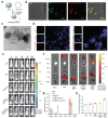
Targeting ability of MNPs@ANG‐EXO in vivo and in vitro. a) Schematic diagram of MNPs conjugated to exosomes after incubation at 4 °C overnight. b) Representative confocal microscopy images of MNPs@ANG‐EXOs, highlighting the colocalization of exosomes (red, PKH26) with the magnetic NPs (green, FITC). Brightfield and merged images are shown. Scale bar, 5 µm. c) TEM image of exosome‐conjugated magnetic nanoparticles. Scale bar, 100 nm. d) Cellular uptake of MNP@ANG‐EXO after 6 h of incubation with LN229 cells. Scale bar, 50 µm (left), 10 µm (right). e) In vivo distribution of saline, MNPs, MNP@EXO, MNP@Lamp2b‐EXO, and MNP@ANG‐EXO in orthotopic DIPG‐bearing mice at 24 h postinjection. f) Ex vivo fluorescence images of the main organs after injection under a magnetic field. g) Quantitative analysis of the fluorescence images (n = 3; ****p < 0.0001, compared with saline treatment group). h) ICP–MS detection of iron ions levels in the brain (n = 3; *p < 0.05, ***p < 0.001, compared with saline treatment group). All of the above data are shown as the mean ± SD.
Figure 5
Figure 5
In vitro enhancement of ferroptosis by MNPs@BQR@ANG‐EXO‐siGPX4. a) Percent of BQR released from the MNPs in solutions with different pH values. b) Viability of LN229 cells after coculture with different concentrations of MNPs, MNP@BQR, MNP@ANG‐EXO‐siGPX4, or MNP@ANG‐EXO‐siGPX4@BQR for 48 h. c) Growth curve based on the OD450 using a CCK‐8 assay in cells after coculture with different NPs in LN229 cells. d) Western blot showing the protein expression of DHODH and GPX4 in LN229 cells after the addition of different NPs. e) MDA levels detected in LN229 cells after coculture with different NPs (n = 3 and were normalized to the level in the control group; ****p < 0.0001, compared with control group). f) GSH levels in LN229 cells treated with different NPs for 48 h (n = 3 and were normalized to the level in the control group; ***p <0.001, compared with control group). g) Flow cytometry analysis of Fe2+ (FerroOrange staining) in LN229 cells incubated with different NPs. And the quantification of mean fluorescence intensity (MFI) values in FerroOrange on LN229 cells (n = 3 and were normalized to the level in the control group; ****p < 0.0001, compared with control group). h) Flow cytometry analysis of ROS (DCFH‐DA staining) in LN229 cells incubated with different NPs. And the quantification of mean fluorescence intensity (MFI) values in ROS on LN229 cells (n = 3 and were normalized to the level in the control group; ****p < 0.0001, compared with control group). All of the above data are shown as the mean ± SD.
Figure 6
Figure 6
Antitumor efficiency of MNP@BQR@ANG‐EXO‐siGPX4. a) Timeline and 3D printed mouse helmet model schematic of the animal experiment. b) Luminescence images of orthotopic LN229‐Luc+ human GBM tumor‐bearing nude mice following different treatments monitored on days 7, 14, and 21. c) Quantitative analysis of the luminescence images (n = 5; ***p < 0.001, compared with saline treatment group). d) Survival curves of the mice in the different groups, n = 5. e). Mouse body weight changes during different treatments, n = 5. f) Representative H&E images and 4‐HNE, GPX4 and DHODH immunochemistry images of the xenograft GBM tumors after the indicated treatment. Scale bar, 10 µm. All of the above data are shown as the mean ± SD.

References

    1. Omuro A., DeAngelis L. M., JAMA, J. Am. Med. Assoc. 2013, 310, 1842. - PubMed
    1. Tan A. C., Ashley D. M., Lopez G. Y., Malinzak M., Friedman H. S., Khasraw M., CA Cancer J. Clin. 2020, 70, 299. - PubMed
    1. Le Rhun E., Preusser M., Roth P., Reardon D. A., van den Bent M., Wen P., Reifenberger G., Weller M., Cancer Treat. Rev. 2019, 80, 101896. - PubMed
    1. a) Sampson J. H., Gunn M. D., Fecci P. E., Ashley D. M., Nat. Rev. Cancer 2019, 20, 12; - PMC - PubMed
    2. b) Lim M., Xia Y., Bettegowda C., Weller M., Nat. Rev. Clin. Oncol. 2018, 15, 422. - PubMed
    1. Stupp R., Taillibert S., Kanner A., Read W., Steinberg D., Lhermitte B., Toms S., Idbaih A., Ahluwalia M. S., Fink K., Di Meco F., Lieberman F., Zhu J. J., Stragliotto G., Tran D., Brem S., Hottinger A., Kirson E. D., Lavy‐Shahaf G., Weinberg U., Kim C. Y., Paek S. H., Nicholas G., Bruna J., Hirte H., Weller M., Palti Y., Hegi M. E., Ram Z., JAMA, J. Am. Med. Assoc. 2017, 318, 2306. - PMC - PubMed

Publication types

Substances