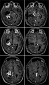
Brain cryptococcoma mimicking a glioblastoma in an immunocompetent patient: A rare case report and comprehensive review
- PMID: 35509529
- PMCID: PMC9062938
- DOI: 10.25259/SNI_1243_2021
Brain cryptococcoma mimicking a glioblastoma in an immunocompetent patient: A rare case report and comprehensive review
Abstract
Background: Cryptococcosis is an invasive fungal infection primarily affecting lungs and potentially spreading to the central nervous. This fungal infection might be misdiagnosed as other infection diseases, such as tuberculosis; granulomatous diseases, like sarcoidosis; and even neoplastic diseases. Some previous reports described cases of cryptococcomas resembling brain tumors. In this paper, we present a very rare presentation of brain cryptococcoma mimicking a malignant glioma. To the best of our knowledge, this is the third case description in the literature.
Case description: A 64-year-old male patient presented at the hospital with a history of progressive frontal headache for 1 month, becoming moderate to severe, associated with visual changes, without nausea or vomiting. No fever was reported. He was a heavy smoker and denied other relevant previous medical data. Neuroimage disclosed a right temporal expansive lesion initially considered a malignant glioma. The patient underwent a right temporal craniotomy and biopsy revealed a cryptococcoma.
Conclusion: Cryptococcomas characteristics in magnetic resonance are quite nonspecific. They should always be included in differential diagnosis of expansive brain lesions, both malignant and benign. Therefore, once cryptococcomas may resemble like other intracranial expansive lesions, biopsy should always be carried out to clarify diagnosis and avoid inadequate treatment and definition of prognosis only based on radiological patterns.
Keywords: Brain neoplasm; Differential diagnosis; Neurocryptococcosis; Surgery.
Copyright: © 2022 Surgical Neurology International.
Conflict of interest statement
There are no conflicts of interest.
Figures


References
-
- Ang SY, Ng VW, Kumar SD, Low SY. Cryptococcosis mimicking lung carcinoma with brain metastases in an immunocompetent patient. J Clin Neurosci. 2017;35:73–5. - PubMed
-
- Chen S, Chen X, Zhang Z, Quan L, Kuang S, Luo X. MRI findings of cerebral cryptococcosis in immunocompetent patients. J Med Imaging Radiat Oncol. 2011;55:52–7. - PubMed
-
- Goldman DL, Khine H, Abadi J, Lindenberg DJ, La P, Niang R, et al. Serologic evidence for Cryptococcus neoformans infection in early childhood. Pediatrics. 2001;107:e66. - PubMed
-
- Khandelwal N, Gupta V, Singh P. Central nervous system fungal infections in tropics. Neuroimaging Clin N Am. 2011;21:859–66. - PubMed
Publication types
LinkOut - more resources
Full Text Sources
