Case Reports
. 2022 Apr 22:13:166.
doi: 10.25259/SNI_1041_2021. eCollection 2022.

Endoscopic third ventriculostomy for noncommunicating hydrocephalus by vertebrobasilar dolichoectasia: A case report

Affiliations
Case Reports

Endoscopic third ventriculostomy for noncommunicating hydrocephalus by vertebrobasilar dolichoectasia: A case report

Kazuho Karasudani et al. Surg Neurol Int. .

Abstract

Background: Vertebrobasilar dolichoectasia (VBD) is a vasculopathy characterized by the elongation, widening, and tortuosity of a cerebral artery. Rarely, hydrocephalus results when the extended basilar artery impairs communication of the cerebral ventricle and cerebrospinal fluid dynamics. We experienced such a case when a patient underwent endoscopic third ventriculostomy (ETV) for noncommunicating hydrocephalus with VBD.

Case description: A 54-year-old man presented with cognitive dysfunction and was diagnosed with VBD by magnetic resonance imaging (MRI). Seven years later, he exhibited subacute impaired consciousness due to acute noncommunicating hydrocephalus, undergoing external ventricular drainage (EVD) that improved consciousness. After EVD removal, the noncommunicating hydrocephalus did not recur; however, 7 months later, subacute consciousness impairment due to noncommunicating hydrocephalus was again observed. MRI showed a significant dilation of both lateral ventricles and ballooning of the third ventricle while the right posterior cerebral artery shifted slightly posteriorly. The patient underwent ETV and clinical symptoms improved. One year after the treatment, MRI observed a patent ETV fenestration and no deleterious changes in clinical symptoms were observed.

Conclusion: ETV can be an effective treatment for the noncommunicating hydrocephalus with VBD when performed with preoperative assessment of vascular anatomy and attention to vascular injury.

Keywords: Countercurrent pulsation; Endoscopic third ventriculostomy; Non-communicating hydrocephalus; Vertebrobasilar dolichoectasia.


Conflict of interest statement

There are no conflicts of interest.

Figures

Figure 1:
Figure 1:
(a-d) Initial MRI (a: T2-weighted image, b: T1-weighted volume isotropic turbo spin-echo acquisition, c: sagittal reconstruction image of MRA, and d: MRA). The basilar artery was dilated and the top of the artery extended upward (b and d). The right posterior cerebral artery extended the floor of the third ventricle upward but did not touch the fornix (c). The enlargement of the ventricles was not observed (a). (e-h) MRI 5 years later (the second examination); (e) T2-weighted image, (f) T1-weighted volume isotropic turbo spin-echo acquisition, (g: sagittal reconstruction image of MRA, h: MRA). The left internal carotid artery was asymptomatically occluded (h). The basilar artery and right posterior cerebral artery extended further upward and the floor of the third ventricle touched the fornix (f and g). The enlargement of the ventricles was not observed (e). (i-l) MRI 1 year and 7 months later (the third examination, just before ETV); (i) T2-weighted image, (j) fluid-attenuated inversion recovery image, (k) sagittal reconstruction image of MRA, and (l) T2 drive image. The bilateral ventricle and the third ventricle were enlarged (I and j). The right posterior cerebral artery extended further upward and touched the fornix while deformities of the midbrain and pons were also observed (k). The floor of the third ventricle was observed to be ballooning (l).
Figure 2:
Figure 2:
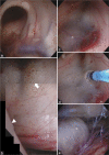
Images of the endoscopic third ventricle ventriculostomy. (a) The foramen of Monro was observed from the right ventricle, which was enlarged. (b) Consolidated images of endoscopic findings in the third ventricle. The floor of the third ventricle extended to the fornix due to the upward extension of the right posterior cerebral artery (white arrowhead). The mamillary body was stretched (white arrow). (c) The basilar artery (asterisk) was confirmed through the floor of the third ventricle. (d) The floor of the third ventricle was fenestrated to avoid injuring the basilar artery. (e) The vascular wall of the basilar artery (asterisk) had white sclerotic changes.
Figure 3:
Figure 3:
Postoperative MRI showed that the ventricles were shrinking (a: T2-weighted image) and the floor of the third ventricle was fenestrated (b: T2 DRIVE image).

References

    1. Ebrahimzadeh K, Bakhtevari MH, Shafizad M, Rezaei O. Hydrocephalus as a rare compilation of vertebrobasilar dolichoectasia: A case report and review of the literature. Surg Neurol Int. 2017;8:256. - PMC - PubMed
    1. Gutierrez J, Sacco RL, Wright CB. Dolichoectasia-an evolving arterial disease. Nat Rev Neurol. 2011;7:41–50. - PubMed
    1. Jagetia A, Patel K, Sachdeva D, Rathore L. Dolichoectatic internal carotid artery presenting as a sellar-suprasellar mass with symptomatic hydrocephalus. Neurol India. 2017;65:681–2. - PubMed
    1. Jiang L, Gao G, Zhou Y. Endoscopic third ventriculostomy and ventriculoperitoneal shunt for patients with non-communicating hydrocephalus: A PRISMA-compliant meta-analysis. Medicine. 2018;97:e12139. - PMC - PubMed
    1. Labidi M, Lavoie P, Lapointe G, Obaid S, Weil AG, Bojanowski MW, et al. Predicting success of endoscopic third ventriculostomy: Validation of the ETV success score in a mixed population of adult and pediatric patients. J Neurosurg. 2015;123:1447–55. - PubMed

Publication types

LinkOut - more resources