. 2022 Apr 25:2022:4117520.
doi: 10.1155/2022/4117520. eCollection 2022.

Seahorse Protein Hydrolysate Ameliorates Proinflammatory Mediators and Cartilage Degradation on Posttraumatic Osteoarthritis with an Obesity Rat Model

Affiliations

Seahorse Protein Hydrolysate Ameliorates Proinflammatory Mediators and Cartilage Degradation on Posttraumatic Osteoarthritis with an Obesity Rat Model

Sabri Sudirman et al. Biomed Res Int. .

Abstract

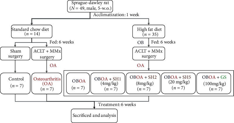
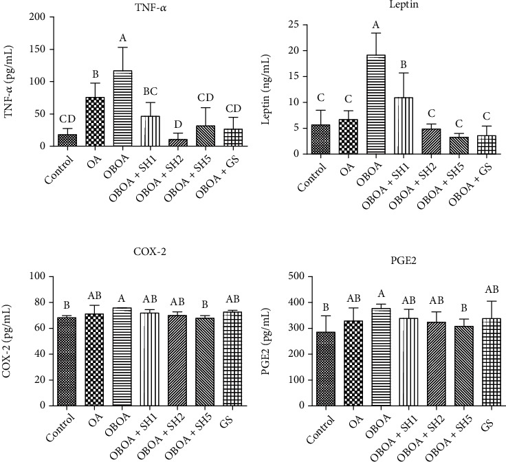
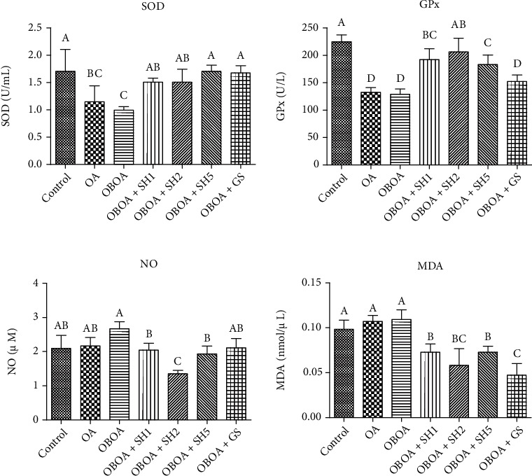
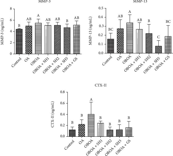
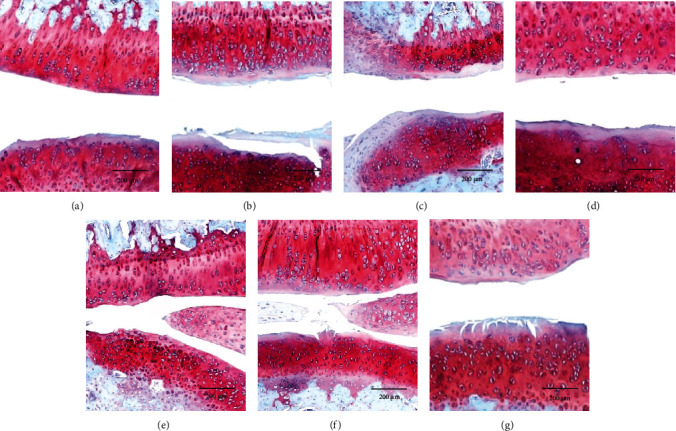
Osteoarthritis (OA) is one of the age-related diseases and is highly present on the knees. Obesity and mechanical injuries as a risk factor of OA are attributed to cartilage disintegration, joint loading, and inflammation. This study is aimed at investigating the effects of seahorse protein hydrolysate (SH) on posttraumatic osteoarthritis in an obesity rat. The OA model was developed by anterior cruciate ligament transection with medial meniscectomy in a high-fat diet- (HFD-) induced obesity rat model. The male Sprague-Dawley rats were fed a HFD for 6 weeks before OA surgery. The OA rats were treated with oral gavage by 4, 8, or 20 mg/kg of body weight of SH for 6 weeks of treatment. The expressions of plasma proinflammatory factors, C-telopeptide of type II collagen, and matrix metalloproteinase- (MMP-) 3 and MMP-13 were reduced by SH treatment. Plasma superoxide dismutase and glutathione peroxidase activities were enhanced by SH. SH also relieved the pain of the knee joint and swelling as well as decreased proteoglycan loss in the knee articular cartilage caused by osteoarthritis. Based on these results, SH suppressed proinflammatory factors and attenuated cartilage degradation and pain in the OA model. Therefore, seahorse protein hydrolysate might be a potential opportunity for improving the development of osteoarthritis.


Conflict of interest statement

All authors declare that they have no conflicts of interest.

Figures

Figure 1
Figure 1
The flowchart of seahorse hydrolysate (SH) treatment on an anterior cruciate ligament transection with medial meniscectomy- (ACLT+MMx-) induced osteoarthritis (OA) in high-fat diet-induced obese (OB) rat models.
Figure 2
Figure 2
Effects of seahorse hydrolysate treatment on plasma tumor necrosis factor-alpha (TNF-α), leptin, cyclooxygenase (COX)-2, and prostaglandin E2 (PGE2) levels in anterior cruciate ligament transection with medial meniscectomy surgery-induced osteoarthritis in high fat diet-induced obesity rats. Data were shown as the mean ± SD (n = 7). The values with different letters (a–d) represent significantly different (p < 0.05) as analyzed by Duncan's multiple range test.
Figure 3
Figure 3
Effects of seahorse hydrolysate treatment on plasma antioxidant activity and oxidative stress markers in anterior cruciate ligament transection on medial meniscectomy surgery-induced osteoarthritis in high fat diet-induced obesity rats. Data were shown as the mean ± SD (n = 7). The values with different letters (a–c) represent significantly different (p < 0.05) as analyzed by Duncan's multiple range test. GPx: glutathione peroxidase; MDA: malondialdehyde; NO: nitric oxide; SOD: superoxide dismutase.
Figure 4
Figure 4
Effects of seahorse hydrolysate treatment on plasma matrix metalloproteinase- (MMP-) 3, MMP-13, and C-terminal cross-linked telopeptide of type II collagen (CTX-II) level in anterior cruciate ligament transection on medial meniscectomy surgery-induced osteoarthritis in high fat diet-induced obesity rats. Data were shown as the mean ± SD (n = 7). The values with different letters (a–c) represent significantly different (p < 0.05) as analyzed by Duncan's multiple range test.
Figure 5
Figure 5
Representative of operated-knee joint cartilage with Safranin-O staining for each group after 6 weeks of treatment. Cartilage (orange to red) and nuclei (black).

Similar articles

Cited by

References

    1. Prieto-Alhambra D., Judge A., Javaid M. K., Cooper C., Diez-Perez A., Arden N. K. Incidence and risk factors for clinically diagnosed knee, hip and hand osteoarthritis: influences of age, gender and osteoarthritis affecting other joints. Annals of the Rheumatic Diseases . 2014;73(9):1659–1664. doi: 10.1136/annrheumdis-2013-203355. - DOI - PMC - PubMed
    1. Loeser R. F., Goldring S. R., Scanzello C. R., Goldring M. B. Osteoarthritis: a disease of the joint as an organ. Arthritis and Rheumatism . 2012;64(6):1697–1707. doi: 10.1002/art.34453. - DOI - PMC - PubMed
    1. Heidari B. Knee osteoarthritis prevalence, risk factors, pathogenesis and features: part I. Caspian Journal of Internal Medicine . 2011;2(2):205–212. - PMC - PubMed
    1. Divella R., De Luca R., Abbate I., Naglieri E., Daniele A. Obesity and cancer: the role of adipose tissue and adipo-cytokines-induced chronic inflammation. Journal of Cancer . 2016;7(15):2346–2359. doi: 10.7150/jca.16884. - DOI - PMC - PubMed
    1. Teeple E., Jay G. D., Elsaid K. A., Fleming B. C. Animal models of osteoarthritis: challenges of model selection and analysis. The AAPS Journal . 2013;15(2):438–446. doi: 10.1208/s12248-013-9454-x. - DOI - PMC - PubMed

Substances