. 2022 Mar;46(2):255-265.
doi: 10.1016/j.jgr.2021.06.011. Epub 2021 Jul 3.

Cardioprotective effect of ginsenoside Rb1 via regulating metabolomics profiling and AMP-activated protein kinase-dependent mitophagy

Affiliations

Cardioprotective effect of ginsenoside Rb1 via regulating metabolomics profiling and AMP-activated protein kinase-dependent mitophagy

Jingui Hu et al. J Ginseng Res. 2022 Mar.

Abstract

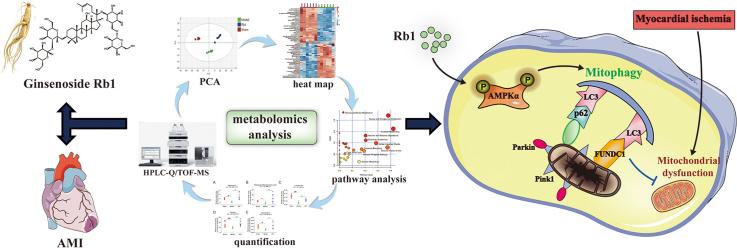
Background: Ginsenoside Rb1, a bioactive component isolated from the Panax ginseng, acts as a remedy to prevent myocardial injury. However, it is obscure whether the cardioprotective functions of Rb1 are related to the regulation of endogenous metabolites, and its potential molecular mechanism still needs further clarification, especially from a comprehensive metabolomics profiling perspective.

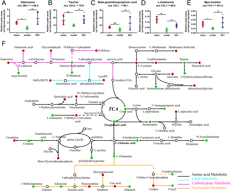
Methods: The mice model of acute myocardial ischemia (AMI) and oxygen glucose deprivation (OGD)-induced cardiomyocytes injury were applied to explore the protective effect and mechanism of Rb1. Meanwhile, the comprehensive metabolomics profiling was conducted by high-performance liquid chromatography and quadrupole time-of-flight mass spectrometry (HPLC-Q/TOF-MS) and a tandem liquid chromatography and mass spectrometry (LC-MS).

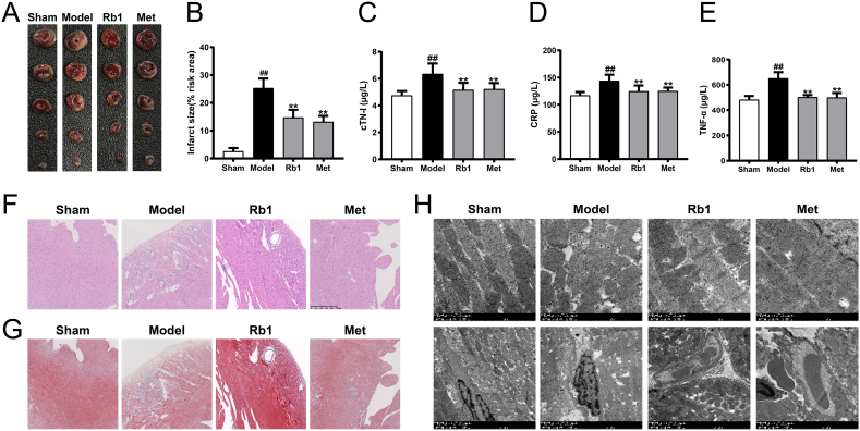
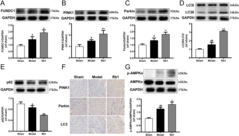
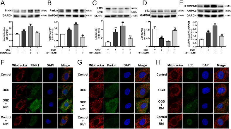
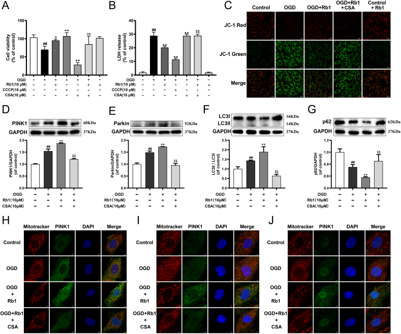
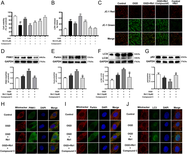
Results: Rb1 treatment profoundly reduced the infarct size and attenuated myocardial injury. The metabolic network map of 65 differential endogenous metabolites was constructed and provided a new inspiration for the treatment of AMI by Rb1, which was mainly associated with mitophagy. In vivo and in vitro experiments, Rb1 was found to improve mitochondrial morphology, mitochondrial function and promote mitophagy. Interestingly, the mitophagy inhibitor partly attenuated the cardioprotective effect of Rb1. Additionally, Rb1 markedly facilitated the phosphorylation of AMP-activated protein kinase α (AMPKα), and AMPK inhibition partially weakened the role of Rb1 in promoting mitophagy.

Conclusions: Ginsenoside Rb1 protects acute myocardial ischemia injury through promoting mitophagy via AMPKα phosphorylation, which might lay the foundation for the further application of Rb1 in cardiovascular diseases.

Keywords: AMPK; Acute myocardial ischemia; Ginsenoside Rb1; Metabolomics; Mitophagy.


Conflict of interest statement

The authors declare no competing financial interest.

Figures

Image 1
Graphical abstract
Fig. 1
Fig. 1
Rb1 effectively protected acute myocardial ischemia-injured mice. (A) Representative images of 2,3,5-triphenyl tetrazolium chloride (TTC) staining. (B) The statistical results of TTC staining. The serum contents of (C) cTN-I, (D) CRP and (E) TNF-α. Representative images of (F) H&E and (G) Masson's trichrome staining (200 × ). Scale bar = 250 μm. (H) Myocardial ultrastructure was detected by transmission electron microscopy (5000 × ). Scale bar = 1 μm. Results were presented as mean ± SD. ##P < 0.01 vs. the sham group; ∗∗P < 0.01 vs. the model group, n = 6.
Fig. 2
Fig. 2
Validation and semi-quantification of selected metabolites, and the metabolic network map of the differential metabolites. The RPA of (A) adenosine, (B) taurine, (C) β-GPA, (D) L-isoleucine and (E) myo-Inositol, in reference to the retention time of reference standards and the peak area of internal standards. (F) The metabolic network map of the differential metabolites. The red dots represent that metabolites in the model group were up-regulated compared with those in the Rb1 group and the down-regulated metabolites were represented by green dots. Results were presented as mean ± SD. #P < 0.05, ##P < 0.01 vs. the sham group; ∗∗P < 0.01 vs. the model group, n = 8.
Fig. 3
Fig. 3
Rb1 significantly promoted mitophagy and upregulated AMPKα phosphorylation in AMI-injured mice. The expression of (A) FUNDC1, (B) PINK1, (C) Parkin, (D) LC3Ⅱ/LC3Ⅰ and (E) p62 were detected using western blot analysis. (F) The expression of PINK1, Parkin and LC3 were detected using immunohistochemistry (400 × ). Scale bar = 50 μm. (G) The expression of p-AMPKα and AMPKα were detected using western blot analysis. All the experiments were performed in triplicate. Results were presented as mean ± SD. #P < 0.05, ##P < 0.01 vs. the sham group; ∗P < 0.05, ∗∗P < 0.01 vs. the model group.
Fig. 4
Fig. 4
Rb1 profoundly promoted mitophagy and AMPKα phosphorylation in OGD-injured H9c2 cardiomyocytes. The expression of (A) PINK1, (B) Parkin, (C) LC3Ⅱ/LC3Ⅰ, (D) p62 and (E) p-AMPKα/AMPKα were detected using western blot analysis. Mitochondrial morphology and the colocalization of the mitochondrial with (F) PINK1, (G) Parkin and (H) LC3 were analyzed using a 63 × oil immersion lens (630 × ). Results were obtained from three independent experiments and were presented as mean ± SD. ##P < 0.01 vs. the control group; ∗P < 0.05, ∗∗P < 0.01 vs. the OGD group.
Fig. 5
Fig. 5
Rb1 attenuated OGD-induced H9c2 cardiomyocytes injury through mitophagy. (A) The cell viability of H9c2 cardiomyocytes. (B) The release of LDH in culture medium of H9c2 cardiomyocytes. (C) Mitochondrial membrane potential (ΔΨm) was assessed by probe JC-1. Red fluorescence represents intact ΔΨm. Green fluorescence represents dissipation of ΔΨm. The expression of (D) PINK1, (E) Parkin, (F) LC3II/LC3I and (G) p62 were detected using western blot analysis. Mitochondrial morphology and the colocalization of the mitochondrial with (H) PINK1, (I) Parkin and (J) LC3 were analyzed using a 63 × oil immersion lens (630 × ). Results were obtained from three independent experiments and were presented as mean ± SD. ##P < 0.01 vs. the control group; ∗P < 0.05, ∗∗P < 0.01 vs. the OGD group; $$P < 0.01 vs. the OGD + Rb1 group.
Fig. 6
Fig. 6
Rb1 effectively improved myocardial ischemia through AMPKα mediated mitophagy. (A) The cell viability of H9c2 cardiomyocytes. (B) The release of LDH in culture medium of H9c2 cardiomyocytes. (C) ΔΨm was assessed by probe JC-1. The expression of (D) PINK1, (E) Parkin, (F) LC3II/LC3I and (G) p62 were detected using western blot analysis. Mitochondrial morphology and the colocalization of the mitochondrial with (H) PINK1, (I) Parkin and (J) LC3 were analyzed using a 63 × oil immersion lens (630 × ). Results were obtained from three independent experiments and were presented as mean ± SD. #P < 0.05, ##P < 0.01 vs. the control group; ∗P < 0.05, ∗∗P < 0.01 vs. the OGD group; $$P < 0.01 vs. the OGD + Rb1 group.

References

    1. Shimokawa H., Yasuda S. Myocardial ischemia: current concepts and future perspectives. J Cardiol. 2008;52:67–78. - PubMed
    1. Herzig S., Shaw R.J. AMPK: guardian of metabolism and mitochondrial homeostasis. Nature Reviews. Molecular Cell Biology. 2018;19:121–135. - PMC - PubMed
    1. Hernandez-Resendiz S., Prunier F., Girao H., Dorn G., Hausenloy D.J. Targeting mitochondrial fusion and fission proteins for cardioprotection. J Cell Mol Med. 2020;24:6571–6585. - PMC - PubMed
    1. Chistiakov D.A., Shkurat T.P., Melnichenko A.A., Grechko A.V., Orekhov A.N. The role of mitochondrial dysfunction in cardiovascular disease: a brief review. Ann Med. 2018;50:121–127. - PubMed
    1. Xu Y., Tang Y., Lu J., Zhang W., Zhu Y., Zhang S., Ma G., Jiang P., Zhang W. PINK1-mediated mitophagy protects against hepatic ischemia/reperfusion injury by restraining NLRP3 inflammasome activation. Free Radic Biol Med. 2020;160:871–886. - PubMed

LinkOut - more resources