. 2022 Apr 25:2022:5497991.
doi: 10.1155/2022/5497991. eCollection 2022.

Licoflavone A Suppresses Gastric Cancer Growth and Metastasis by Blocking the VEGFR-2 Signaling Pathway

Affiliations

Licoflavone A Suppresses Gastric Cancer Growth and Metastasis by Blocking the VEGFR-2 Signaling Pathway

Gong Hongxia et al. J Oncol. .

Abstract

Objectives: Licoflavone A (LA) is a natural flavonoid compound derived from the root of Glycyrrhiza. This study investigated the antitumor effect and underlying molecular mechanisms of LA against gastric cancer (GC) in vitro and in vivo.

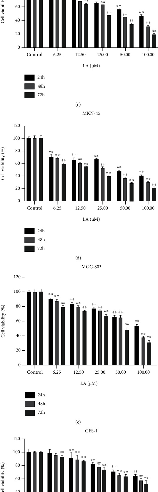
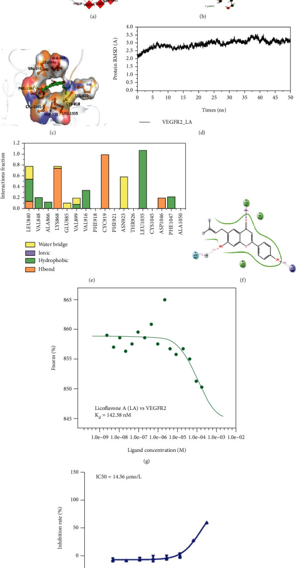
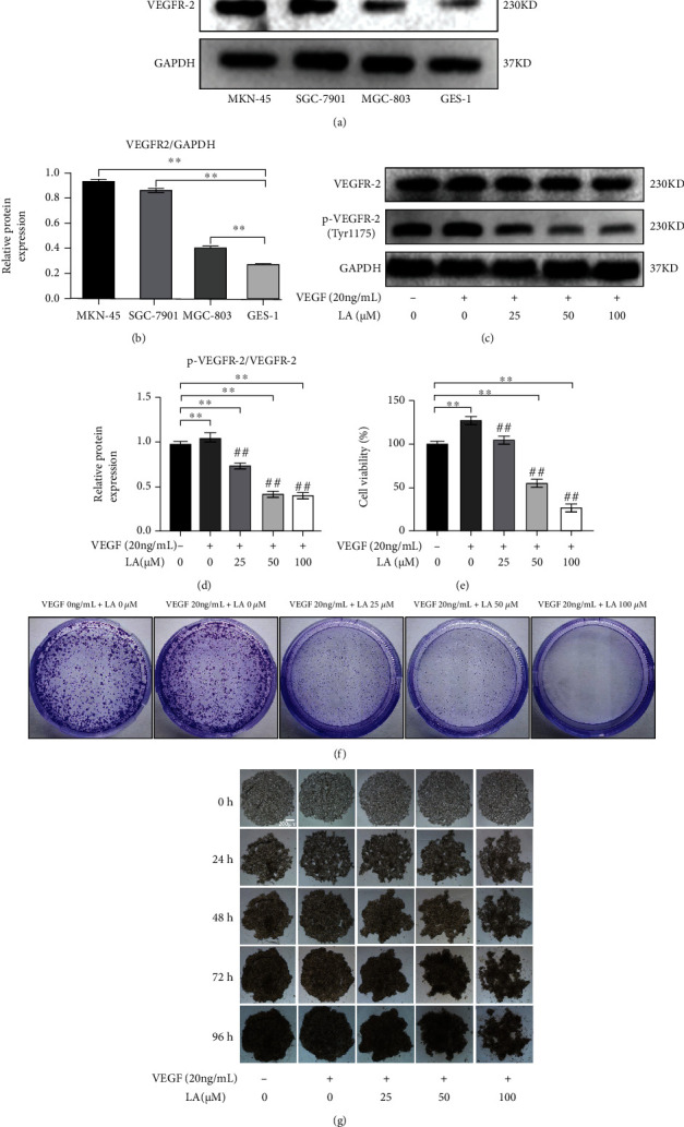
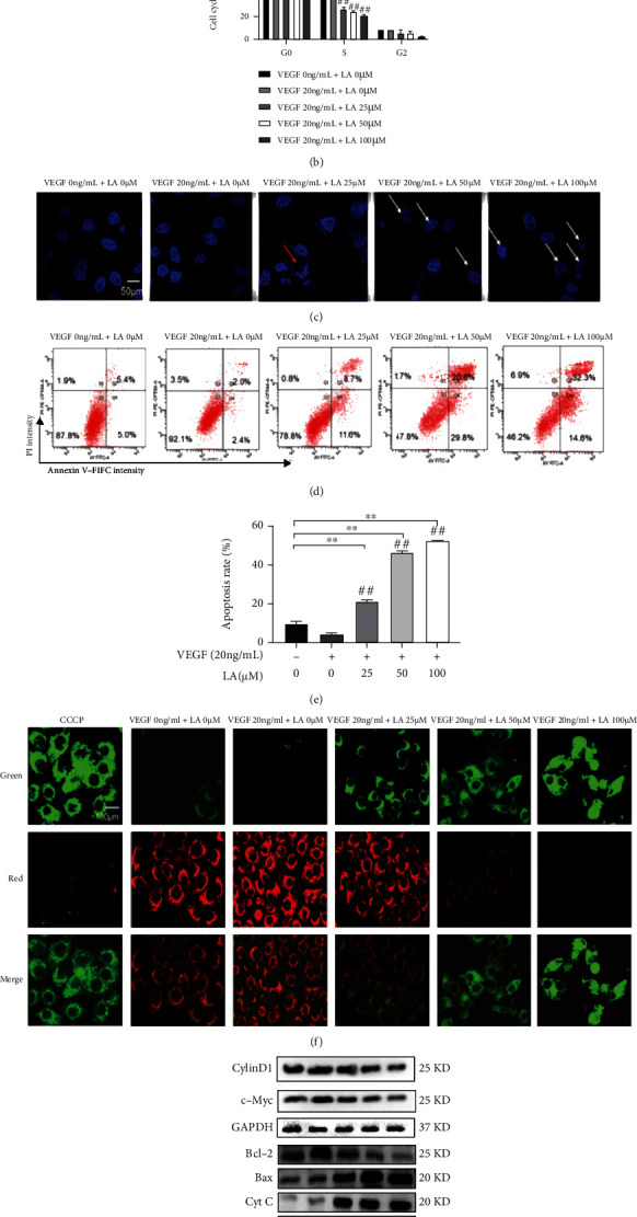
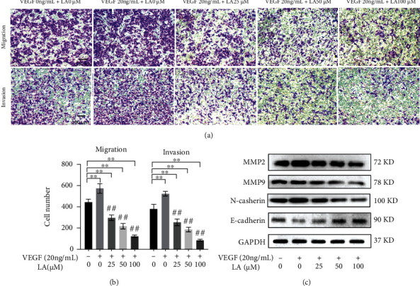
Materials and methods: A CCK8 assay was used to measure the antiproliferative activity of LA in human GC SGC-7901, MKN-45, MGC-803 cells, and human GES-1 cells. Target prediction and protein-protein interaction (PPI) analysis were used to identify the potential molecular targets of LA. The binding pattern of LA to VEGFR-2 was analyzed by molecular docking and molecular dynamic (MD). The affinity of LA for VEGFR-2 was determined by microscale thermophoresis (MST). The protein tyrosine kinase activity of VEGFR-2 in the presence of LA was determined by an enzyme activity test. The effect of LA on the proliferation of VEGF-stimulated MKN-45 cells was measured with CCK8 assays, clone formation assays, and 3D microsphere models. Hoechst 33342 staining, FCM, MMP, and WB assays were used to investigate the ability of LA to block cell cycle and promote apoptosis of VEGF-stimulated MKN-45 cells. Transwell matrix assays were used to measure migration and invasion, and WB assays were used to measure EMT.

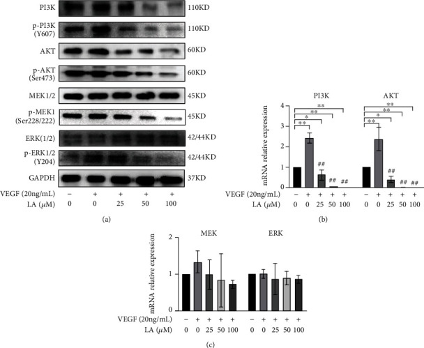
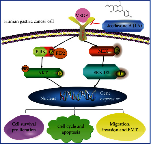
Results: LA inhibited the proliferation of SGC-7901, MKN-45, and MGC-803 cells and VEGF-stimulated MKN-45 cells. VEGFR-2 was identified as the target of LA. LA could also block cell cycle, induce apoptosis, and inhibit migration, invasion, and EMT of VEGF-stimulated MKN-45 cells. Functional analyses further revealed that the cytotoxic effect of LA on VEGF-stimulated MKN-45 cells potentially involved the PI3K/AKT and MEK/ERK signaling pathways.

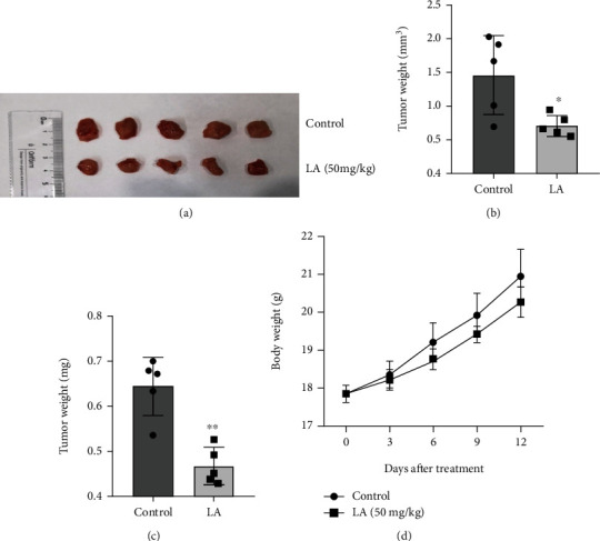
Conclusions: This study demonstrates that LA has anti-GC potency in vitro and in vivo. LA affects the proliferation, cycle, apoptosis, migration, invasion, and EMT by targeting VEGFR-2 and blocks the PI3K/AKT and MEK/ERK signaling pathways in VEGF-stimulated MKN-45 cells.


Conflict of interest statement

The authors declare that there is no conflict of interest regarding the publication of this paper.

Figures

Figure 1
Figure 1
Licoflavone A (LA) suppressed GC cell proliferation in vitro. (a) The root of Glycyrrhiza glabra. (b) The chemical structure of LA. (c, d, e, and f) Human SGC-7901, MKN-45, MGC-803, and GES-1 cells were treated with LA (0, 6.25, 12.5, 25, 50, and 100 μM) for 24, 48, and 72 h, respectively (n = 5). Cell viability was measured by CCK8 assay. Cell viability is represented in percentage relative absorbance compared to the controls. Values are given as the mean ± standard error of the mean (SEM) of three independent experiments. p < 0.05 vs. control and ∗∗p < 0.01 vs. control.
Figure 2
Figure 2
Protein-protein interaction (PPI) analysis and validation of LA targets. (a) The bubble size represents the number of genes enriched in this pathway, the bubble color difference represents the level of gene enrichment, and the circle size represents the number of targets in the pathway. The redder the color, the smaller the p value. (b) A 2D diagram of the interaction between LA and VEGFR-2 (PBD: 4AGD). Dashed lines indicate hydrogen bonds, and eyelashes indicate hydrophobicity. (c) A 3D diagram of the interaction between LA and VEGFR-2 (PBD: 4AGD). The LA structure is shown in green. The amino acids associated with the A-chain binding pocket of 4AGD are indicated in purple. The amino acids associated with the B-chain binding pocket of 4AGD are indicated in indigo. Yellow dashed lines indicate the length of the hydrogen bonds. (d) Stability analysis of modeled proteins using the Desmond module (Schrodinger suite) for VEGFR-2 and LA protein interactions. (e) Histogram and percentage of interaction in molecular dynamics simulation of VEGFR-2-LA. (f) Simulation interaction diagram showing interactions of LA with crucial amino acids of the VEGFR-2 protein. (g) Results of microscale thermophoresis for LA vs. VEGFR-2. (h) Enzyme inhibitory activity of VEGFR-2.
Figure 3
Figure 3
LA arrests the proliferation of VEGF-stimulated MKN-45 cells. (a) The expression of VEGFR-2 protein in SGC-7901, MKN-45, MGC-803, and GES-1 cells. (b) Relative VEGFR-2 expression levels were quantified in SGC-7901, MKN-45, MGC-803, and GES-1 cells. GSE-1 is the reference. (c) The p-VEGFR-2 expression level of LA-treated VEGF-stimulated MKN-45 cell. (d) Relative p-VEGFR-2 protein expression levels were quantified by normalization to GAPDH values as the mean of three independent experiments. (e) The proliferation of VEGF-stimulated MKN-45 cells was measured by the CCK8 assay. (f) LA inhibited the colony formation of VEGF-stimulated MKN-45 cells. (g) 3D microsphere was observed at 0, 24, 48, 72, and 96 h, respectively. Bar = 200 μm. Values are given as the mean ± standard error of the mean (SEM) experiments. n = 3. p < 0.05 vs. control and ∗∗p < 0.01 vs. control. #p < 0.05 vs. VEGF 20 ng/mL + LA 0 μM and ##p < 0.01 vs. VEGF 20 ng/mL + LA 0 μM.
Figure 4
Figure 4
LA arrested cells at G1 phase and induced apoptosis of VEGF-stimulated MKN-45 cells. (a) Flow cytometry assay of cell cycle distribution. (b) The quantitation of cell cycle distribution. The data are presented as mean ± SD (n = 3). (c) The Hoechst 33342-stained nuclei were imaged under laser confocal microscopy. Bar = 50 μm. Representative photographs of the morphological changes observed. The red arrow indicates apoptotic bodies and the white arrows indicate shrunken and deformed nuclei of apoptotic cells. (d) Apoptosis of LA-treated VEGF-stimulated MKN-45 cells was determined by flow cytometric analysis. (e) The quantitation of cell cycle distribution. The data are presented as mean ± SD (n = 3). (f) A JC-10 kit was used to measure the mitochondrial membrane potential of VEGF-stimulated MKN-45 cells by laser confocal microscopy. CCCP staining results were used as the positive control. JC-10 is the monomer with green fluorescence, while JC-10 is the polymer with red fluorescence. Bar = 100 μm. (g) Cyclin D1, c-Myc, Bcl-2, Bax, Cyt C, caspase 9, and cleaved-caspase 3 protein expression levels were detected by western blot.
Figure 5
Figure 5
LA suppressed the migration and invasion capacities of VEGF-stimulated MKN-45 cells. VEGF-stimulated MKN-45 cells were treated with LA for 72 h. (a) Transwell assays without or with Matrigel were carried out to evaluate the migration and invasion capacity. (b) The numbers of migrated and invaded cells were quantified. Scar bar = 100 μm. The data are presented as mean ± SD (n = 3). p < 0.05 vs. VEGF 0 ng/mL + LA 0 μM and ∗∗p < 0.01 vs. VEGF 0 ng/mL + LA 0 μM. #p < 0.05 vs. VEGF 20 ng/mL + LA 0 μM and ##p < 0.01 vs. VEGF 20 ng/mL + LA 0 μM. (c) Western blot analysis of EMT-regulated proteins MMP2, MMP9, E-cadherin, and N-cadherin.
Figure 6
Figure 6
LA suppressed GC growth and metastasis in VEGF-stimulated MKN-45 cells through PI3K/AKT and MEK/ERK signaling pathway activation. (a) Western blot analysis of PI3K, p-PI3K, AKT, p-AKT, MEK, p-MEK, ERK, and p-ERK in LA-treated VEGF-stimulated MKN-45 cells. (b and c) RT-PCR analysis of PI3K, AKT, MEK, and ERK mRNA relative expression in LA-treated VEGF-stimulated MKN-45 cells. The data are presented as mean ± SD (n = 3). p < 0.05 vs. VEGF 0 ng/mL + LA 0 μM and ∗∗p < 0.01 vs. VEGF 0 ng/mL + LA 0 μM. #p < 0.05 vs. VEGF 20 ng/mL + LA 0 μM, ##p < 0.01 vs. VEGF 20 ng/mL + LA 0 μM.
Figure 7
Figure 7
LA inhibited the growth of GC cells in vivo. (a) Images of the xenograft tumors of the control and LA treatment group. (b and c) Tumor volume and tumor weight were measured. (d) Body weights of mice were recorded every 3 days after indicated treatment. The data are presented as mean ± SD (n = 5). p < 0.05 vs. control. ∗∗p < 0.01 vs. control. The data are presented as mean ± SD (n = 5).
Figure 8
Figure 8
The schematic representation of the proposed model. LA inhibits activation of VEGFR2, blocking downstream PI3K/AKT and MEK/ERK signaling pathways, which represses GC cell proliferation and metastasis, and induces cycle arrest as well as apoptosis.

References

    1. Latest global cancer data: cancer burden rises to 19.3 million new cases and 10.0 million cancer deaths in 2020. https://www.iarc.fr/fr/news-events/latest-global-cancer-data-cancer-burd...
    1. Zhao T. Q., Zhao Y. D., Liu X. Y., et al. Discovery of 6-chloro-2-(propylthio)-8,9-dihydro-7H-purines containing a carboxamide moiety as potential selective anti-lung cancer agents. European Journal of Medicinal Chemistry . 2018;151:327–338. doi: 10.1016/j.ejmech.2018.03.084. - DOI - PubMed
    1. Jie G. A., Km A., Lei Z. B., Lokeshwar B. Spice up your food for cancer prevention: cancer chemo-prevention by natural compounds from common dietary spices. Evolutionary Diversity as a Source for Anticancer Molecules . 2021:275–308. doi: 10.1016/B978-0-12-821710-8.00013-8. - DOI
    1. Liu H., Wang J., Zhou W., Wang Y., Yang L. Systems approaches and polypharmacology for drug discovery from herbal medicines: an example using licorice. Journal of Ethnopharmacology . 2013;146(3):773–793. doi: 10.1016/j.jep.2013.02.004. - DOI - PubMed
    1. Mj A., Sz A., Sy A., et al. An "essential herbal medicine"--licorice: a review of phytochemicals and its effects in combination preparations. Journal of Ethnopharmacology . 2020;249:p. 112439. doi: 10.1016/j.jep.2019.112439. - DOI - PubMed

LinkOut - more resources