. 2022 Feb 21;9(1):e158.
doi: 10.14440/jbm.2022.379. eCollection 2022.

Development of a reproducible porcine model of infected burn wounds

Affiliations

Development of a reproducible porcine model of infected burn wounds

Sayf Al-Deen Said et al. J Biol Methods. .

Abstract

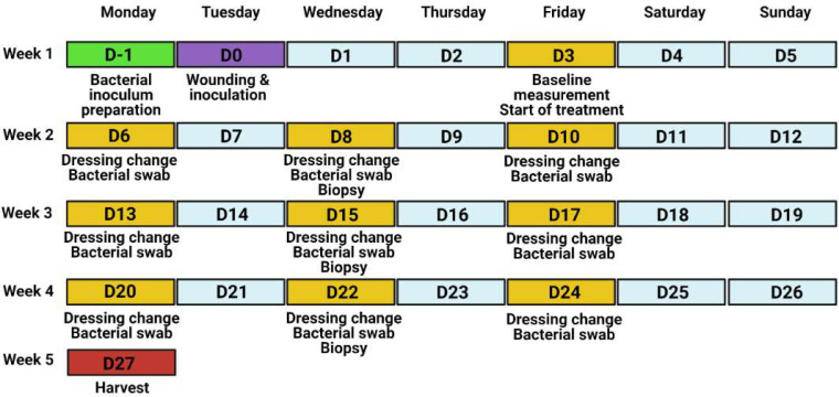
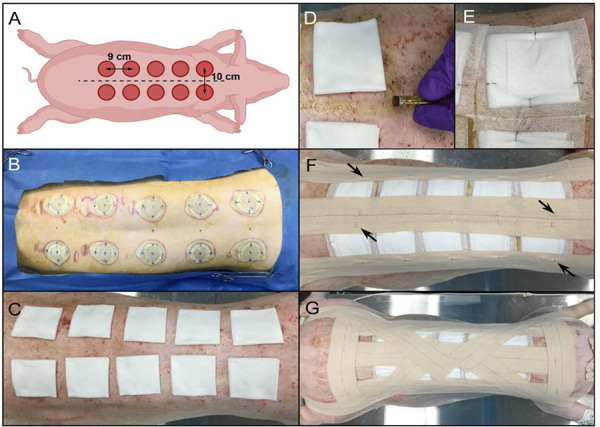
Severe burns are traumatic and physically debilitating injuries with a high rate of mortality. Bacterial infections often complicate burn injuries, which presents unique challenges for wound management and improved patient outcomes. Currently, pigs are used as the gold standard of pre-clinical models to study infected skin wounds due to the similarity between porcine and human skin in terms of structure and immunological response. However, utilizing this large animal model for wound infection studies can be technically challenging and create issues with data reproducibility. We present a detailed protocol for a porcine model of infected burn wounds based on our experience in creating and evaluating full thickness burn wounds infected with Staphylococcus aureus on six pigs. Wound healing kinetics and bacterial clearance were measured over a period of 27 d in this model. Enumerated are steps to achieve standardized wound creation, bacterial inoculation, and dressing techniques. Systematic evaluation of wound healing and bacterial colonization of the wound bed is also described. Finally, advice on animal housing considerations, efficient bacterial plating procedures, and overcoming common technical challenges is provided. This protocol aims to provide investigators with a step-by-step guide to execute a technically challenging porcine wound infection model in a reproducible manner. Accordingly, this would allow for the design and evaluation of more effective burn infection therapies leading to better strategies for patient care.

Keywords: burn wounds; infection; porcine model; protocol; wound healing.


Figures

Figure 1.
Figure 1.
Experimental timeline of the porcine wound infection model.
Figure 2.
Figure 2.
Wound placement design and dressing steps. A. Diagram of wound placement. Pig is measured from the scapulae to the iliac crests (dotted lines) to determine optimal number and spacing of wounds paravertebrally, 5 cm from midline. B. Appearance of skin post-burn creation. Note the blue dots marking the spine and wound placement to ensure even spacing of the wounds. C. Wounds covered with 2” × 2” non-stick gauze pads, with fold towards midline. D. Application of tincture of benzoin around the edges of the gauze pads. E. Telfa dressing and gauze pad secured with skin staples. The staples should oppose each other and be perpendicular to the gauze edge. F. Application of Elastikon® strips along the spine and on each side of the torso to reinforce the Telfa bandages. Staples are applied along the strips to anchor the strips to the dressings (black arrows). G. Steri-drape dressings cover the prior dressing layers and are secured by Elastikon® strips along the spine, lateral to the dressings, and perpendicularly across the shoulders and hips. Staples are added to the perpendicular strips and down the central strip. An additional Elastikon® strip is placed from the center of the back, across the shoulder, under the neck, and across the other shoulder. Two final Elastikon® strips are applied from the center of the back and over the hip and butt.
Figure 3.
Figure 3.
Wound healing assessment by wound closure and histologic scoring. A-C. H&E stained wound biopsy tissue illustrating the progression of dermal regeneration after burn. A. D3 biopsy showing an abundance of adipocytes in the hypodermis with minimal to no inflammatory cell infiltration. B. D10 biopsy showing moderate inflammatory cell infiltration in the hypodermis, with numerous polymorphic neutrophils (black arrows). C. D22 biopsy showing the disappearance of the dermal adipocytes, abundance of fibroblasts (black arrows) and collagen deposition (white arrows). D. Dermis score progression (n = 20 wounds) [13]. E. Average wound area change over time compared to individual wound baseline measurements (n = 36 wounds; mean ± SEM).
Figure 4.
Figure 4.
Assessment of bacterial colonization in burn wounds. A-C. Culture assessment of surface bacterial colonization of burn wounds. A. Dilution plate overview. An undiluted sample is aliquoted into column 1 of a sterile 96 well plate (100 μl, orange) in duplicate (e.g., rows A&B = wound 1, D&E = wound 2, G&H = wound 3). Sterile PBS is added to the remaining wells in each sample row (80 μl, blue). Serial dilutions are made by transferring 20 μl sample to the neighboring well to the right (e.g., column 1 to column 2, then column 2 to column 3, etc.). B. Appearance of bacterial agar plates for dilutions 1–6 after spot plating and overnight growth. Using a multi-channel pipette, 5 μl from each well shown in (A) were spotted onto duplicate square agar plates and incubated overnight. C. Quantification of the average viable CFU/wound determined by culture assessment over the duration of the experiment (n = 36 wounds; mean ± SEM). D. Visualization of bacterial colonization by modified Gram stain of biopsy tissue samples from Day 3 and Day 6 post-wounding and inoculation. Insets show a higher magnification to highlight the surface colonization on Day 3 and invasive colonization on Day 6.

References

    1. Jeschke MG, van Baar ME, Choudhry MA, Chung KK, Gibran NS, Logsetty S. Burn injury. Nat Rev Dis Primers. 2020. Feb;6(1):11. https://doi.org/10.1038/s41572-020-0145-5 10.1038/s41572-020-0145-5 PMID: - DOI - PMC - PubMed
    1. Seaton M, Hocking A, Gibran NS. Porcine models of cutaneous wound healing. ILAR J. 2015;56(1):127–38. https://doi.org/10.1093/ilar/ilv016 10.1093/ilar/ilv016 PMID: - DOI - PubMed
    1. Moins-Teisserenc H, Cordeiro DJ, Audigier V, Ressaire Q, Benyamina M, Lambert J, et al. . Severe Altered Immune Status After Burn Injury Is Associated With Bacterial Infection and Septic Shock. Front Immunol. 2021;12:586195. https://doi.org/10.3389/fimmu.2021.586195 10.3389/fimmu.2021.586195 PMID: - DOI - PMC - PubMed
    1. Church D, Elsayed S, Reid O, Winston B, Lindsay R. Burn wound infections. Clin Microbiol Rev. 2006;19(2):403-34. https://doi.org/10.1128/CMR.19.2.403-434.2006 10.1128/CMR.19.2.403-434.2006 PMID: - DOI - PMC - PubMed
    1. Lachiewicz AM, Hauck CG, Weber DJ, Cairns BA, van Duin D. Bacterial Infections After Burn Injuries: Impact of Multidrug Resistance. Clin Infect Dis. 2017;65(12):2130-6. https://doi.org/10.1093/cid/cix682 10.1093/cid/cix682 PMID: - DOI - PMC - PubMed

LinkOut - more resources