Review
. 2022 Mar 19:19:17.
doi: 10.25259/CMAS_02_12_2021. eCollection 2022.

Serous fluids and hematolymphoid disorders

Affiliations
Review

Serous fluids and hematolymphoid disorders

Ali Gabali. Cytojournal. .

Abstract

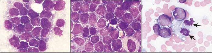
Diagnosing hematolymphoid neoplasm by evaluating fine-needle aspiration (FNA) cytology sample is controversial and requires experience and clinical skills. This concept becomes more challenging when evaluating hematolymphoid neoplasm in body fluid. Differentiating between low-grade lymphoma and reactive lymphocytes is often difficult by morphology alone as reactive lymphoid cells may acquire activation morphology from being exposed to different cytokines within the body fluid. However, in most cases there are specific features that may aid in differentiating small reactive from non-reactive lymphocytes including the round shape of the nucleus, the absence of visible nucleoli and the presence of fine clumped chromatin. In large cell lymphoma and leukemia cells involvement of body fluid this concept becomes less challenging. Large cell lymphoma and leukemia cells tend to have large size nuclei, less mature chromatin, and visible nucleoli with and without cytoplasmic vacuoles. However, to reach accurate diagnosis and subclassification, the utilizing of flow cytometry, to confirm monoclonality, and other ancillary studies such immunocytochemistry, cytogenetics and molecular studies is needed. This review article will be incorporated finally as one of the chapters in CMAS (CytoJournal Monograph/Atlas Series) #2. It is modified slightly from the chapter by the initial authors in the first edition of Diagnostic Cytopathology of Serous Fluids.

Keywords: Body Fluid; Large Cell; Leukemia; Lymphoma; Small Cell.


Figures

Figure 1:
Figure 1:
(a) Follicular lymphoma lymphocyte with characteristic cleaved or notched nucleus (arrow). (b) CLL cells in pleural fluid Cytospin preparation. (c) Bloody pleural fluid in a patient with CLL. Contamination with peripheral blood must be considered before diagnosing a patient as having pleural fluid involvement with CLL.
Figure 2:
Figure 2:
Burkitt lymphoma, ascitic fluid; monomorphous population of medium-sized cells with prominent cytoplasmic vacuolization. Note the mitoses in C (arrows). [DQ stain].
Figure 3:
Figure 3:
(a,b) DQ-stained cytology smears from ascitic fluid showing large atypical lymphoid cells later proved to be diffuse large B-cell lymphoma. (c,d) PAP-stained cytology smears showing same cells as in a and b. (e) Histology section from same patient with malignant effusion showing primary colonic diffuse large B-cell lymphoma.
Figure 4:
Figure 4:
HHV-8 positive primary effusion lymphoma (ascitic fluid). The specimen shows monomorphic, dispersed, medium to large cells with moderate amount of basophilic cytoplasm with scant cytoplasmic vacuoles. Nuclei are round to slightly irregular with fine chromatin and variable prominence of nucleoli. [Wright–Giemsa stain.]
Figure 5:
Figure 5:
(a) Chest radiograph showing cardiomegaly in a patient with severe dyspnea and massive malignant pericardial effusion. (b) Pericardial fluid cytology, in patient from (a), presenting with primary cardiac lymphoma and isolated massive pericardial effusion. This case turned out to be diffuse large B-cell lymphoma.
Figure 6:
Figure 6:
Neutrophils with intracellular bacteria (arrow) in neutrophilic peritonitis.
Figure 7:
Figure 7:
(a) PAP-stained cytology smear from ascitic fluid showing megakaryocyte (arrow). (b) Tissue section from subsequent biopsy showing megakaryocytes in extramedullary hematopoiesis. (c) Tissue section with factor VIII immunohistochemistry staining of megakaryocyte.
Figure 8:
Figure 8:
Clusters of blasts and immature myeloid cells with cytoplasmic granules (arrows) and irregular nuclei in myeloid sarcoma.

References

    1. DeMay RM. Chicago: ASCP Press; 1999. Practical Principles of Cytopathology.
    1. Suh YK, Shabaik A, Meurer WT, Shin SS. Lymphoid cell aggregates: a useful clue in the fine-needle aspiration diagnosis of follicular lymphomas. Diagn Cytopathol. 1997;17:467–71. doi: 10.1002/(SICI)1097-0339(199712)17:6<467::AID-DC17>3.0.CO;2-K. - DOI - PubMed
    1. Dunphy CH, Collins B, Ramos R, Grosso LE. Secondary pleural involvement by an AIDS-related anaplastic large cell (CD30+) lymphoma simulating metastatic adenocarcinoma. Diagn Cytopathol. 1998;18:113–7. doi: 10.1002/(SICI)1097-0339(199802)18:2<113::AID-DC6>3.0.CO;2-G. - DOI - PubMed
    1. Beaty MW, Fetsch P, Wilder AM, Marincola F, Abati A. Effusion cytology of malignant melanoma. A morphologic and immunocytochemical analysis including application of the MART-1 antibody. Cancer. 1997;81:57–63. doi: 10.1002/(SICI)1097-0142(19970225)81:1<57::AID-CNCR12>3.0.CO;2-B. - DOI - PubMed
    1. Hallman JR, Geisinger KR. Cytology of fluids from pleural, peritoneal and pericardial cavities in children. A comprehensive survey. Acta Cytol. 1994;38:209–17. - PubMed

LinkOut - more resources