

Ascending aorta thoracic endovascular aortic repair for infected pseudoaneurysm
- PMID: 35510219
- PMCID: PMC9058959
- DOI: 10.1016/j.jvscit.2022.02.005
Ascending aorta thoracic endovascular aortic repair for infected pseudoaneurysm
Abstract
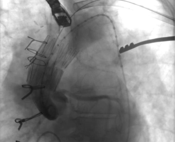
A 70-year-old woman with a bioprosthetic aortic valve replacement for aortic valve endocarditis complicated by recurrent endocarditis and requiring homograft aortic root replacement 10 years earlier had presented at 1 month after her admission for pseudomonal bacteremia with right-sided chest pain. An aortic pseudoaneurysm, identified on computed tomography, was treated with an ascending aorta thoracic endovascular aortic repair using two overlapping abdominal aortic stent grafts in the ascending aorta. Postoperative and follow-up imaging demonstrated exclusion of the pseudoaneurysm with stable positioning of the stent grafts. Ascending aorta thoracic endovascular aortic repair can be performed safely with good short-term results in patients presenting with infected pseudoaneurysms of the ascending aorta.
Keywords: Ascending aorta; Infected pseudoaneurysm; TEVAR.
© 2022 The Author(s).
Figures


Similar articles
-
Endovascular Repair for Ascending Aortic Graft Side Branch Pseudoaneurysm: A Report of Two Cases.EJVES Vasc Forum. 2022 Apr 13;55:48-51. doi: 10.1016/j.ejvsvf.2022.03.009. eCollection 2022. EJVES Vasc Forum. 2022. PMID: 35515008 Free PMC article.
-
Back-Table Modified Stent-Graft for Endovascular Repair of Ascending Aorta.J Endovasc Ther. 2021 Dec;28(6):888-896. doi: 10.1177/15266028211028201. Epub 2021 Jun 30. J Endovasc Ther. 2021. PMID: 34190632
-
Ascending Aorta Endovascular Repair of a Symptomatic Penetrating Atherosclerotic Ulcer with a Custom-Made Endograft.Ann Vasc Surg. 2018 Feb;47:280.e1-280.e4. doi: 10.1016/j.avsg.2017.08.027. Epub 2017 Sep 7. Ann Vasc Surg. 2018. PMID: 28890066
-
Thoracic endovascular aortic repair for the ascending aorta: experience and pitfalls.J Vis Surg. 2018 May 9;4:92. doi: 10.21037/jovs.2018.03.01. eCollection 2018. J Vis Surg. 2018. PMID: 29963381 Free PMC article. Review.
-
Update on Trials & Devices for Endovascular Management of the Ascending Aorta and Arch.Tech Vasc Interv Radiol. 2021 Jun;24(2):100756. doi: 10.1016/j.tvir.2021.100756. Epub 2021 Jul 26. Tech Vasc Interv Radiol. 2021. PMID: 34602266 Review.
Cited by
-
Endovascular repair of a ruptured ascending aortic pseudoaneurysm with concomitant pericardiocentesis.J Vasc Surg Cases Innov Tech. 2025 Mar 5;11(3):101775. doi: 10.1016/j.jvscit.2025.101775. eCollection 2025 Jun. J Vasc Surg Cases Innov Tech. 2025. PMID: 40230833 Free PMC article.
-
Minimally Invasive Endovascular Repair for Nondissected Ascending Aortic Disease: A Systematic Review.Emerg Med Int. 2023 Sep 19;2023:5592622. doi: 10.1155/2023/5592622. eCollection 2023. Emerg Med Int. 2023. PMID: 37767197 Free PMC article. Review.
-
Endovascular repair of ascending aortic pathologies in patients unfit for open surgery: case series and literature review.J Vasc Surg Cases Innov Tech. 2024 Feb 14;10(3):101455. doi: 10.1016/j.jvscit.2024.101455. eCollection 2024 Jun. J Vasc Surg Cases Innov Tech. 2024. PMID: 38510094 Free PMC article.
-
Staged Hybrid Management of a Mycotic Ascending Aortic Pseudoaneurysm.Ann Thorac Surg Short Rep. 2024 Jun 13;2(4):725-728. doi: 10.1016/j.atssr.2024.05.021. eCollection 2024 Dec. Ann Thorac Surg Short Rep. 2024. PMID: 39790573 Free PMC article.
References
-
- Oderich G.S., Panneton J.M., Bower T.C., Cherry K.J., Rowland C.M., Noel A.A., et al. Infected aortic aneurysms: aggressive presentation, complicated early outcome, but durable results. J Vasc Surg. 2001;34:900–908. - PubMed
-
- Haidar G.M., Hicks T.D., Strosberg D.S., El-Sayed H.F., Davies M.G. “In situ” endografting in the treatment of arterial and graft infections. J Vasc Surg. 2017;65:1824–1829. - PubMed
Publication types
LinkOut - more resources
Full Text Sources
