

The diagnostic utility of unenhanced computed tomography of the brain and D-dimer levels in acute cerebral venous sinus thrombosis: A quantitative study
- PMID: 35510244
- PMCID: PMC9062933
- DOI: 10.25259/JCIS_76_2021
The diagnostic utility of unenhanced computed tomography of the brain and D-dimer levels in acute cerebral venous sinus thrombosis: A quantitative study
Abstract
Objectives: (1) To calculate the sensitivity and specificity of the Hounsfield Unit (HU), the HU to hematocrit (H:H) ratio, and the D-dimer level in the diagnosis of acute CVST. (2) To assess the D-dimer level's linear relationship with the HU and the H:H ratio.
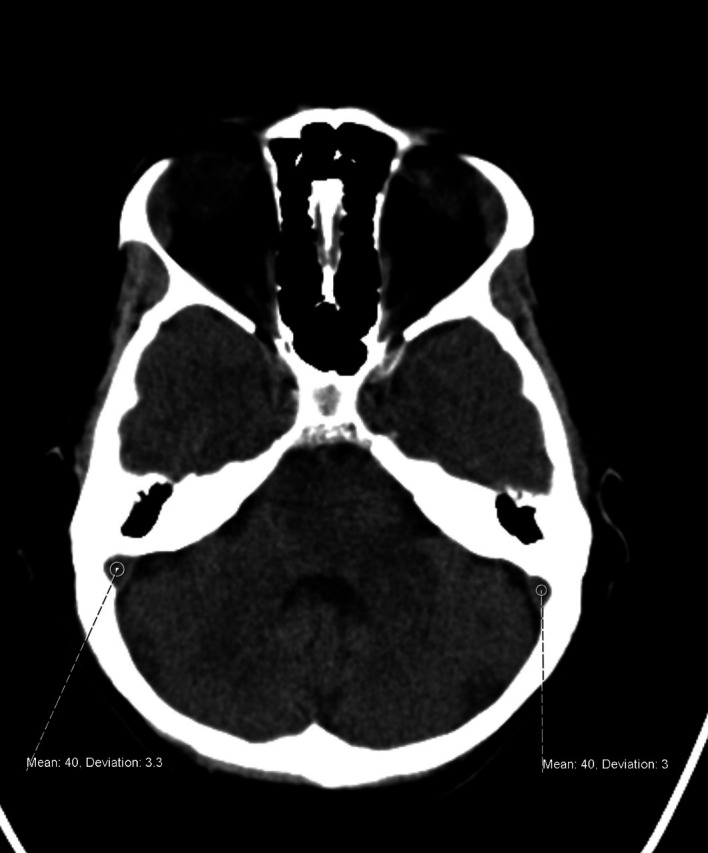
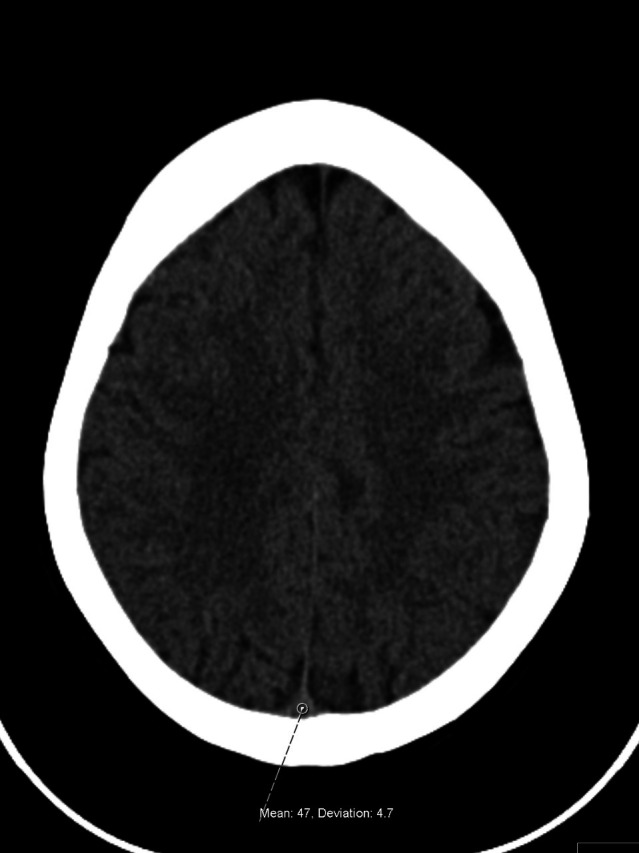
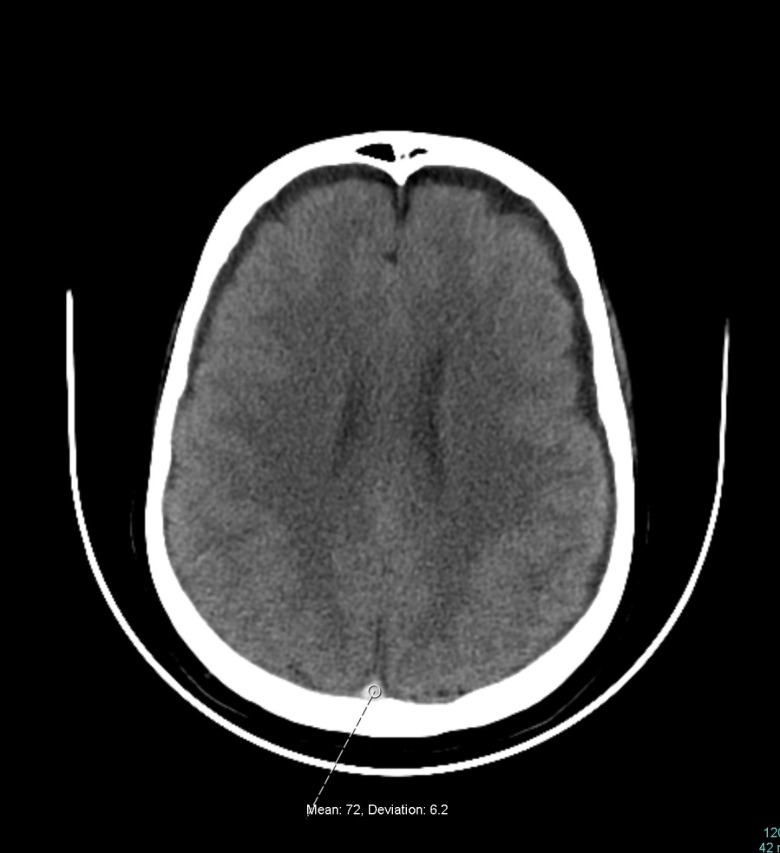
Materials and methods: A single-center retrospective case-control study was conducted from 2005 to 2020. The inclusion criteria for the thrombosed and control groups were specified. A region of interest (ROI) was plotted on the respective sinuses to calculate the HU. The H:H ratio was calculated by dividing the HU value by the hematocrit value. The receiver operating characteristic curve was used to calculate the sensitivity and specificity of the HU and the H:H ratio at different cutoff values. The Pearson correlation was used to assess the linear relationship between the D-dimer level and the HU and H:H ratio.
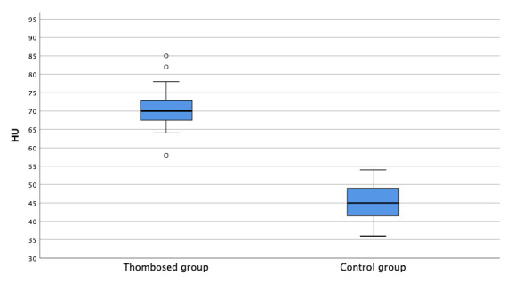
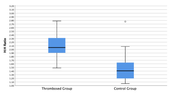
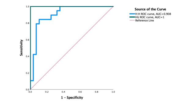
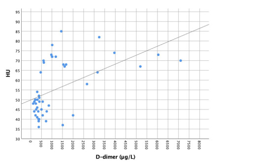
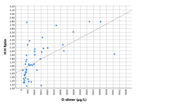
Results: There were 19 patients in the thrombosed group and 28 patients in the control group. There were significant differences in the mean HU (71 ± 6.3 vs. 45 ± 4.8, P < 0.001) and the mean H:H ratio (2.11 ± 0.38 vs. 1.46 ± 0.63,P < 0.001). An optimal HU value of 56 yielded 100% sensitivity and specificity. An H:H value of 1.48 yielded a sensitivity of 100% and a specificity of 65%, an H:H ratio of 1.77 demonstrated a sensitivity of 85% and a specificity of 90%, and an H:H ratio of 1.88 yielded a sensitivity of 79% and a specificity of 93%. D-dimer levels had a 95% and 71% sensitivity and specificity, respectively. There was a significant moderately positive linear correlation between the D-dimer level and the HU (r = 0.52, P < 0.001) and the H:H ratio (r = 0.61, P < 0.001).
Conclusion: Unenhanced CT of the brain can be a valuable objective diagnostic tool for acute CVST diagnosis. Hounsfield blood density and its normalized ratio with hematocrit are positively correlated with D-dimer levels, which may indicate active blood coagulation in a cerebral venous sinus.
Keywords: Acute cerebral venous sinus thrombosis; D-dimer; Hounsfield unit; Hounsfield:Hematocrit ratio.
© 2022 Published by Scientific Scholar on behalf of Journal of Clinical Imaging Science.
Figures
Similar articles
-
Utility of Hounsfield unit and hematocrit values in the diagnosis of acute venous sinus thrombosis in unenhanced brain CTs in the pediatric population.Pediatr Radiol. 2019 Feb;49(2):234-239. doi: 10.1007/s00247-018-4273-y. Epub 2018 Oct 16. Pediatr Radiol. 2019. PMID: 30327829
-
Diagnostic Accuracy of Hounsfield Unit Value and Hounsfield Unit to Hematocrit Ratio in Predicting Cerebral Venous Sinus Thrombosis: A Retrospective Case-Control Study.Cureus. 2024 Apr 3;16(4):e57567. doi: 10.7759/cureus.57567. eCollection 2024 Apr. Cureus. 2024. PMID: 38707168 Free PMC article.
-
Are cerebral veins hounsfield unit and H: H ratio calculating in unenhanced CT eligible to diagnosis of acute cerebral vein thrombosis?J Res Med Sci. 2019 Sep 30;24:83. doi: 10.4103/jrms.JRMS_1027_18. eCollection 2019. J Res Med Sci. 2019. PMID: 31620182 Free PMC article.
-
Determining the Diagnostic Value of Venous Sinus Density Indices in Non-Contrast Brain CT Scan for Early Diagnosis of Cerebral Venous Sinus Thrombosis.Brain Behav. 2025 Feb;15(2):e70324. doi: 10.1002/brb3.70324. Brain Behav. 2025. PMID: 39935195 Free PMC article.
-
Diagnostic Value of Hounsfield Unit and Hematocrit Levels in Cerebral Vein Thrombosis in the Emergency Department.J Emerg Med. 2021 Sep;61(3):234-240. doi: 10.1016/j.jemermed.2021.07.016. Epub 2021 Aug 21. J Emerg Med. 2021. PMID: 34429219
Cited by
-
The accuracy of the Hounsfield unit in pulmonary embolism diagnostics.Clin Exp Emerg Med. 2024 Sep;11(3):295-303. doi: 10.15441/ceem.23.113. Epub 2024 Jan 29. Clin Exp Emerg Med. 2024. PMID: 38286507 Free PMC article.
-
Analysis of the Risk Factors for Elevated D-Dimer Level After Breast Cancer Surgery: A Multicenter Study Based on Nursing Follow-Up Data.Front Oncol. 2022 Jul 19;12:772726. doi: 10.3389/fonc.2022.772726. eCollection 2022. Front Oncol. 2022. PMID: 35928882 Free PMC article.
References
-
- Ferro JM, Canhao P, Stam J, Bousser MG, Barinagarrementeria F, ISCVT Investigators. Prognosis of cerebral vein and dural sinus thrombosis: Results of the International Study on Cerebral Vein and Dural Sinus Thrombosis (ISCVT) Stroke. 2004;35:664–70. doi: 10.1161/01.STR.0000117571.76197.26. - DOI - PubMed
LinkOut - more resources
Full Text Sources