

Verification of surface electromyographic activity of the oblique externus abdominis using ultrasound shear wave elastography
- PMID: 35510415
- PMCID: PMC9069374
- DOI: 10.14814/phy2.15295
Verification of surface electromyographic activity of the oblique externus abdominis using ultrasound shear wave elastography
Abstract
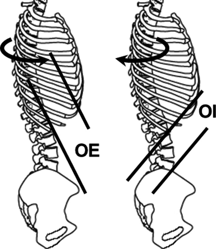
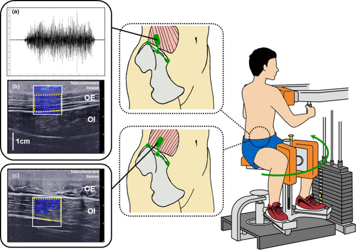
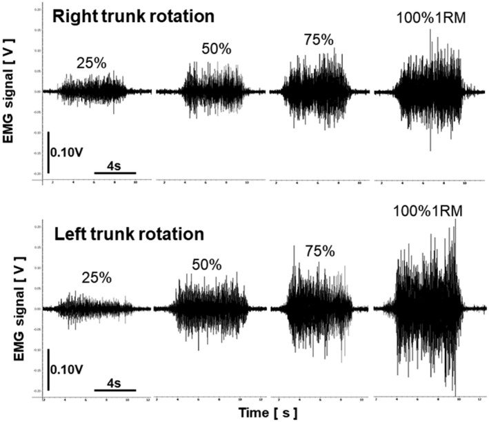
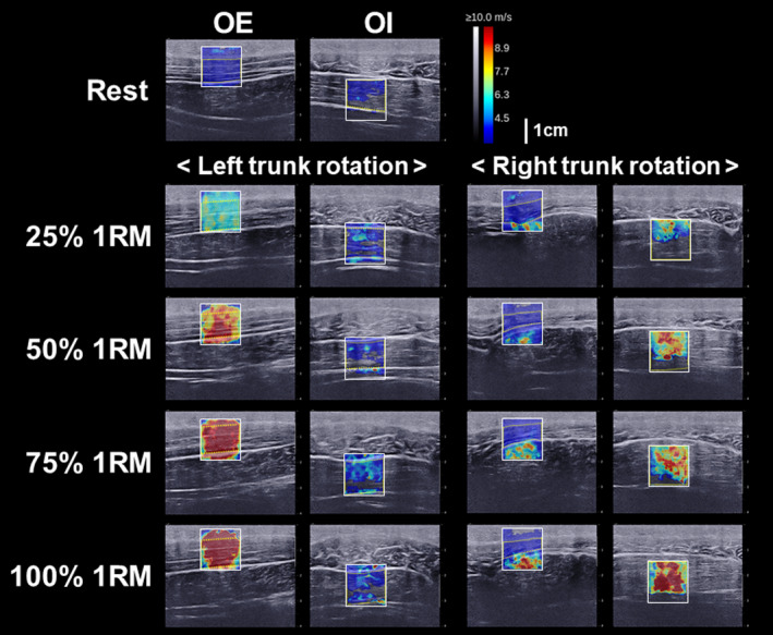
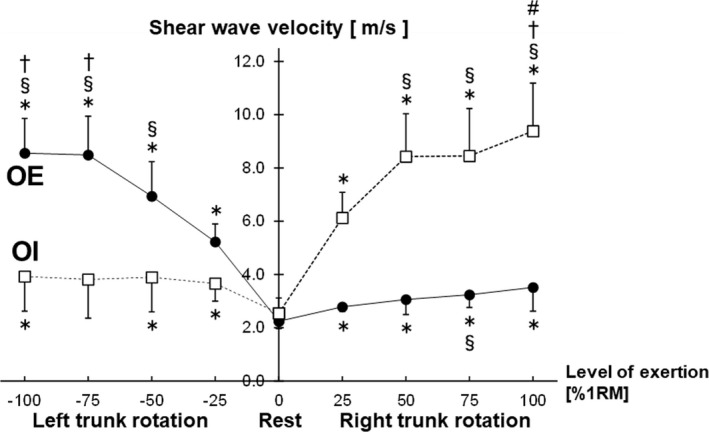
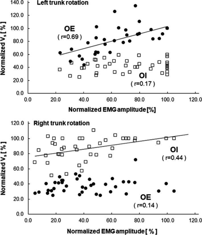
This study used ultrasound shear wave elastography (SWE) to revalidate whether surface electromyographic (EMG) electrodes placed on the oblique externus abdominis (OE) can detect only the OE activity without the confounding activity of the underlying oblique internus abdominis (OI). During left and right trunk rotations, the EMG activity was acquired using surface EMG electrodes placed on the right OE. Shear wave velocity (Vs ) values of the right OE and OI were acquired using SWE. The EMG activity during the left and right trunk rotations significantly increased as the level of exertion increased (25%, 50%, 75%, and 100% of the one-repetition maximum [1RM]). The Vs of the right OE was significantly different only between 25% and 75% 1RM in the right trunk rotation, but significantly increased from 25% to 75% 1RM during the left trunk rotation. The Vs of the right OI during the right trunk rotation significantly increased with increased levels of exertion, except between 50% and 75% 1RM. The results for the Vs of the OE and OI in the right trunk rotation suggest that surface EMG electrodes placed on the OE would detect not only the antagonistic OE activity but also the agonistic OI activity.
Keywords: abdominal muscles; oblique internus abdominis; signal crosstalk; surface electromyography; trunk rotation.
© 2022 The Authors. Physiological Reports published by Wiley Periodicals LLC on behalf of The Physiological Society and the American Physiological Society.
Conflict of interest statement
The authors declare that they have no conflicts of interest associated with this manuscript.
Figures

Similar articles
-
Association between expiratory mouth pressure and abdominal muscle activity in healthy young males.Eur J Appl Physiol. 2024 Jul;124(7):2139-2151. doi: 10.1007/s00421-024-05430-5. Epub 2024 Mar 1. Eur J Appl Physiol. 2024. PMID: 38427101
-
Differential activity of regions of transversus abdominis during trunk rotation.Eur Spine J. 2005 May;14(4):393-400. doi: 10.1007/s00586-004-0799-9. Epub 2004 Nov 30. Eur Spine J. 2005. PMID: 15940481 Free PMC article.
-
Abdominal Muscle Activation During Common Modifications of the Trunk Curl-up Exercise.J Strength Cond Res. 2021 Feb 1;35(2):428-435. doi: 10.1519/JSC.0000000000002439. J Strength Cond Res. 2021. PMID: 29319600
-
Application of Ultrasonography in the Assessment of Abdominal and Lumbar Trunk Muscle Activity in Participants With and Without Low Back Pain: A Systematic Review.J Manipulative Physiol Ther. 2019 Sep;42(7):541-550. doi: 10.1016/j.jmpt.2019.05.003. J Manipulative Physiol Ther. 2019. PMID: 31864437
-
Electromyographic Analyses of Trunk Musculature after Stroke: An Integrative Review.Top Stroke Rehabil. 2022 Jul;29(5):366-381. doi: 10.1080/10749357.2021.1940725. Epub 2021 Jul 18. Top Stroke Rehabil. 2022. PMID: 34275435 Review.
Cited by
-
Association between expiratory mouth pressure and abdominal muscle activity in healthy young males.Eur J Appl Physiol. 2024 Jul;124(7):2139-2151. doi: 10.1007/s00421-024-05430-5. Epub 2024 Mar 1. Eur J Appl Physiol. 2024. PMID: 38427101
References
-
- Andonian, P. , Viallon, M. , Le Goff, C. , de Bourguignon, C. , Tourel, C. , Morel, J. , Giardini, G. , Gergelé, L. , Millet, G. P. , & Croisille, P. (2016). Shear‐wave elastography assessments of quadriceps stiffness changes prior to, during and after prolonged exercise: A longitudinal study during an extreme mountain ultra‐marathon. PLoS One, 11(8), e0161855. 10.1371/journal.pone.0161855 - DOI - PMC - PubMed
Publication types
MeSH terms
LinkOut - more resources
Full Text Sources
