

Novel application of absorbable gelatine sponge combined with polyurethane film for dermal reconstruction of wounds with bone or tendon exposure
- PMID: 35510525
- PMCID: PMC9797930
- DOI: 10.1111/iwj.13832
Novel application of absorbable gelatine sponge combined with polyurethane film for dermal reconstruction of wounds with bone or tendon exposure
Abstract
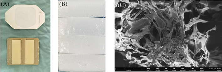
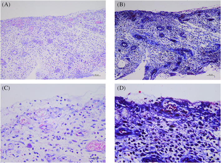
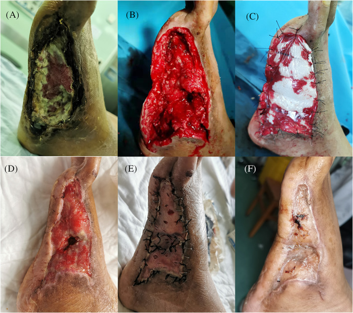
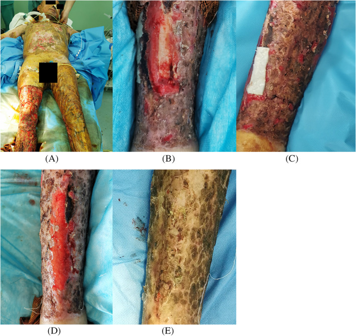
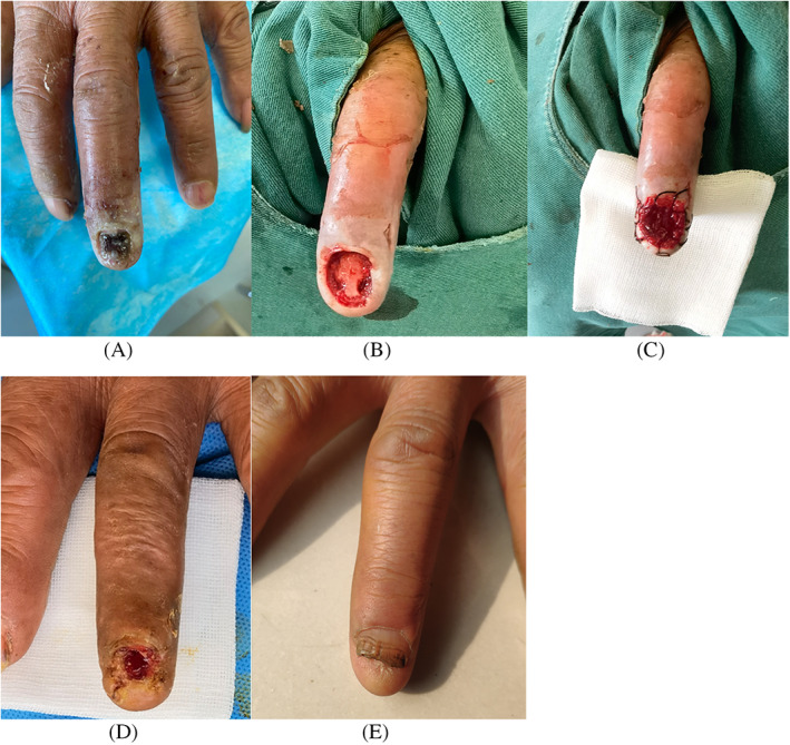
Trauma, burns, and diabetes result in nonhealing wounds that can cause bone or tendon exposure, a significant health threat. The use of an artificial regeneration template combined with skin grafting as an alternative method to highly invasive flap surgery has been shown to be an effective way to cover full-thickness skin defects with bone or tendon exposure for both functional and aesthetic recovery. However, artificial regeneration templates, such as Pelnac, are overwhelmingly expensive, limiting their clinical use. Here, we demonstrate for the first time that polyurethane film combined with absorbable gelatine sponge, affordable materials widely used for haemostasis, are effective for dermal reconstruction in wounds with bone or tendon exposure. The absorbable gelatine sponge combined with polyurethane film was applied to eight patients, all resulting in adequate granulation that fully covered the exposed bone or tendon. The outcome of absorbable gelatine sponge combined with polyurethane film application indicates that this approach is a potential novel and cost-effective dermal reconstruction strategy for the treatment of severe wounds with bone or tendon exposure.
Keywords: absorbable gelatine sponge; artificial dermis; full-thickness skin defect; polyurethane film; wound healing.
© 2022 The Authors. International Wound Journal published by Medicalhelplines.com Inc (3M) and John Wiley & Sons Ltd.
Conflict of interest statement
All authors hereby declare that they have no conflicts of interest.
Figures
Similar articles
-
Negative-pressure wound therapy combined with artificial dermis (Terudermis) followed by split-thickness skin graft might be an effective treatment option for wounds exposing tendon and bone: A retrospective observation study.Medicine (Baltimore). 2021 Apr 9;100(14):e25395. doi: 10.1097/MD.0000000000025395. Medicine (Baltimore). 2021. PMID: 33832132 Free PMC article.
-
[Clinical effect of bi-layered artificial dermis and autologous skin graft in repairing bone and/or tendon exposed wounds].Zhonghua Shao Shang Za Zhi. 2020 Mar 20;36(3):179-186. doi: 10.3760/cma.j.cn501120-20191119-00437. Zhonghua Shao Shang Za Zhi. 2020. PMID: 32241043 Chinese.
-
Exposed tibial bone after burns: Flap reconstruction versus dermal substitute.Burns. 2016 Mar;42(2):e31-7. doi: 10.1016/j.burns.2015.08.013. Epub 2015 Sep 12. Burns. 2016. PMID: 26376411
-
Novosorb® Biodegradable Temporising Matrix (BTM) and its Applications.Surg Technol Int. 2023 Sep 15;42:47-52. Surg Technol Int. 2023. PMID: 37053370 Review.
-
Systematic Review and Meta-analysis of Biodegradable Temporizing Matrix Application for Complex Wound Reconstruction.J Burn Care Res. 2025 Jan 24;46(1):82-89. doi: 10.1093/jbcr/irae081. J Burn Care Res. 2025. PMID: 38733573
Cited by
-
An Advanced Review: Polyurethane-Related Dressings for Skin Wound Repair.Polymers (Basel). 2023 Nov 1;15(21):4301. doi: 10.3390/polym15214301. Polymers (Basel). 2023. PMID: 37959982 Free PMC article. Review.
-
Human Placenta-Derived Mesenchymal Stem Cells Combined With Artificial Dermal Scaffold Enhance Wound Healing in a Tendon-Exposed Wound of a Rabbit Model.Cell Transplant. 2024 Jan-Dec;33:9636897241228922. doi: 10.1177/09636897241228922. Cell Transplant. 2024. PMID: 38334047 Free PMC article.
References
-
- Harrison T, Kindred J, Marks‐Maran D. Reducing avoidable harm caused by pressure ulcers. Br J Nurs. 2013;22(6):S4 S6, S8 passim. - PubMed
-
- Yeong EK, Yu YC, Chan ZH, Roan TL. Is artificial dermis an effective tool in the treatment of tendon‐exposed wounds? J Burn Care Res. 2013;34(1):161‐167. - PubMed
-
- Soejima K, Nozaki M, Sasaki K, Takeuchi M, Negishi N. Reconstruction of burn deformity using artificial dermis combined with thin split‐skin grafting. Burns. 1997;23(6):501‐504. - PubMed
MeSH terms
Substances
Grants and funding
LinkOut - more resources
Full Text Sources
